Foreign object segmentation in chest x-rays through anatomy-guided shape insertion
Publication
Metrics
AI Quick Summary
This paper proposes a method for instance segmentation of foreign objects in chest X-rays using synthetic data generated via anatomy-guided shape insertions and augmentations, achieving performance comparable to fully supervised models with significantly fewer manual annotations.
Paper Preview
Abstract
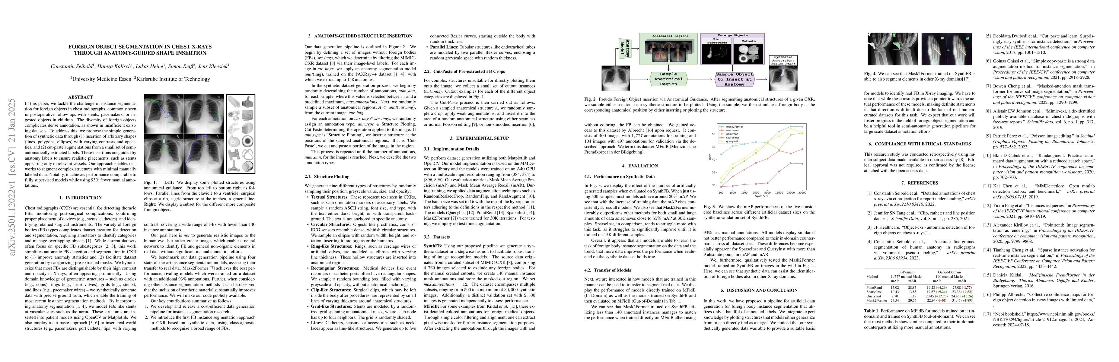
In this paper, we tackle the challenge of instance segmentation for foreign objects in chest radiographs, commonly seen in postoperative follow-ups with stents, pacemakers, or ingested objects in children. The diversity of foreign objects complicates dense annotation, as shown in insufficient existing datasets. To address this, we propose the simple generation of synthetic data through (1) insertion of arbitrary shapes (lines, polygons, ellipses) with varying contrasts and opacities, and (2) cut-paste augmentations from a small set of semi-automatically extracted labels. These insertions are guided by anatomy labels to ensure realistic placements, such as stents appearing only in relevant vessels. Our approach enables networks to segment complex structures with minimal manually labeled data. Notably, it achieves performance comparable to fully supervised models while using 93\% fewer manual annotations.
AI Key Findings
Get AI-generated insights about this paper's methodology, results, significance, and more — seven facets brought into focus.
Authors
PDF Preview
Related Papers
No references found for this paper.
Discussion 0