Academic Profile
Statistics
Similar Authors
Papers on arXiv
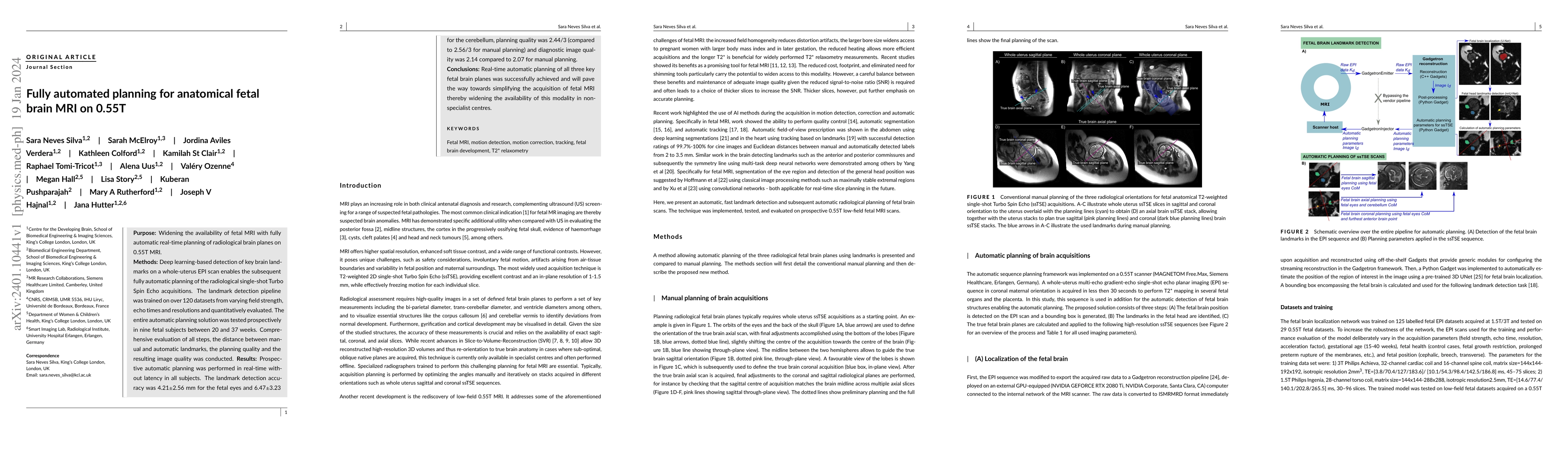
Purpose: Widening the availability of fetal MRI with fully automatic real-time planning of radiological brain planes on 0.55T MRI. Methods: Deep learning-based detection of key brain landmarks on a ...
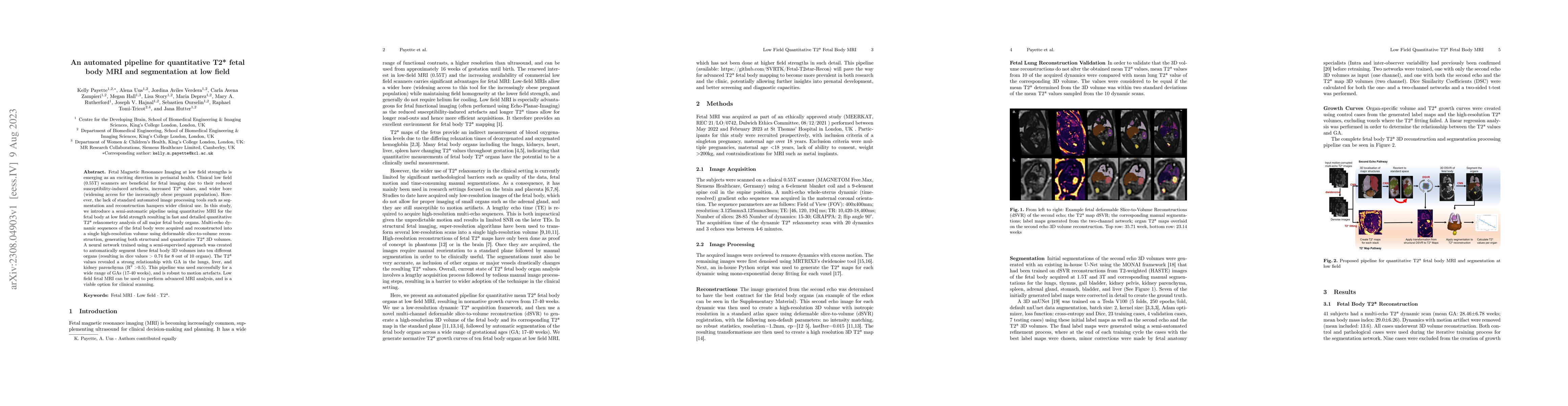
Fetal Magnetic Resonance Imaging at low field strengths is emerging as an exciting direction in perinatal health. Clinical low field (0.55T) scanners are beneficial for fetal imaging due to their re...
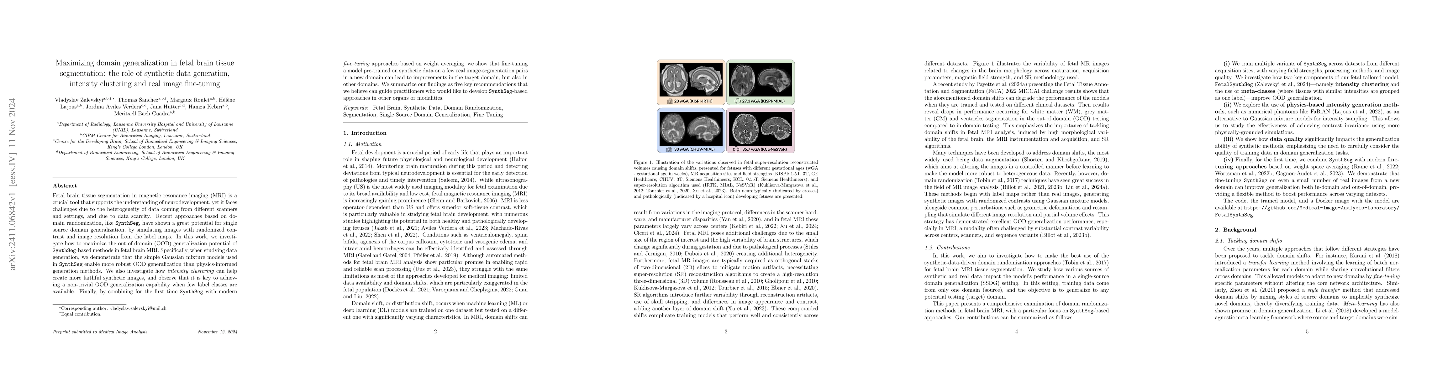
Fetal brain tissue segmentation in magnetic resonance imaging (MRI) is a crucial tool that supports the understanding of neurodevelopment, yet it faces challenges due to the heterogeneity of data comi...
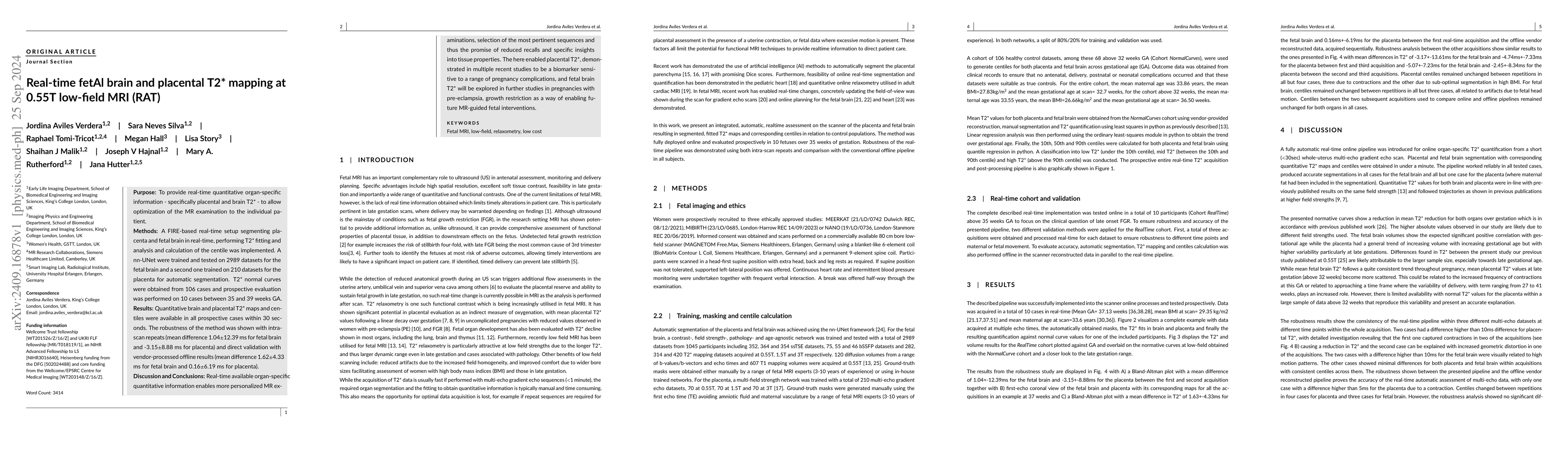
Purpose: To provide real-time quantitative organ-specific information - specifically placental and brain T2* - to allow optimization of the MR examination to the individual patient. Methods: A FIRE-...
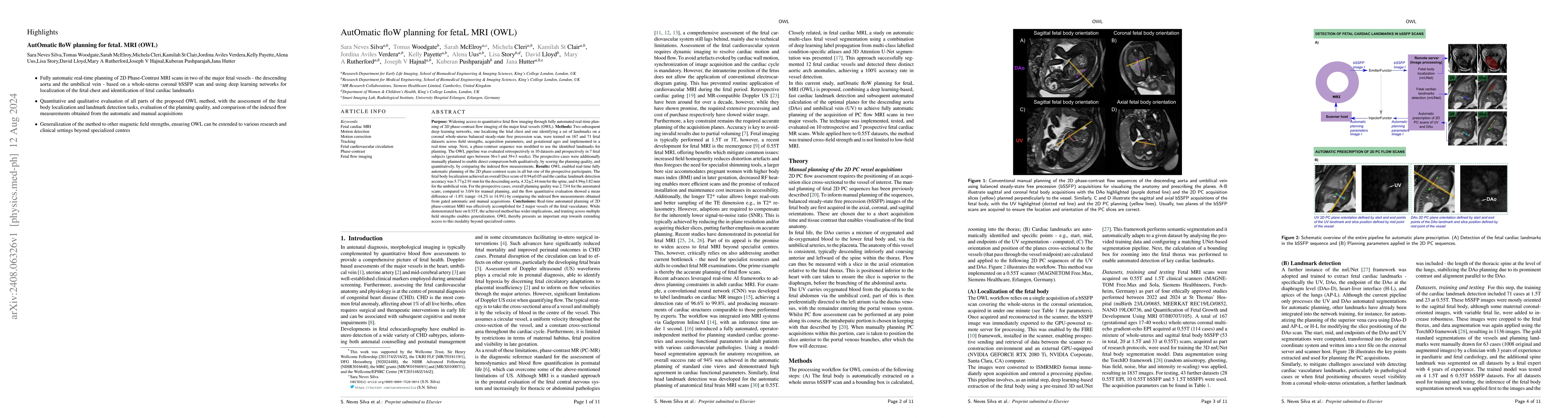
Two subsequent deep learning networks, one localizing the fetal chest and one identifying a set of landmarks on a coronal whole-uterus balanced steady-state free precession scan, were trained on 167 a...
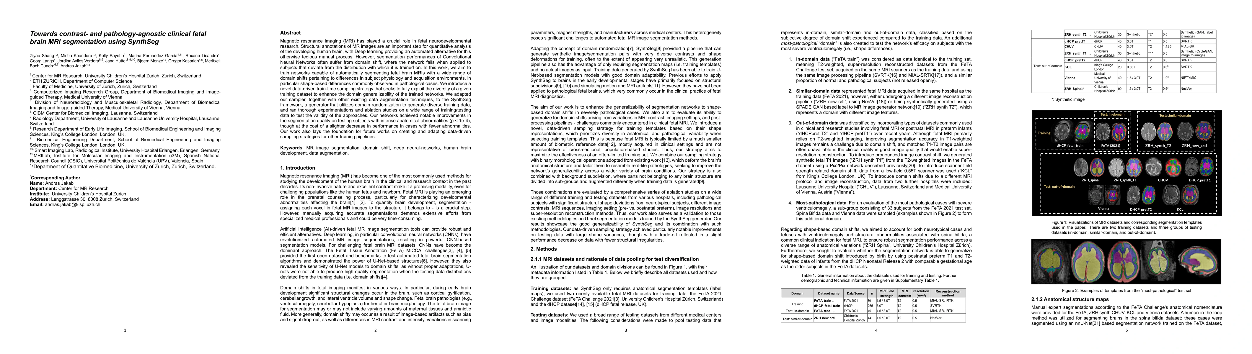
Magnetic resonance imaging (MRI) has played a crucial role in fetal neurodevelopmental research. Structural annotations of MR images are an important step for quantitative analysis of the developing h...
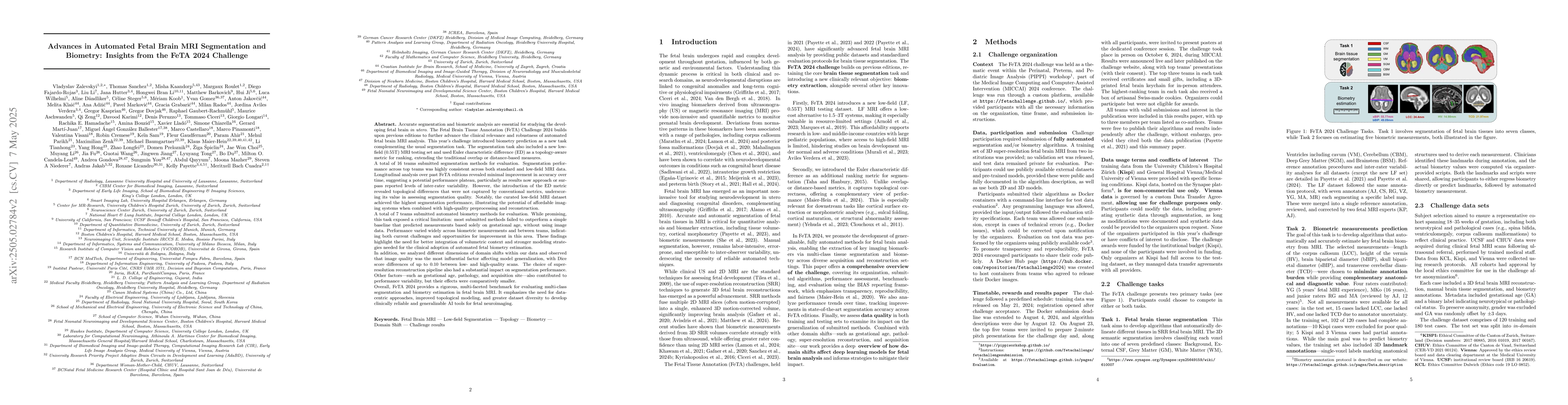
Accurate fetal brain tissue segmentation and biometric analysis are essential for studying brain development in utero. The FeTA Challenge 2024 advanced automated fetal brain MRI analysis by introducin...
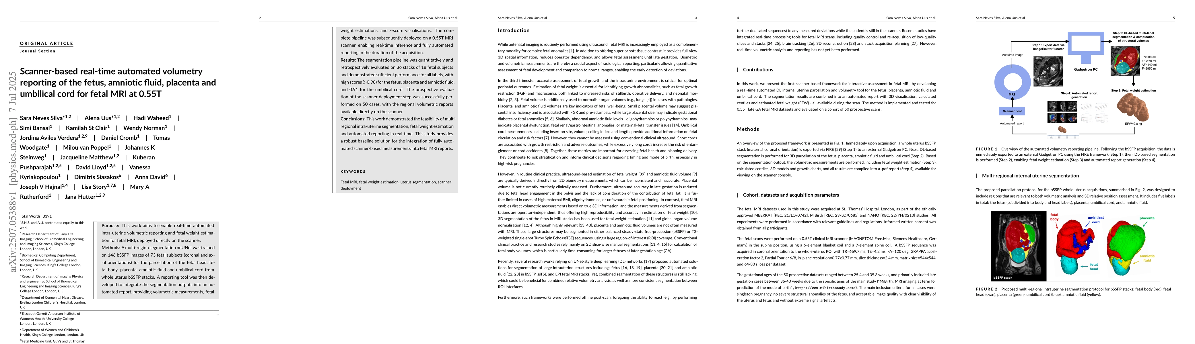
Purpose: This work aims to enable real-time automated intra-uterine volumetric reporting and fetal weight estimation for fetal MRI, deployed directly on the scanner. Methods: A multi-region segmentati...
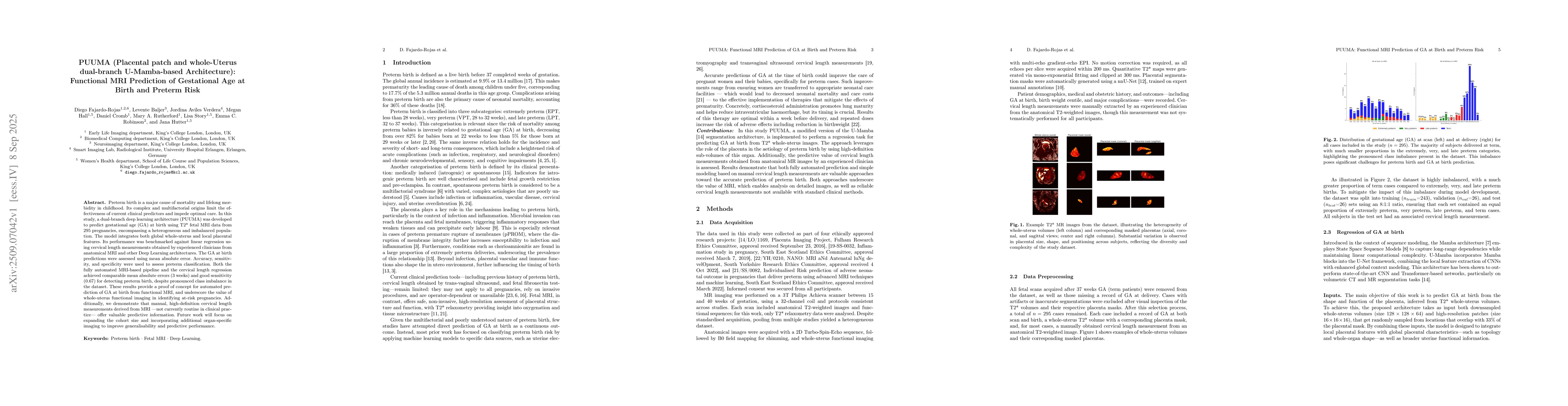
Preterm birth is a major cause of mortality and lifelong morbidity in childhood. Its complex and multifactorial origins limit the effectiveness of current clinical predictors and impede optimal care. ...
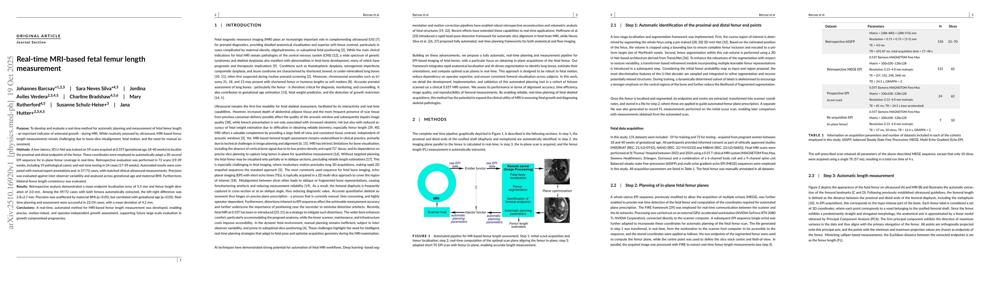
Purpose: To develop and evaluate a real-time method for automatic planning and measurement of fetal femur length - an important indicator of antenatal growth - during MRI. While routinely assessed by ...
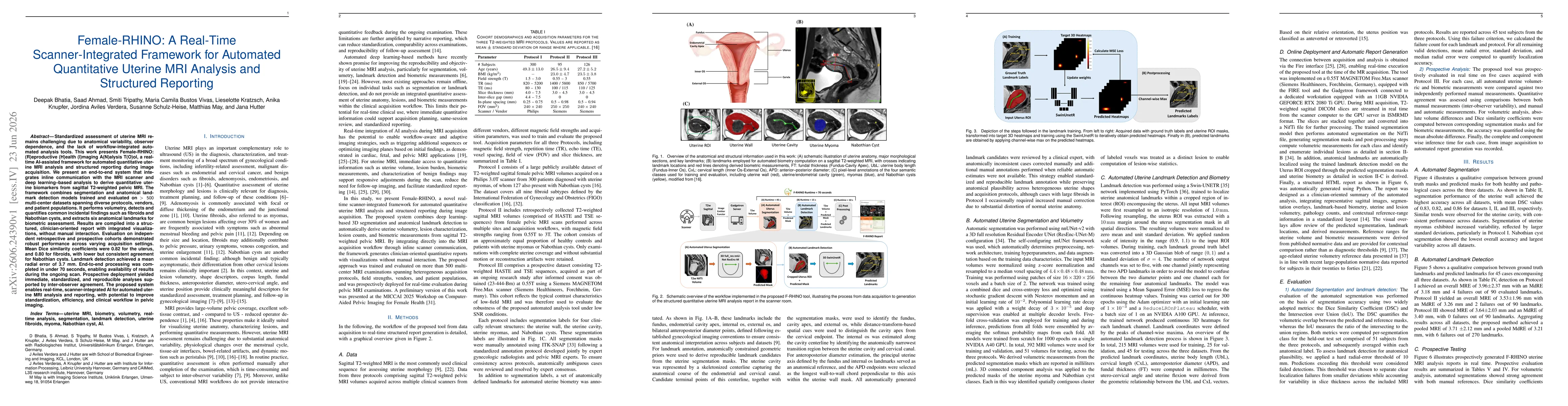
Standardized assessment of uterine MRI remains challenging due to anatomical variability, observer dependence, and the lack of workflow-integrated automated analysis tools. This work presents Female-R...