Summary
Purpose: This work aims to enable real-time automated intra-uterine volumetric reporting and fetal weight estimation for fetal MRI, deployed directly on the scanner. Methods: A multi-region segmentation nnUNet was trained on 146 bSSFP images of 73 fetal subjects (coronal and axial orientations) for the parcellation of the fetal head, fetal body, placenta, amniotic fluid and umbilical cord from whole uterus bSSFP stacks. A reporting tool was then developed to integrate the segmentation outputs into an automated report, providing volumetric measurements, fetal weight estimations, and z-score visualisations. The complete pipeline was subsequently deployed on a 0.55T MRI scanner, enabling real-time inference and fully automated reporting in the duration of the acquisition. Results: The segmentation pipeline was quantitatively and retrospectively evaluated on 36 stacks of 18 fetal subjects and demonstrated sufficient performance for all labels, with high scores (>0.98) for the fetus, placenta and amniotic fluid, and 0.91 for the umbilical cord. The prospective evaluation of the scanner deployment step was successfully performed on 50 cases, with the regional volumetric reports available directly on the scanner. Conclusions: This work demonstrated the feasibility of multi-regional intra-uterine segmentation, fetal weight estimation and automated reporting in real-time. This study provides a robust baseline solution for the integration of fully automated scanner-based measurements into fetal MRI reports.
AI Key Findings
Generated Sep 03, 2025
Methodology
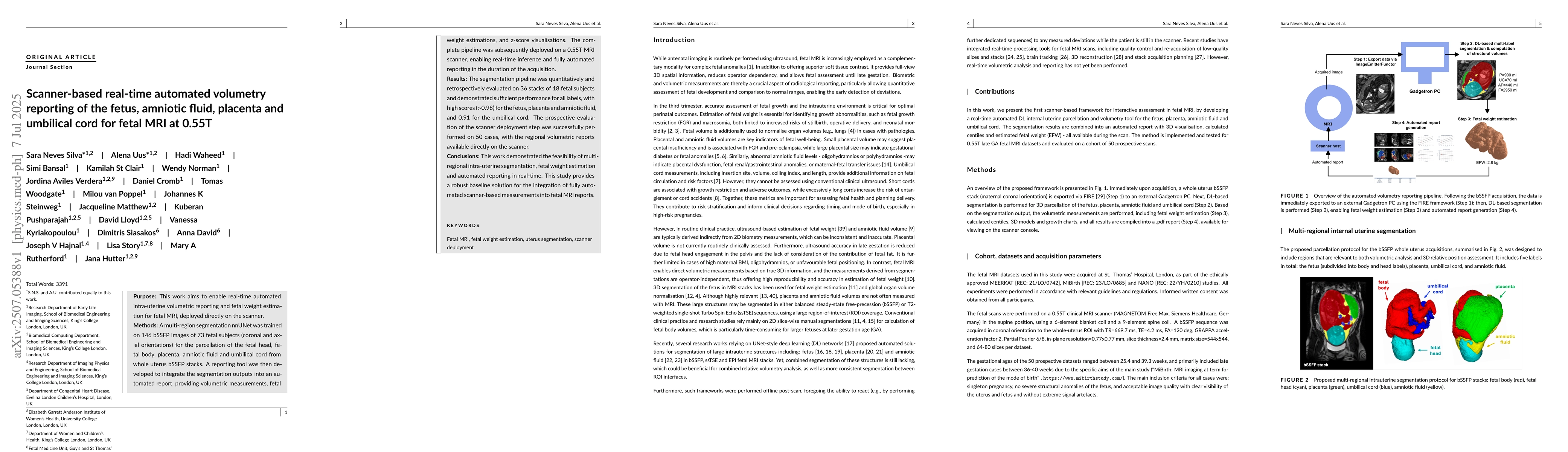
This study developed a multi-region segmentation nnUNet trained on 146 bSSFP images from 73 fetal subjects for parcellation of fetal head, body, placenta, amniotic fluid, and umbilical cord from whole uterus bSSFP stacks. A reporting tool was then integrated to provide volumetric measurements, fetal weight estimations, and z-score visualizations, deployed on a 0.55T MRI scanner for real-time inference and automated reporting.
Key Results
- The segmentation pipeline demonstrated high performance (>0.98) for the fetus, placenta, and amniotic fluid, and 0.91 for the umbilical cord.
- Prospective evaluation of scanner deployment was successfully performed on 50 cases, with regional volumetric reports available directly on the scanner.
- Fetal weight estimation was computed from total fetal label volume using classical formulas derived by Baker et al. and Kacem et al.
Significance
This research provides a robust baseline solution for integrating fully automated scanner-based measurements into fetal MRI reports, potentially aiding in early identification of potential anomalies and supporting clinical decision-making.
Technical Contribution
The development of a real-time, automated intra-uterine volumetry reporting pipeline for fetal MRI, directly integrated into the scanner environment using the FIRE framework.
Novelty
This is the first pipeline to combine segmentation of the fetus, placenta, umbilical cord, and amniotic fluid in one network, along with corresponding normative ranges specific to the late GA range and 0.55T fetal MRI.
Limitations
- The testing and training cohort were predominantly from the late GA range, singleton pregnancies, normal fetal anatomy, and from the same 0.55T acquisition protocol.
- Further retraining of the model on early GA datasets, fetal structural anomalies, and higher-field strength protocols is required for translation to clinical practice.
- Generation of normative growth models for the whole duration of the second and third trimesters is also needed.
Future Work
- Extending the GA range, optimizing the pipeline for 1.5T and 3T, and further sub-parcellating fetal brain and body ROIs.
- Integration of automated image quality control and image restoration measures to correct motion and intensity artifacts.
Paper Details
PDF Preview
Citation Network
Current paper (gray), citations (green), references (blue)
Display is limited for performance on very large graphs.
Similar Papers
Found 4 papersFully automated planning for anatomical fetal brain MRI on 0.55T
Sarah McElroy, Raphael Tomi-Tricot, Joseph V Hajnal et al.
Real-time fetAl brain and placental T2* mapping at 0.55T low-field MRI (RAT)
Raphael Tomi-Tricot, Joseph V Hajnal, Shaihan J Malik et al.
No citations found for this paper.
Comments (0)