Academic Profile
Statistics
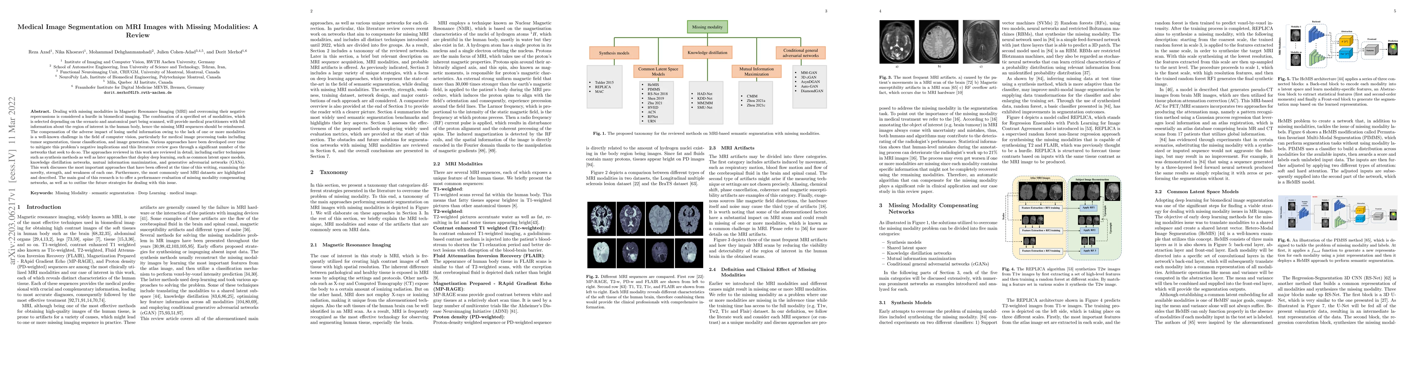
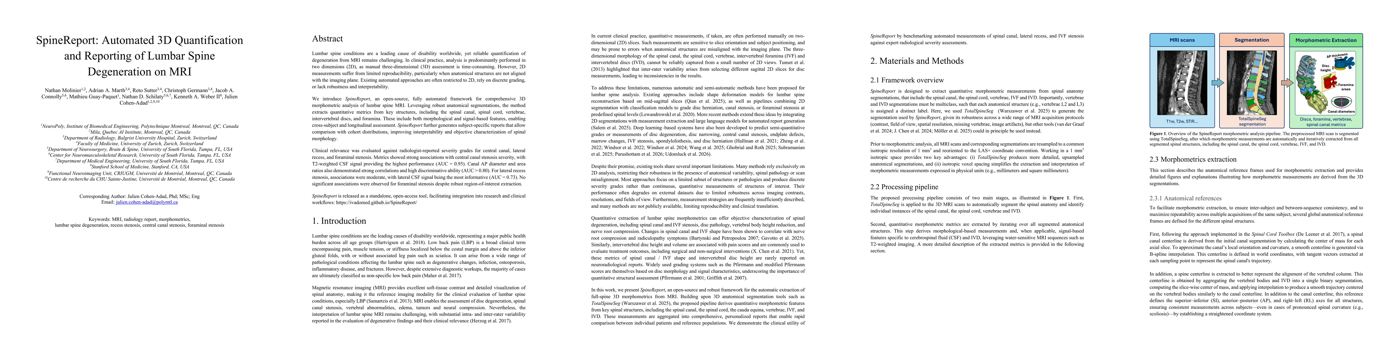
Similar Authors
Papers on arXiv
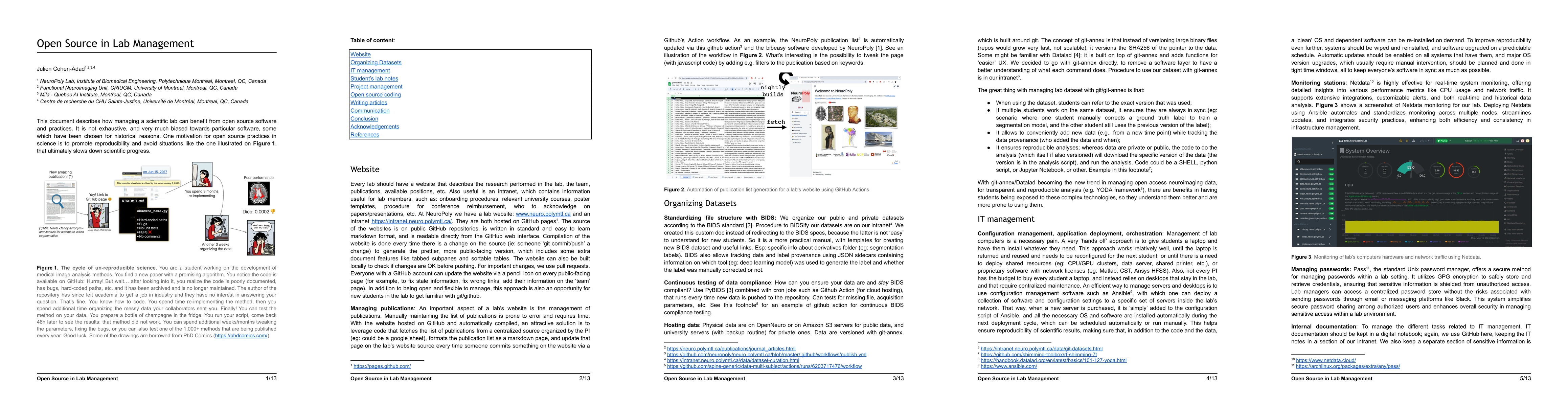
This document explores the advantages of integrating open source software and practices in managing a scientific lab, emphasizing reproducibility and the avoidance of pitfalls. It details practical ...
Precise identification of spinal nerve rootlets is relevant to delineate spinal levels for the study of functional activity in the spinal cord. The goal of this study was to develop an automatic met...
BACKGROUND: Functional Magnetic Resonance Imaging (fMRI) is based on the Blood Oxygenation Level Dependent contrast and has been exploited for the indirect study of the neuronal activity within both...
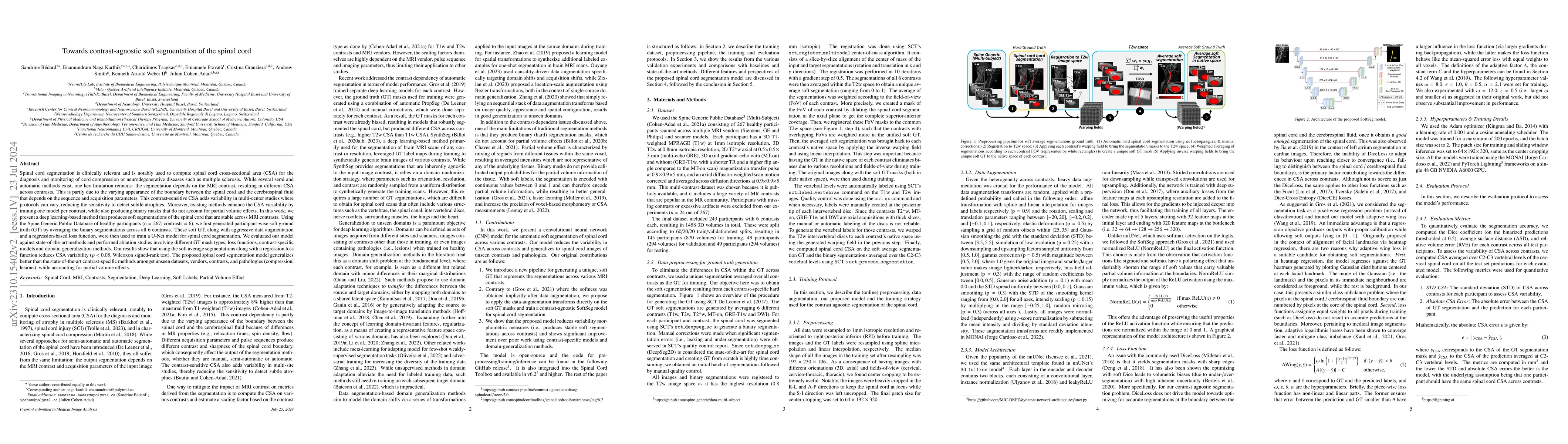
Spinal cord segmentation is clinically relevant and is notably used to compute spinal cord cross-sectional area (CSA) for the diagnosis and monitoring of cord compression or neurodegenerative diseas...
The Brain Imaging Data Structure (BIDS) is a community-driven standard for the organization of data and metadata from a growing range of neuroscience modalities. This paper is meant as a history of ...
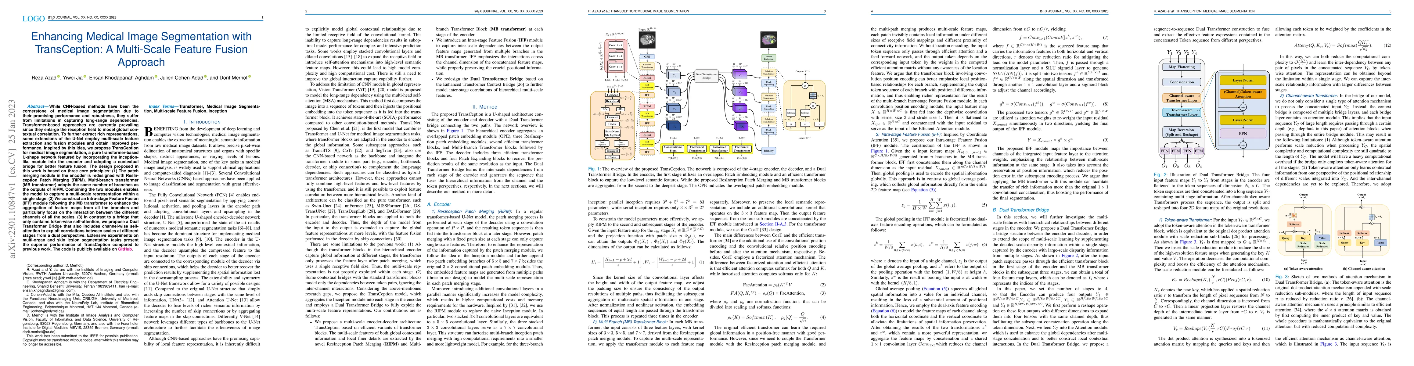
While CNN-based methods have been the cornerstone of medical image segmentation due to their promising performance and robustness, they suffer from limitations in capturing long-range dependencies. ...
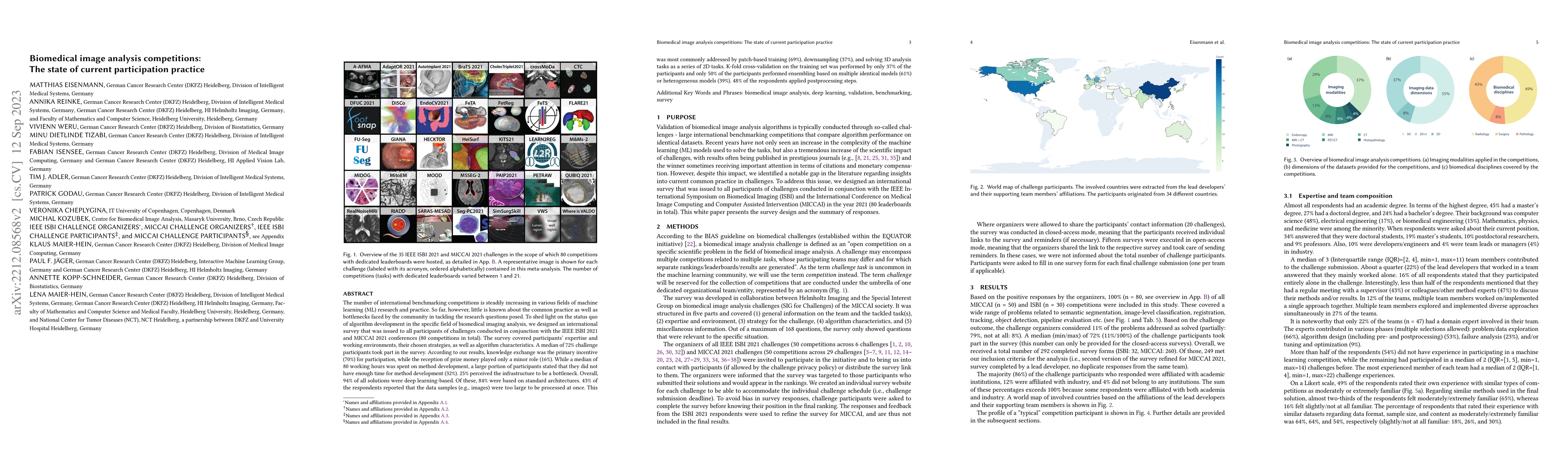
The number of international benchmarking competitions is steadily increasing in various fields of machine learning (ML) research and practice. So far, however, little is known about the common pract...
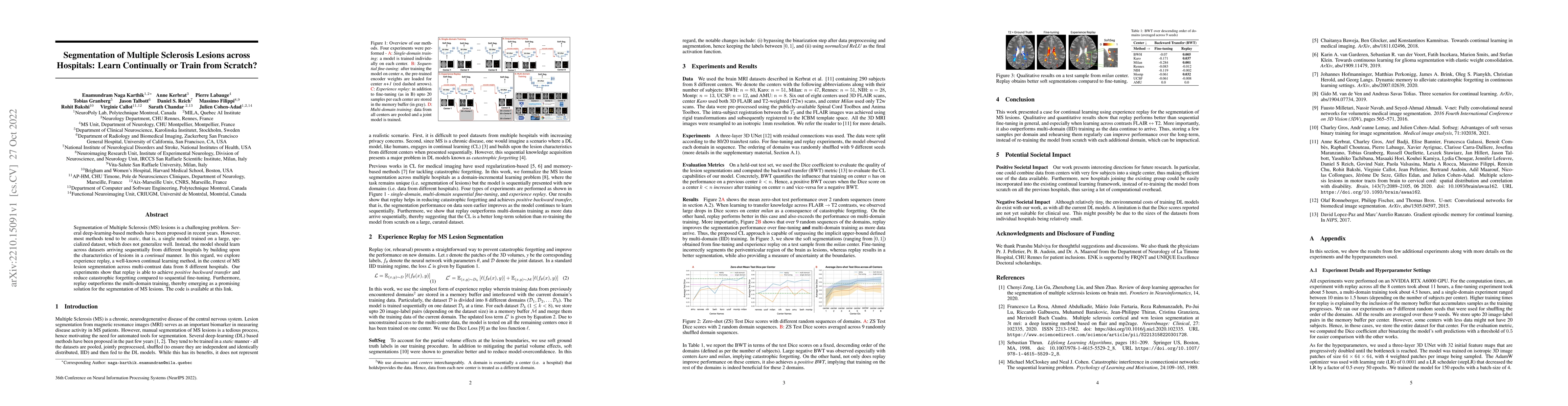
Segmentation of Multiple Sclerosis (MS) lesions is a challenging problem. Several deep-learning-based methods have been proposed in recent years. However, most methods tend to be static, that is, a ...
Convolutional neural networks (CNNs) have been the consensus for medical image segmentation tasks. However, they suffer from the limitation in modeling long-range dependencies and spatial correlatio...
Accurate and automatic segmentation of intervertebral discs from medical images is a critical task for the assessment of spine-related diseases such as osteoporosis, vertebral fractures, and interve...
Dealing with missing modalities in Magnetic Resonance Imaging (MRI) and overcoming their negative repercussions is considered a hurdle in biomedical imaging. The combination of a specified set of mo...
Medical tasks are prone to inter-rater variability due to multiple factors such as image quality, professional experience and training, or guideline clarity. Training deep learning networks with ann...
The spinal cord (SC), which conveys information between the brain and the peripheral nervous system, plays a key role in various neurological disorders such as multiple sclerosis (MS) and amyotrophi...
Labeling vertebral discs from MRI scans is important for the proper diagnosis of spinal related diseases, including multiple sclerosis, amyotrophic lateral sclerosis, degenerative cervical myelopath...
Multiple sclerosis is an inflammatory disorder of the central nervous system. Quantitative MRI has huge potential to provide intrinsic and normative values of tissue properties useful for diagnosis,...
Background: Magnetic field inhomogeneities generate important geometric distortions in reconstructed echo-planar images. Various procedures were proposed for correcting these distortions on brain im...
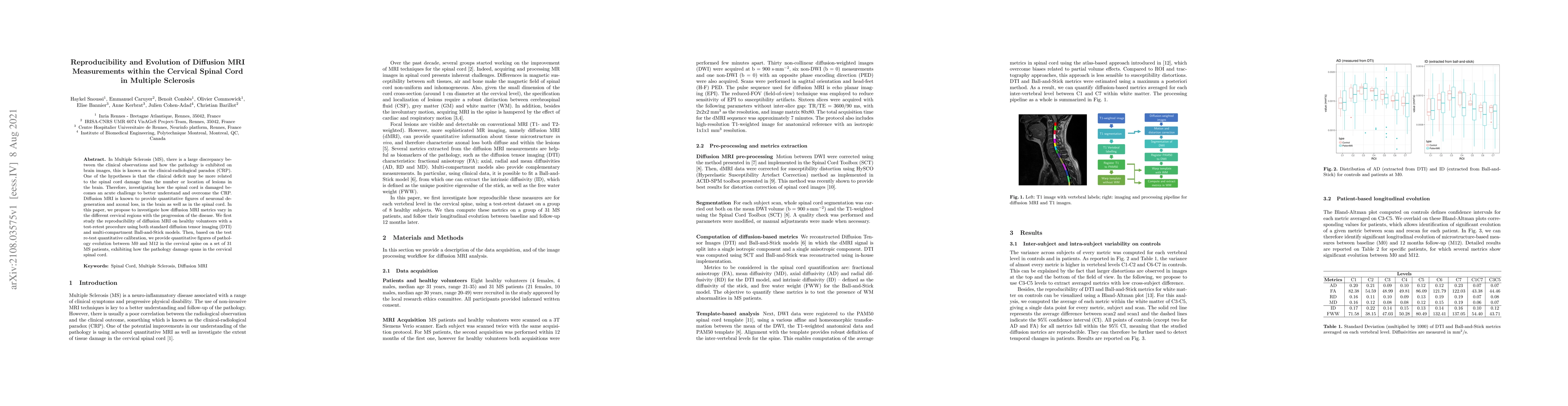
In Multiple Sclerosis (MS), there is a large discrepancy between the clinical observations and how the pathology is exhibited on brain images, this is known as the clinical-radiological paradox (CRP...
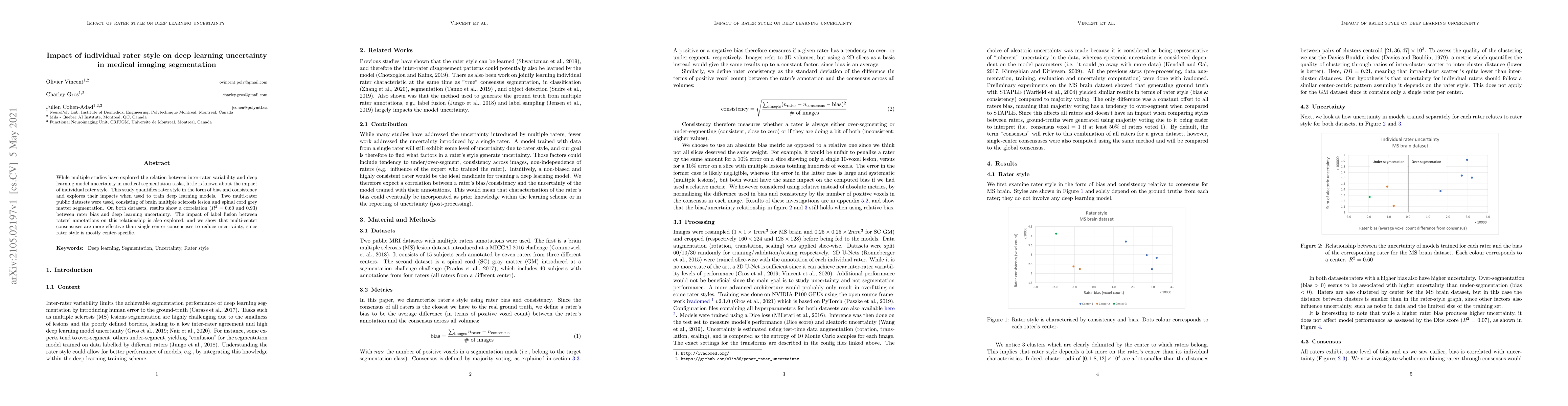
While multiple studies have explored the relation between inter-rater variability and deep learning model uncertainty in medical segmentation tasks, little is known about the impact of individual ra...
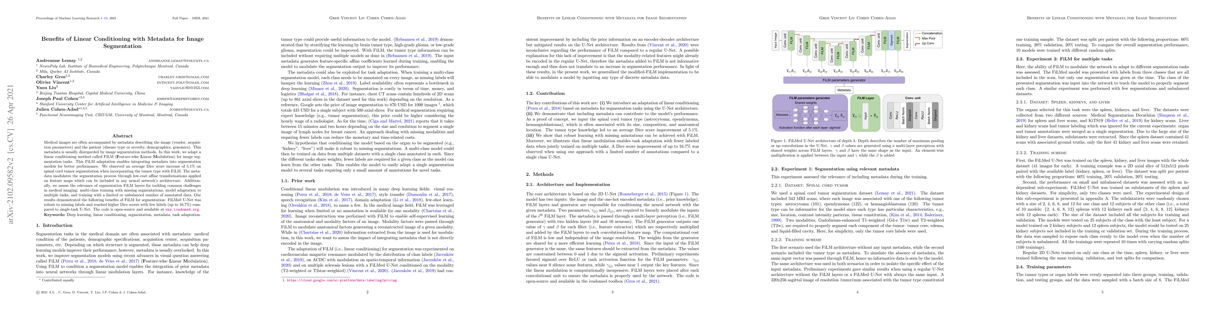
Medical images are often accompanied by metadata describing the image (vendor, acquisition parameters) and the patient (disease type or severity, demographics, genomics). This metadata is usually di...
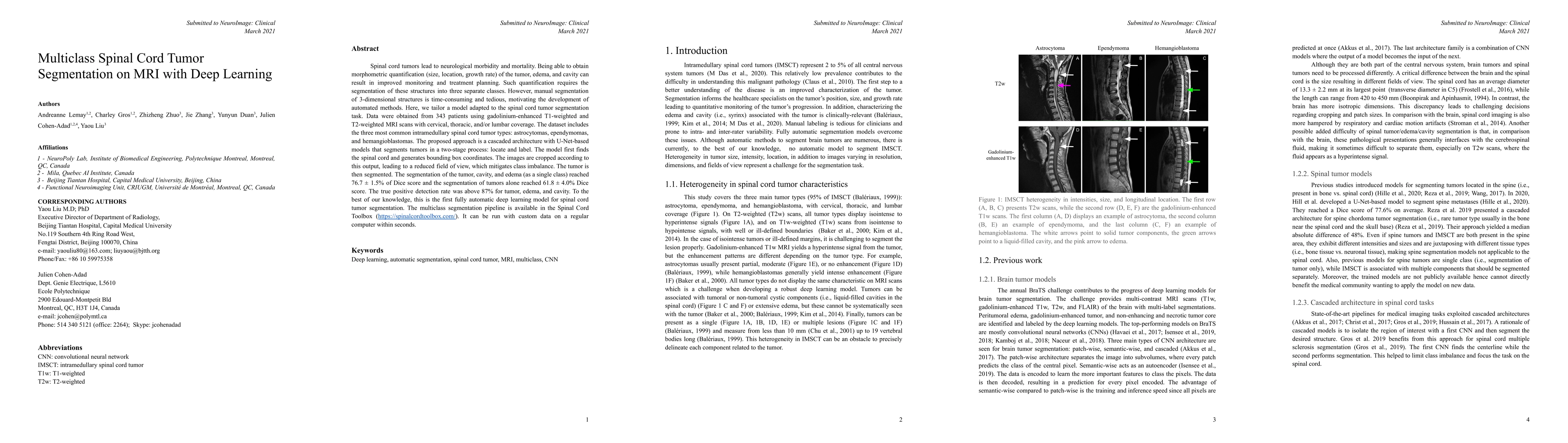
Spinal cord tumors lead to neurological morbidity and mortality. Being able to obtain morphometric quantification (size, location, growth rate) of the tumor, edema, and cavity can result in improved...
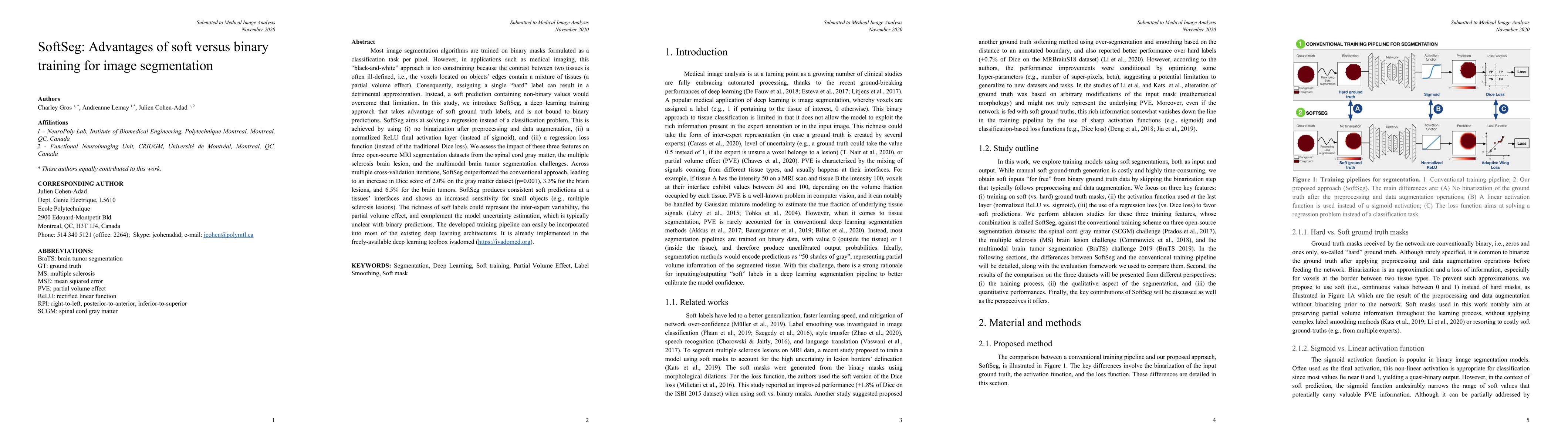
Most image segmentation algorithms are trained on binary masks formulated as a classification task per pixel. However, in applications such as medical imaging, this "black-and-white" approach is too...
ivadomed is an open-source Python package for designing, end-to-end training, and evaluating deep learning models applied to medical imaging data. The package includes APIs, command-line tools, docu...
The semantic image segmentation task consists of classifying each pixel of an image into an instance, where each instance corresponds to a class. This task is a part of the concept of scene understa...
Model quantization is leveraged to reduce the memory consumption and the computation time of deep neural networks. This is achieved by representing weights and activations with a lower bit resolutio...
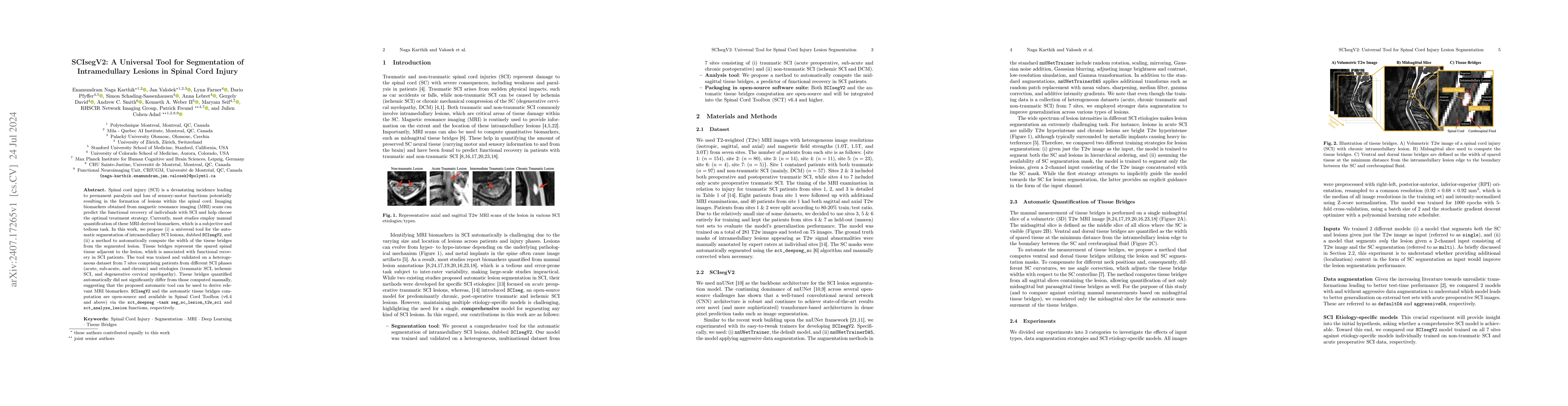
Spinal cord injury (SCI) is a devastating incidence leading to permanent paralysis and loss of sensory-motor functions potentially resulting in the formation of lesions within the spinal cord. Imaging...
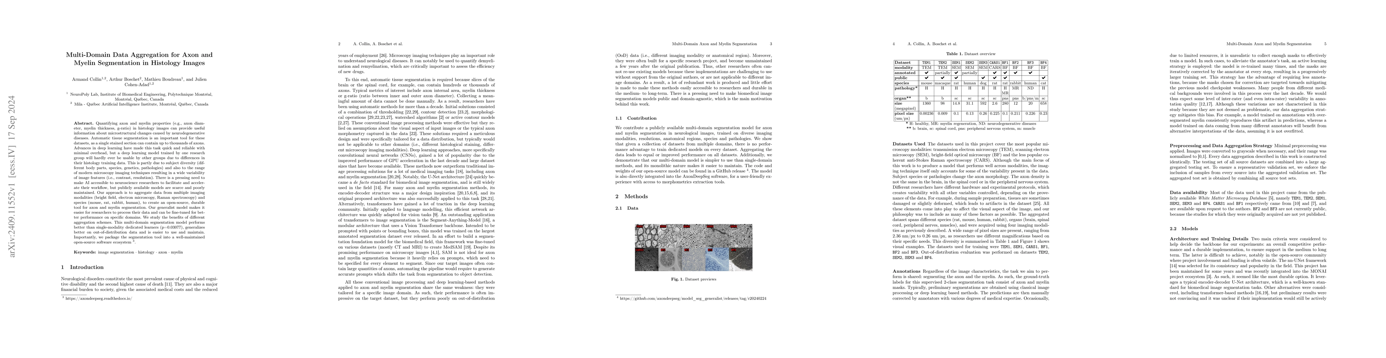
Quantifying axon and myelin properties (e.g., axon diameter, myelin thickness, g-ratio) in histology images can provide useful information about microstructural changes caused by neurodegenerative dis...
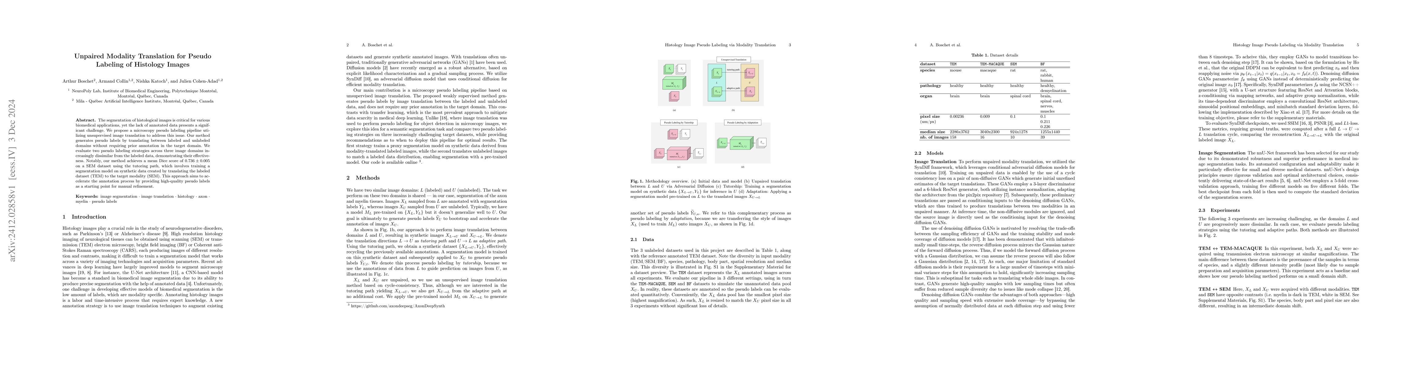
The segmentation of histological images is critical for various biomedical applications, yet the lack of annotated data presents a significant challenge. We propose a microscopy pseudo labeling pipeli...
Preclinical diffusion MRI (dMRI) has proven value in methods development and validation, characterizing the biological basis of diffusion phenomena, and comparative anatomy. While dMRI enables in vivo...
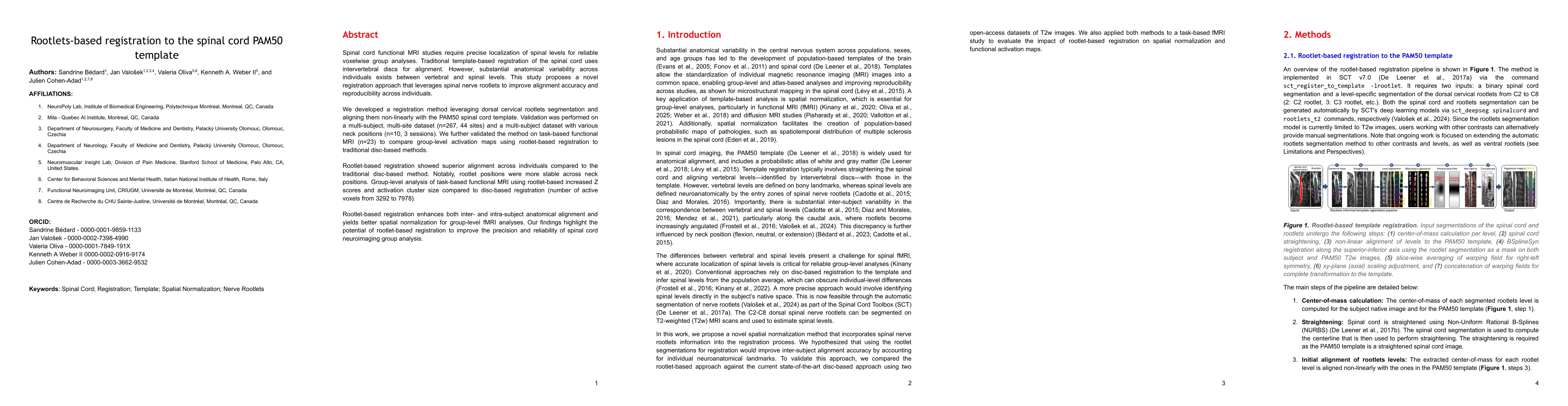
Spinal cord functional MRI studies require precise localization of spinal levels for reliable voxelwise group analyses. Traditional template-based registration of the spinal cord uses intervertebral d...
Morphometric measures derived from spinal cord segmentations can serve as diagnostic and prognostic biomarkers in neurological diseases and injuries affecting the spinal cord. While robust, automatic ...
Purpose: To develop a deep learning method for the automatic segmentation of spinal nerve rootlets on various MRI scans. Material and Methods: This retrospective study included MRI scans from two open...
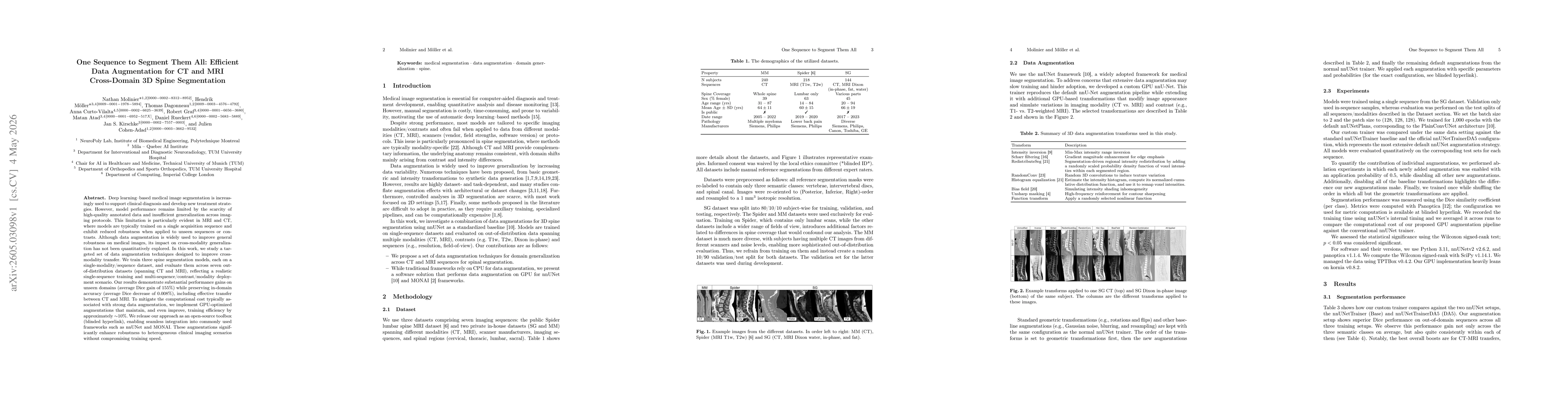
Deep learning-based medical image segmentation is increasingly used to support clinical diagnosis and develop new treatment strategies. However, model performance remains limited by the scarcity of hi...
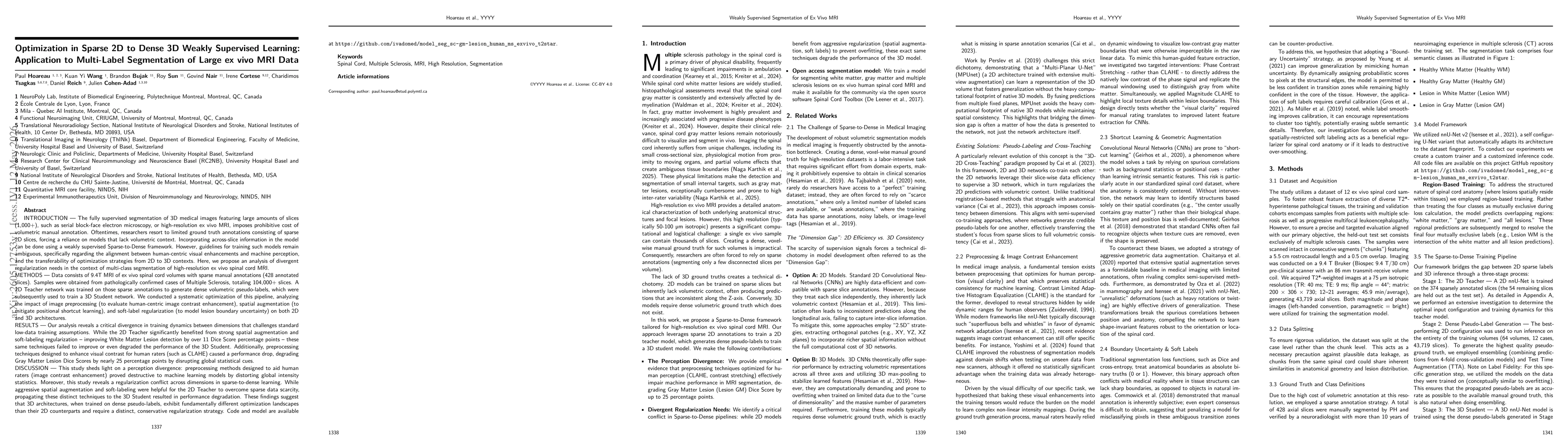
INTRODUCTION | Fully supervised 3D segmentation of high-resolution ex vivo MRI is limited by the prohibitive cost of volumetric annotation, forcing reliance on sparse 2D slices. Weakly supervised Spar...
Lumbar spine conditions are a leading cause of disability worldwide, yet reliable quantification of degeneration from MRI remains challenging. In clinical practice, analysis is predominantly performed...