Publication

Published: Sep 17, 2025
Categories: q-bio.TO, eess.IV, physics.med-ph

Metrics

Source: ArXiv

Quick Actions

View PDF

Quick Answers

What methodology did the authors use?

The study developed RootletSeg, a deep learning model for segmenting spinal cord nerve rootlets from T1w, T2w, and MP2RAGE-UNIT1 MRI scans. It utilized a combination of open-source datasets, isotropic MRI scans, and a self-configuring deep learning framework (nnU-Net) for automated segmentation and spinal level inference. More in Methodology →

What are the key results?

RootletSeg achieved accurate segmentation of spinal cord nerve rootlets across multiple MRI contrasts. — The model demonstrated the ability to infer spinal levels directly from MRI data, establishing a correlation between vertebral and spinal levels. More in Key Results →

Why is this work significant?

This research provides a valuable tool for neuroimaging analysis, enabling more precise spinal cord segmentation and level inference. It has potential applications in lesion classification, neuromodulation therapy, and fMRI group analysis, advancing clinical and research applications in spinal cord studies. More in Significance →

What are the main limitations?

The model was trained and tested only on healthy participants in the C2-T1 region, limiting its applicability to pathological cases. — The study focused on cervical spinal cord analysis without distinguishing between genders or age groups, which may affect generalizability. More in Limitations →

Paper Preview

Abstract

Purpose: To develop a deep learning method for the automatic segmentation of spinal nerve rootlets on various MRI scans. Material and Methods: This 
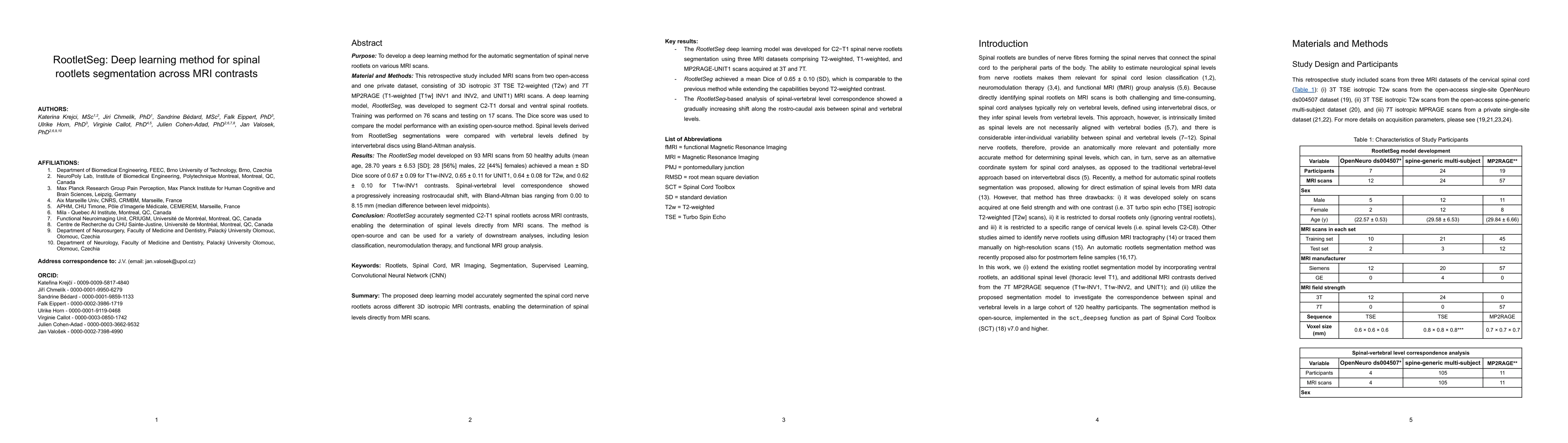
retrospective study included MRI scans from two open-access and one private dataset, consisting of 3D isotropic 3T TSE T2-weighted (T2w) and 7T MP2RAGE (T1-weighted [T1w] INV1 and INV2, and UNIT1) MRI scans. A deep learning model, RootletSeg, was developed to segment C2-T1 dorsal and ventral spinal rootlets. Training was performed on 76 scans and testing on 17 scans. The Dice score was used to compare the model performance with an existing open-source method. Spinal levels derived from RootletSeg segmentations were compared with vertebral levels defined by intervertebral discs using Bland-Altman analysis. Results: The RootletSeg model developed on 93 MRI scans from 50 healthy adults (mean age, 28.70 years $\pm$ 6.53 [SD]; 28 [56%] males, 22 [44%] females) achieved a mean $\pm$ SD Dice score of 0.67 $\pm$ 0.09 for T1w-INV2, 0.65 $\pm$ 0.11 for UNIT1, 0.64 $\pm$ 0.08 for T2w, and 0.62 $\pm$ 0.10 for T1w-INV1 contrasts. Spinal-vertebral level correspondence showed a progressively increasing rostrocaudal shift, with Bland-Altman bias ranging from 0.00 to 8.15 mm (median difference between level midpoints). Conclusion: RootletSeg accurately segmented C2-T1 spinal rootlets across MRI contrasts, enabling the determination of spinal levels directly from MRI scans. The method is open-source and can be used for a variety of downstream analyses, including lesion classification, neuromodulation therapy, and functional MRI group analysis.

AI Key Findings

Generated Sep 30, 2025

Methodology — What approach did the authors take?

The study developed RootletSeg, a deep learning model for segmenting spinal cord nerve rootlets from T1w, T2w, and MP2RAGE-UNIT1 MRI scans. It utilized a combination of open-source datasets, isotropic MRI scans, and a self-configuring deep learning framework (nnU-Net) for automated segmentation and spinal level inference.

Key Results — What are the main findings?

  • RootletSeg achieved accurate segmentation of spinal cord nerve rootlets across multiple MRI contrasts.
  • The model demonstrated the ability to infer spinal levels directly from MRI data, establishing a correlation between vertebral and spinal levels.
  • The study found slight differences in rostrocaudal spinal level lengths compared to previous MRI and post-mortem studies, potentially due to population variability and acquisition protocols.

Significance — Why does this research matter?

This research provides a valuable tool for neuroimaging analysis, enabling more precise spinal cord segmentation and level inference. It has potential applications in lesion classification, neuromodulation therapy, and fMRI group analysis, advancing clinical and research applications in spinal cord studies.

Technical Contribution — What is the technical contribution?

The technical contribution includes the development of RootletSeg as an open-source, deep learning model integrated into the Spinal Cord Toolbox (SCT), enabling automated segmentation of spinal cord nerve rootlets and direct spinal level inference from MRI data.

Novelty — What is new about this work?

This work introduces a novel approach for directly inferring spinal levels from MRI data using deep learning, which differs from previous methods that relied on anatomical landmarks or manual segmentation. The integration with the Spinal Cord Toolbox enhances its accessibility and utility for neuroimaging research.

Limitations — What are the limitations of this study?

  • The model was trained and tested only on healthy participants in the C2-T1 region, limiting its applicability to pathological cases.
  • The study focused on cervical spinal cord analysis without distinguishing between genders or age groups, which may affect generalizability.

Future Work — What did the authors propose for future work?

  • Extending the model to include pathological cases and thoraco-lumbar regions for broader applicability.
  • Developing more robust methods for spinal level inference in complex regions with overlapping rootlets.
  • Investigating the impact of different MRI acquisition protocols on segmentation accuracy and spinal level inference.

Paper Details

Paper ID: 2509.16255
Comments: 26 pages, 6 figures, 4 tables
Categories:
q-bio.TO eess.IV physics.med-ph

How to Cite This Paper

BibTeX
@article{eippert2025rootletseg,
  title         = {RootletSeg: Deep learning method for spinal rootlets segmentation across
  MRI contrasts},
  author        = {Eippert, Falk and Valosek, Jan and Chmelik, Jiri and others},
  year          = {2025},
  eprint        = {2509.16255},
  archivePrefix = {arXiv},
  primaryClass  = {q-bio.TO},
}
APA 7
Eippert, F., Valosek, J., Chmelik, J., Cohen-Adad, J., Krejci, K., Bédard, S., Horn, U., & Callot, V. (2025). RootletSeg: Deep learning method for spinal rootlets segmentation across
  MRI contrasts. arXiv. https://arxiv.org/abs/2509.16255
MLA 9
Eippert, Falk, et al. "RootletSeg: Deep learning method for spinal rootlets segmentation across
  MRI contrasts." arXiv, 2025, arxiv.org/abs/2509.16255.

Similar Papers

Found 4 papers

Segmentation of spinal rootlets across MRI contrasts with RootletSeg.

Eippert, Falk, Cohen-Adad, Julien, Krejčí, Kateřina et al.

2026-05-02

Automatic Segmentation of the Spinal Cord Nerve Rootlets

Jan Valosek, Theo Mathieu, Raphaelle Schlienger et al.

2024-05-02

Multiclass Spinal Cord Tumor Segmentation on MRI with Deep Learning

Andreanne Lemay, Julien Cohen-Adad, Yaou Liu et al.

2021-03-31

Comments (0)

This website uses cookies to ensure you get the best experience. Learn more