Automatic segmentation of spinal multiple sclerosis lesions: How to generalize across MRI contrasts?
Publication
Metrics
AI Quick Summary
Researchers used a technique called FiLM to improve segmentation of spinal multiple sclerosis lesions on MRI images, achieving 0.72 Dice score performance, highlighting the need to address inter-rater variability as a key bottleneck in this task.
Paper Preview
Abstract
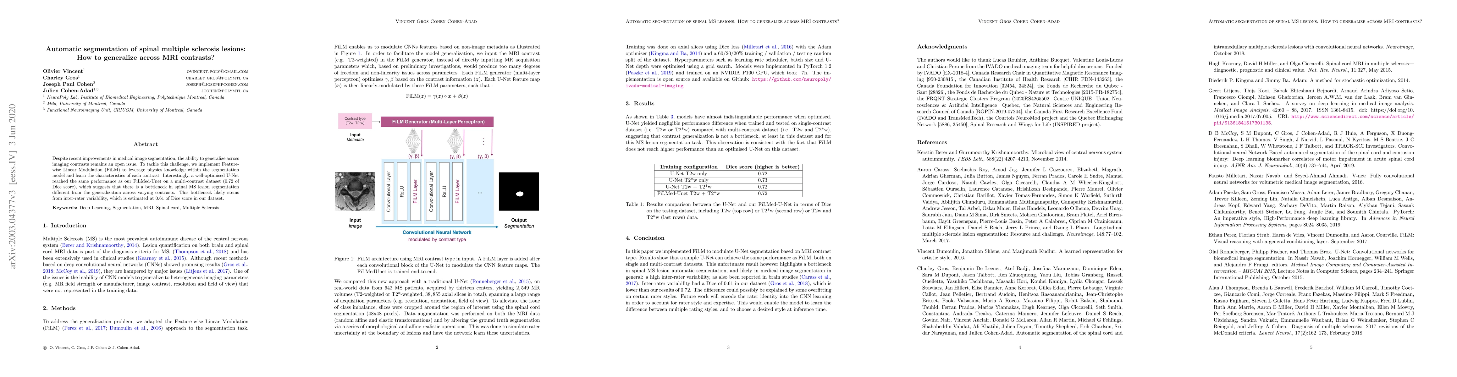
Despite recent improvements in medical image segmentation, the ability to generalize across imaging contrasts remains an open issue. To tackle this challenge, we implement Feature-wise Linear Modulation (FiLM) to leverage physics knowledge within the segmentation model and learn the characteristics of each contrast. Interestingly, a well-optimised U-Net reached the same performance as our FiLMed-Unet on a multi-contrast dataset (0.72 of Dice score), which suggests that there is a bottleneck in spinal MS lesion segmentation different from the generalization across varying contrasts. This bottleneck likely stems from inter-rater variability, which is estimated at 0.61 of Dice score in our dataset.
AI Key Findings
Get AI-generated insights about this paper's methodology, results, significance, and more — seven facets brought into focus.
Impact
Paper Details
PDF Preview
Key Terms
Citation Network
Current paper (gray), citations (green), references (blue)
Display is limited for performance on very large graphs.
Discussion 0