ArxivLens
P
Researcher

Paola Borri

10 papers on arXiv

10 arXiv Papers
6 Total Publications
Profile

Academic Profile

Name P. F. Borri
Total Papers 6
Scholar Profile View on Semantic Scholar
Metrics

Statistics

10 arXiv Papers
6 Total Publications
Publications

Papers on arXiv

Thumbnail for Differential Dynamic Microscopy can be applied to Differential
  Interference Contrast images despite shadowing effects
arXiv
Differential Dynamic Microscopy can be applied to Differential Interference Contrast images despite shadowing effects

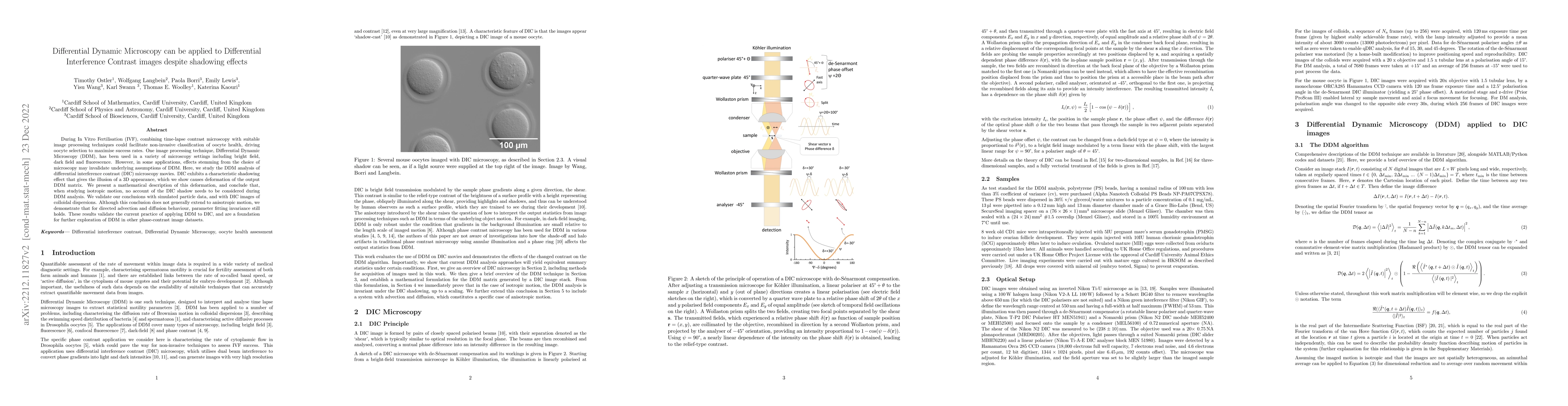
During In Vitro Fertilisation (IVF), combining time-lapse contrast microscopy with suitable image processing techniques could facilitate non-invasive classification of oocyte health, driving oocyte ...

Thumbnail for Correlative extinction and single fluorophore bleaching microscopy for
  ligand quantification on gold nanoparticles
arXiv
Correlative extinction and single fluorophore bleaching microscopy for ligand quantification on gold nanoparticles

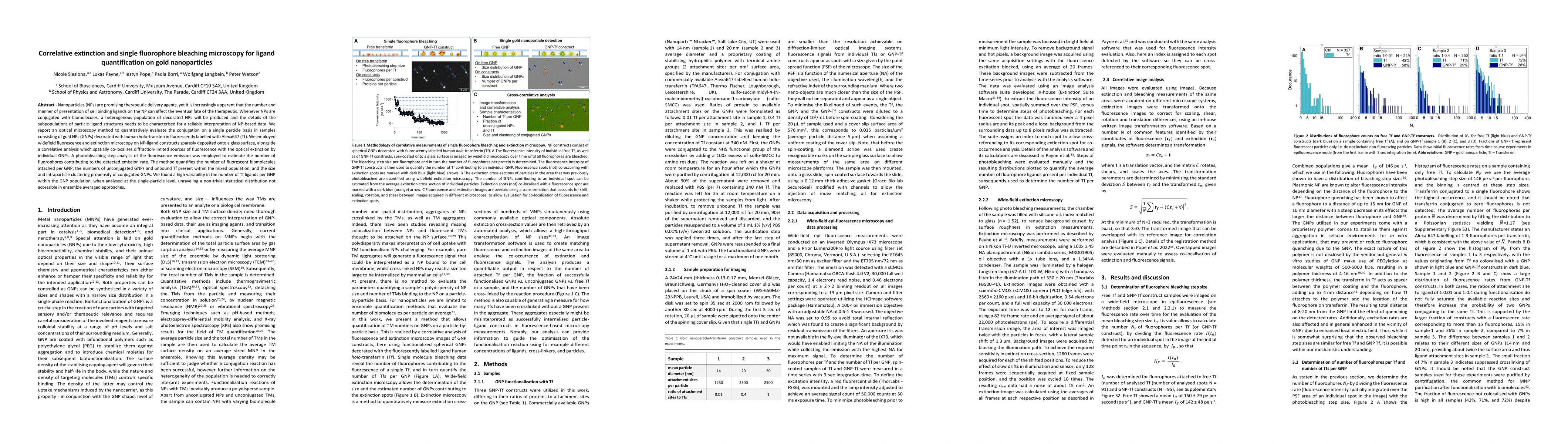
Nanoparticles (NPs) are promising therapeutic delivery agents, yet it is increasingly apparent that the number and manner of presentation of cell binding ligands on the NP can affect the eventual fa...

Thumbnail for Correlative light electron microscopy using small gold nanoparticles as
  single probes
arXiv
Correlative light electron microscopy using small gold nanoparticles as single probes

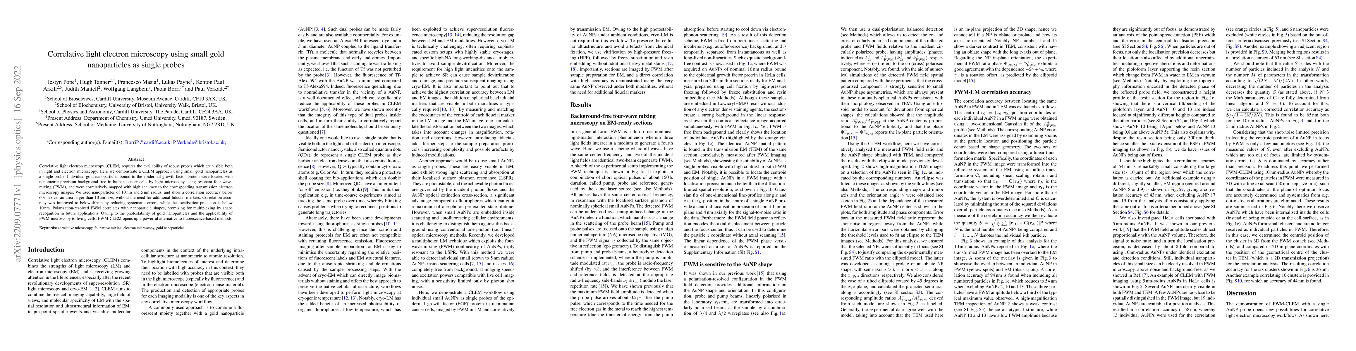
Correlative light electron microscopy (CLEM) requires the availability of robust probes which are visible both in light and electron microscopy. Here we demonstrate a CLEM approach using small gold ...

Thumbnail for Quantitatively linking morphology and optical response of individual
  silver nanohedra
arXiv
Quantitatively linking morphology and optical response of individual silver nanohedra

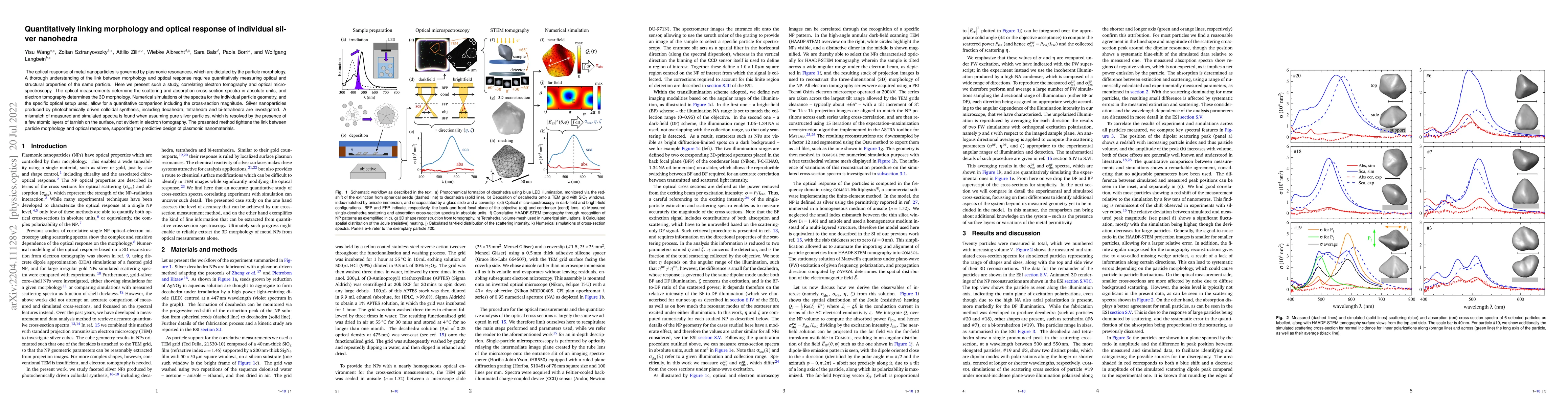
The optical response of metal nanoparticles is governed by plasmonic resonances, which are dictated by the particle morphology. A thorough understanding of the link between morphology and optical re...

Thumbnail for Sizing individual dielectric nanoparticles with quantitative
  differential interference contrast microscopy
arXiv
Sizing individual dielectric nanoparticles with quantitative differential interference contrast microscopy

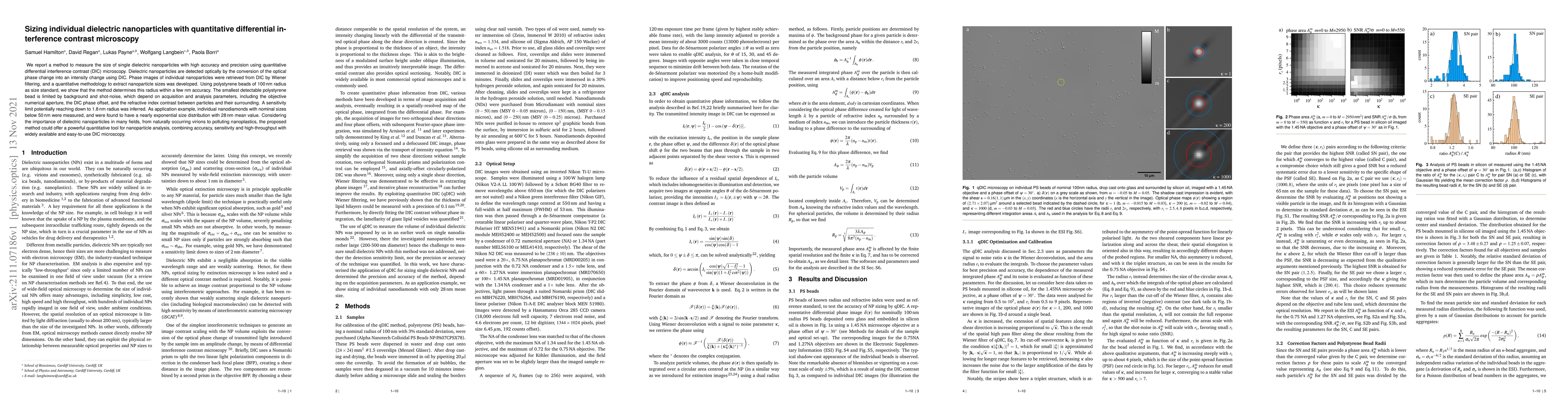
We report a method to measure the size of single dielectric nanoparticles with high accuracy and precision using quantitative differential interference contrast (DIC) microscopy. Dielectric nanopart...

Thumbnail for Simultaneous microscopic imaging of thickness and refractive index of
  thin layers by heterodyne interferometric reflectometry (HiRef)
arXiv
Simultaneous microscopic imaging of thickness and refractive index of thin layers by heterodyne interferometric reflectometry (HiRef)

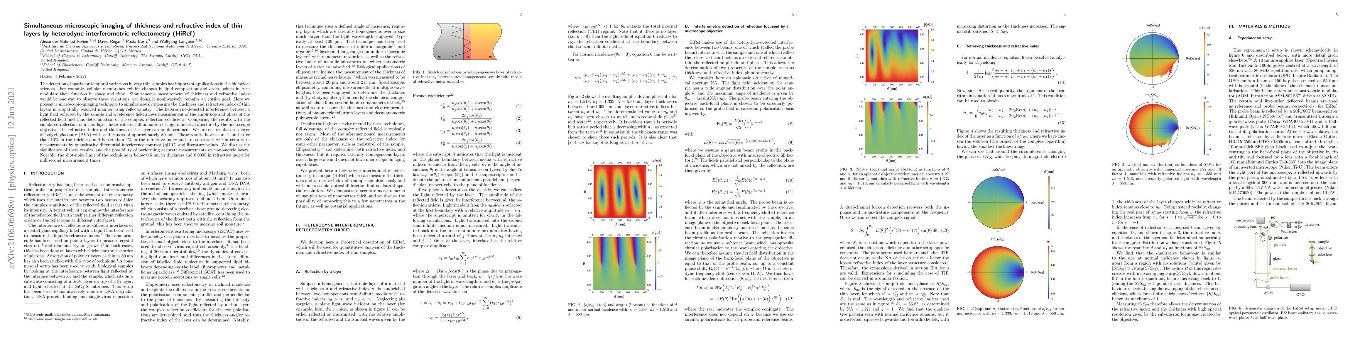
The detection of spatial or temporal variations in very thin samples has important applications in the biological sciences. For example, cellular membranes exhibit changes in lipid composition and o...

Thumbnail for uFLIM -- Unsupervised analysis of FLIM-FRET microscopy data
arXiv
uFLIM -- Unsupervised analysis of FLIM-FRET microscopy data

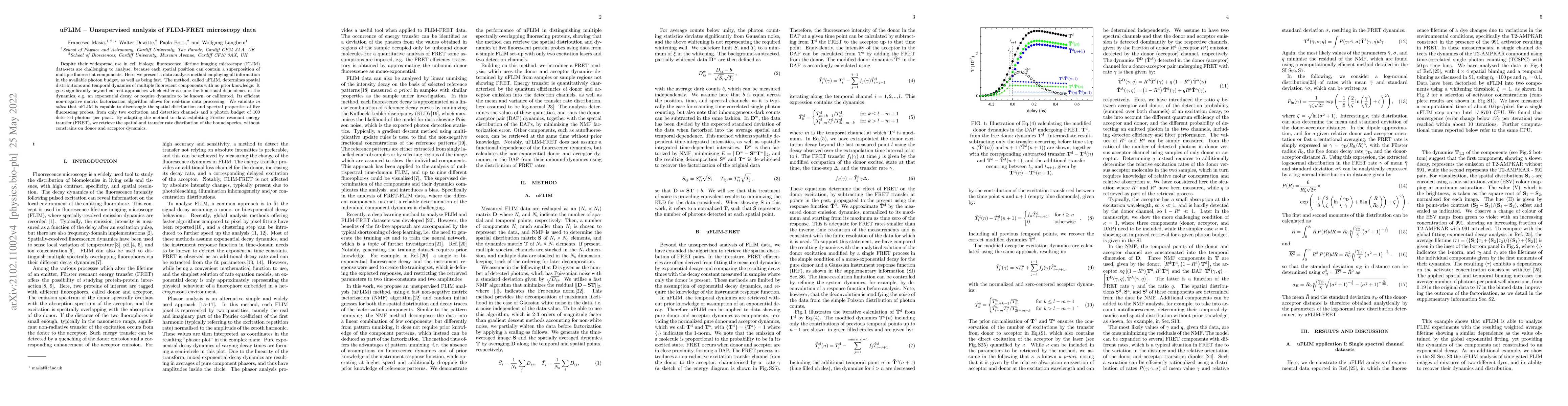
Despite their widespread use in cell biology, fluorescence lifetime imaging microscopy (FLIM) data-sets are challenging to analyse, because each spatial position can contain a superposition of multi...

Thumbnail for Genetically encoding stimulated Raman-scattering probes for cell imaging using infrared fluorescent proteins
arXiv
Genetically encoding stimulated Raman-scattering probes for cell imaging using infrared fluorescent proteins

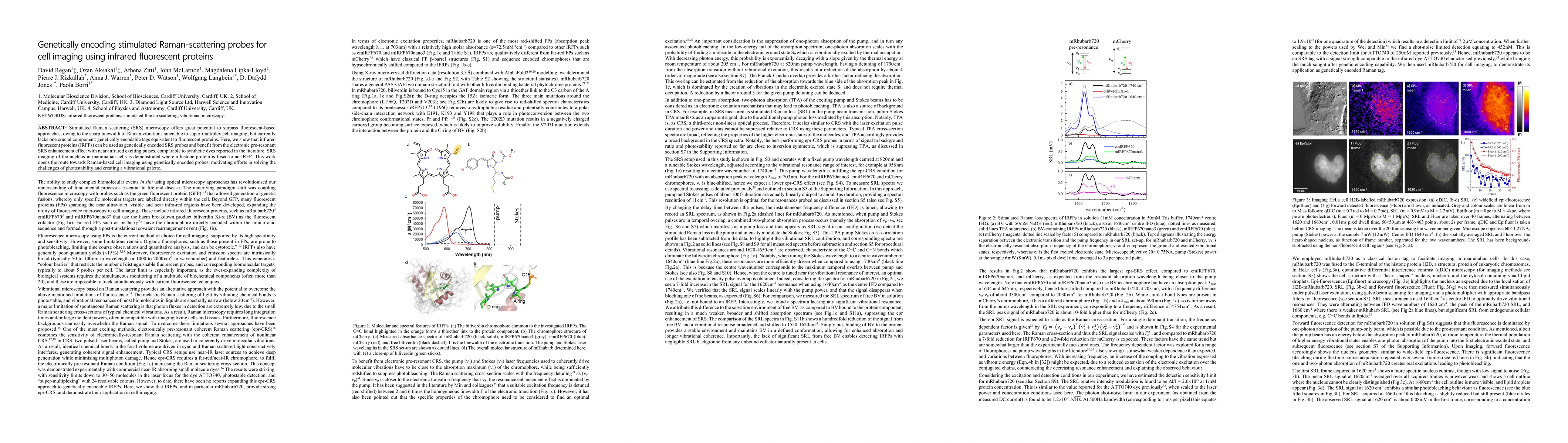
Stimulated Raman scattering (SRS) microscopy offers great potential to surpass fluorescent-based approaches, owing to the sharp linewidth of Raman vibrations amenable to super-multiplex cell imaging, ...

Thumbnail for Sub-nanometer 3D morphometric precision of polarisation-resolved wide-field optical extinction microscopy determines the roundness of individual gold nanospheres
arXiv
Sub-nanometer 3D morphometric precision of polarisation-resolved wide-field optical extinction microscopy determines the roundness of individual gold nanospheres

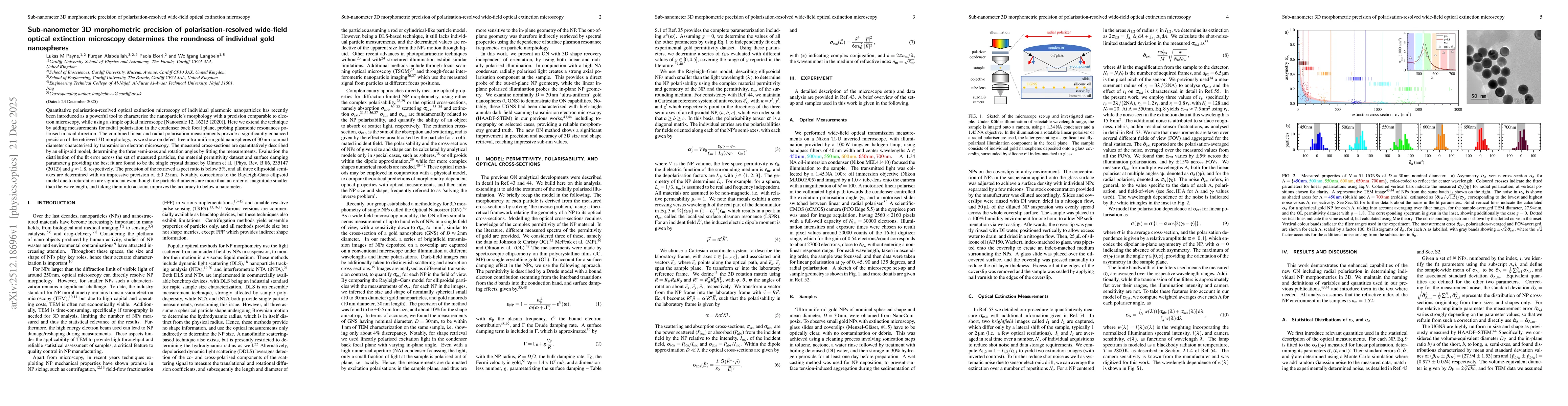
Quantitative polarisation-resolved optical extinction microscopy of individual plasmonic nanoparticles has recently been introduced as a powerful tool to characterise the nanoparticle's morphology wit...

Thumbnail for Understanding the mechanisms of supported lipid membrane reshaping into tubular networks using quantitative DIC microscopy
arXiv
Understanding the mechanisms of supported lipid membrane reshaping into tubular networks using quantitative DIC microscopy

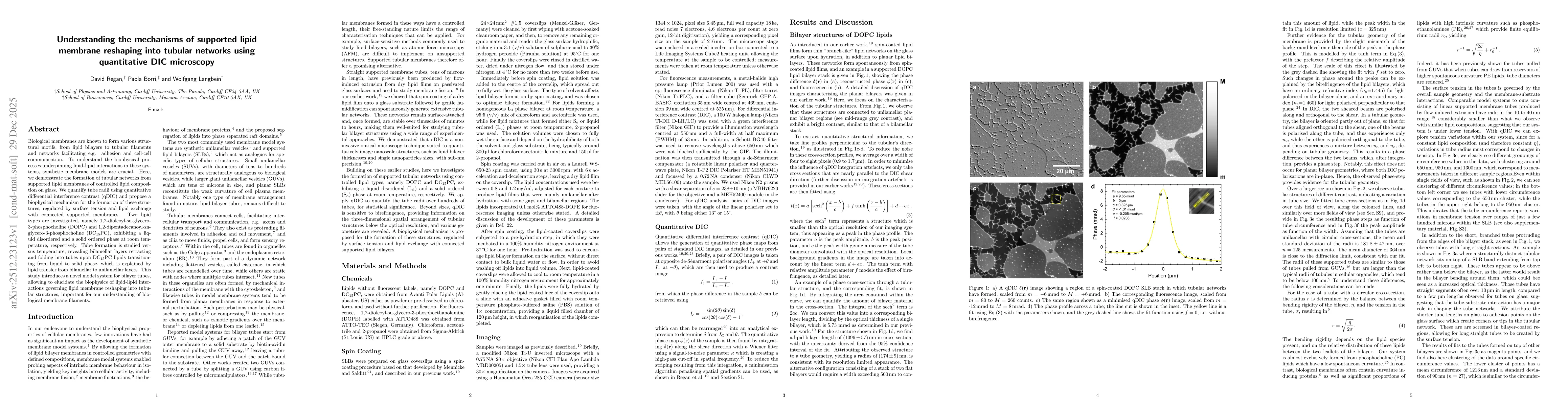
Biological membranes are known to form various structural motifs, from lipid bilayers to tubular filaments and networks facilitating e.g. adhesion and cell-cell communication. To understand the biophy...

This website uses cookies to ensure you get the best experience. Learn more