Publication
Metrics
AI Quick Summary
This paper presents a quantitative optical microscopy method combining widefield fluorescence and extinction microscopy to characterize the conjugation of fluorescently labeled human holo-transferrin on gold nanoparticles. The study reveals significant variability in ligand presentation on individual nanoparticles, highlighting the importance of single-particle analysis over ensemble approaches.
Paper Preview
Abstract
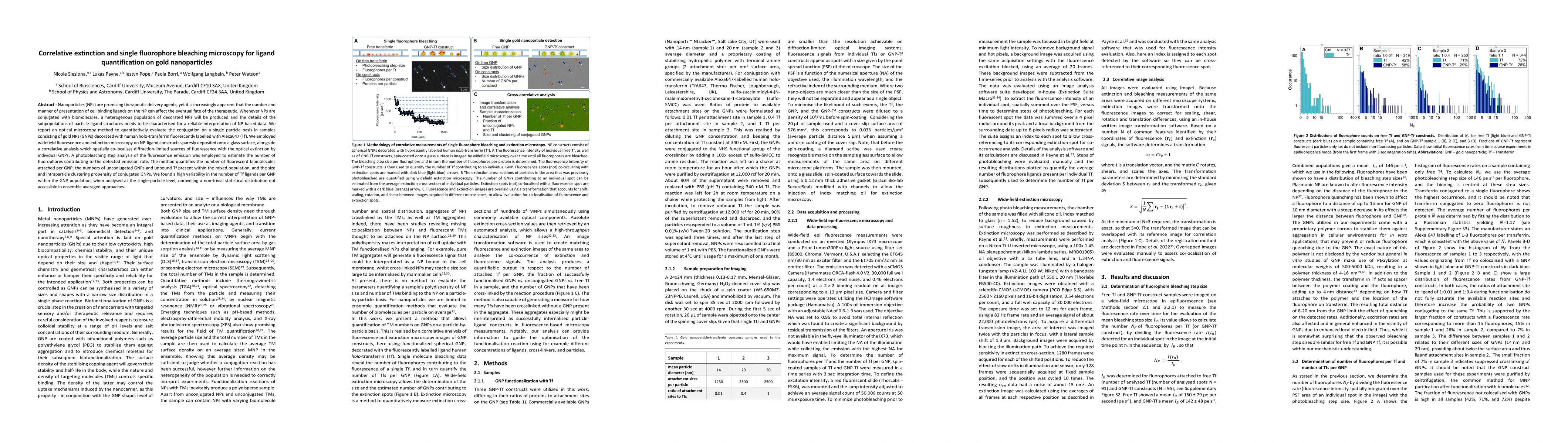
Nanoparticles (NPs) are promising therapeutic delivery agents, yet it is increasingly apparent that the number and manner of presentation of cell binding ligands on the NP can affect the eventual fate of the therapeutic. Whenever NPs are conjugated with biomolecules, a heterogenous population of decorated NPs will be produced and the details of the subpopulations of particle-ligand structures needs to be characterised for a reliable interpretation of NP-based data. We report an optical microscopy method to quantitatively evaluate the conjugation on a single particle basis in samples consisting of gold NPs (GNPs) decorated with human holo-transferrin fluorescently labelled with Alexa647 (Tf). We employed widefield fluorescence and extinction microscopy on NP-ligand constructs sparesly deposited onto a glass surface, alongside a correlative analysis which spatially co-localises diffraction-limited sources of fluorescence with the optical extinction by individual GNPs. A photobleaching step analysis of the fluorescence emission was employed to estimate the number of fluorophores contributing to the detected emission rate. The method quantifies the number of fluorescent biomolecules attached per GNP, the numbers of unconjugated GNPs and unbound Tf present within the mixed population, and the size and intraparticle clustering propensity of conjugated GNPs. We found a high variability in the number of Tf ligands per GNP within the GNP population, when analysed at the single-particle level, unraveling a non-trivial statistical distribution not accessible in ensemble averaged approaches
AI Key Findings
Get AI-generated insights about this paper's methodology, results, significance, and more — seven facets brought into focus.
Impact
Paper Details
Authors
PDF Preview
Key Terms
Citation Network
Current paper (gray), citations (green), references (blue)
Display is limited for performance on very large graphs.
Discussion 0