Prabhakar Pradhan

18 papers on arXiv

Academic Profile

Name: P. Pradhan
Total Papers: 2

Statistics

arXiv Papers 18
Total Publications 2

Papers on arXiv

Thumbnail for Detection of cancer stages through fractal dimension analysis of tissue
  microarrays (TMA) via optical transmission microscopy

Detection of cancer stages through fractal dimension analysis of tissue microarrays (TMA) via optical transmission microscopy

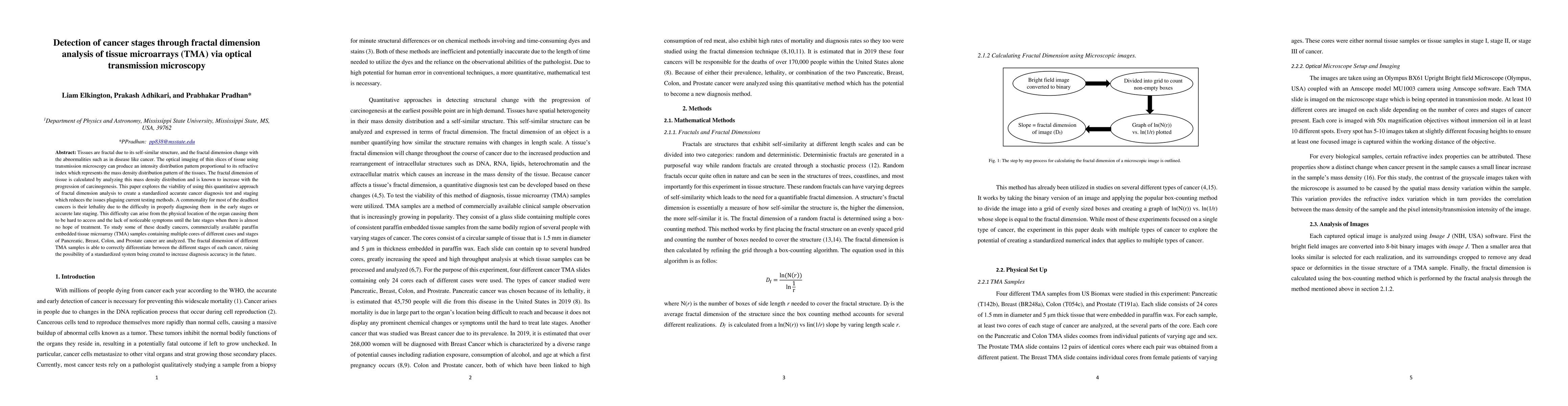
Tissues are fractal due to its self-similar structure, and the fractal dimension change with the abnormalities such as in disease like cancer. The optical imaging of thin slices of tissue using tran...

Thumbnail for Photonics probing of DNA specific spatial mass density fluctuations in
  gut cell nuclei due to total body irradiation via confocal imaging

Photonics probing of DNA specific spatial mass density fluctuations in gut cell nuclei due to total body irradiation via confocal imaging

Abnormalities within cells result in nanoscale structural alterations can be characterized via confocal imaging and quantification of these alterations. Accidental or deliberate exposure to total bo...

Thumbnail for Photonics study of probiotic treatment on brain cells exposed to chronic
  alcoholism using molecular specific nuclear light localization properties via
  confocal imaging

Photonics study of probiotic treatment on brain cells exposed to chronic alcoholism using molecular specific nuclear light localization properties via confocal imaging

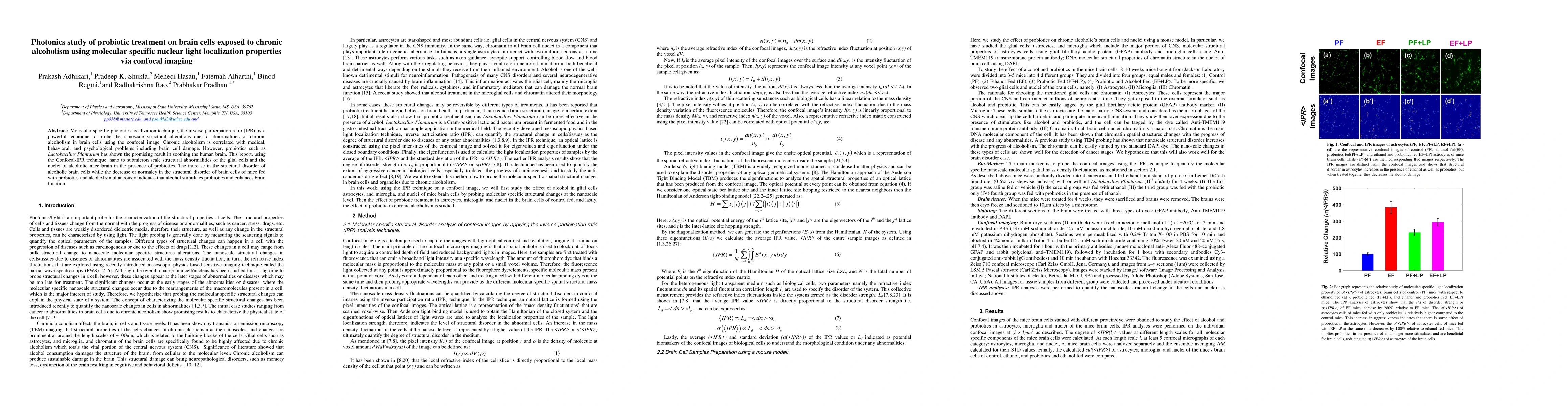
Molecular specific photonics localization technique, the inverse participation ratio (IPR), is a powerful technique to probe the nanoscale structural alterations due to abnormalities or chronic alco...

Thumbnail for Optical probing of pups brain tissue and molecular specific nuclear
  nano-structural alterations due to fetal alcoholism via dual spectroscopic
  approach

Optical probing of pups brain tissue and molecular specific nuclear nano-structural alterations due to fetal alcoholism via dual spectroscopic approach

Mesoscopic physics-based dual spectroscopic imaging techniques, partial wave spectroscopy (PWS) and inverse participation ratio (IPR), are used to quantify the nano to submicron scales structural al...

Thumbnail for Nanoscale structural alterations in cancer cells to assess
  anti-cancerous drug effectiveness in cancer treatment using TEM imaging

Nanoscale structural alterations in cancer cells to assess anti-cancerous drug effectiveness in cancer treatment using TEM imaging

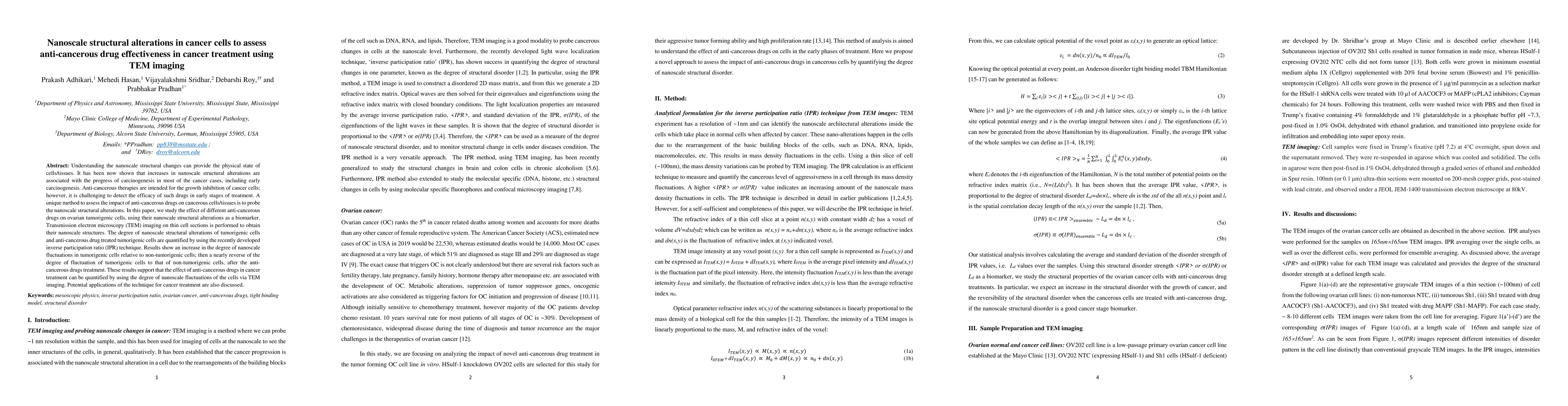
Understanding the nanoscale structural changes can provide the physical state of cells/tissues. It has been now shown that increases in nanoscale structural alterations are associated with the progr...

Thumbnail for Optical detection of structural properties of tumor tissues generated by
  xenografting of drug-sensitive and drug-resistant cancer cells using partial
  wave spectroscopy (PWS)

Optical detection of structural properties of tumor tissues generated by xenografting of drug-sensitive and drug-resistant cancer cells using partial wave spectroscopy (PWS)

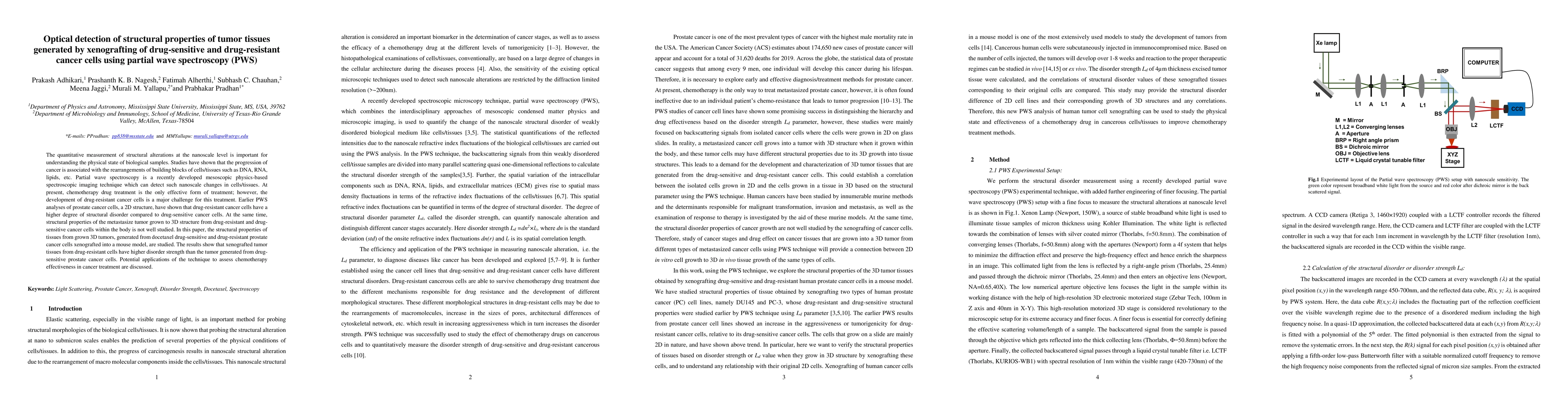
The quantitative measurement of structural alterations at the nanoscale level is important for understanding the physical state of biological samples. Studies have shown that the progression of canc...

Thumbnail for Optical probing of fractal and multifractal connection to structural
  disorder in weakly optical disordered media: Application to cancer detection

Optical probing of fractal and multifractal connection to structural disorder in weakly optical disordered media: Application to cancer detection

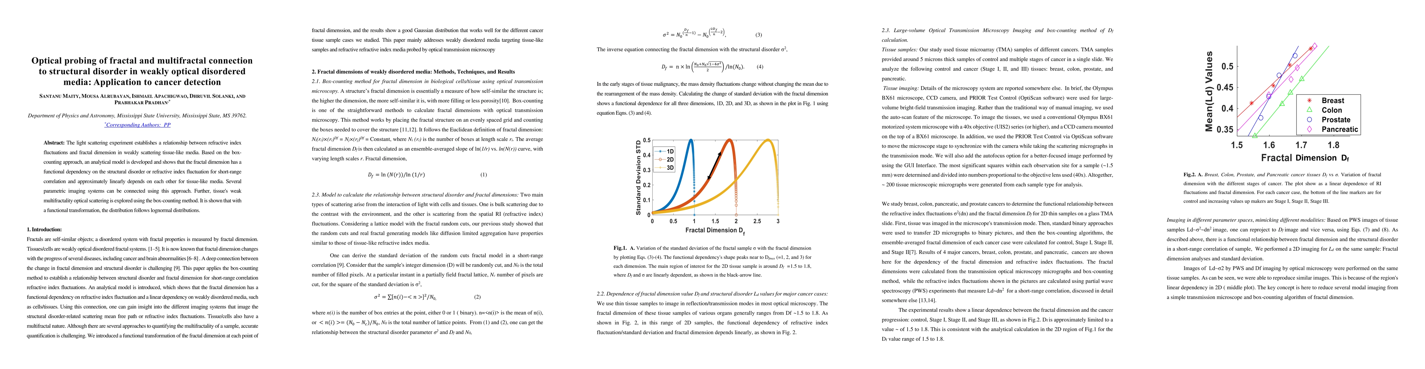
The light scattering experiment establishes a relationship between refractive index fluctuations and fractal dimension in weakly scattering tissue-like media. Based on the box-counting approach, an an...

Thumbnail for Optical detection of the spatial structural alteration in the human
  brain tissues and cells and DNA and chromatin due to Parkinsons disease

Optical detection of the spatial structural alteration in the human brain tissues and cells and DNA and chromatin due to Parkinsons disease

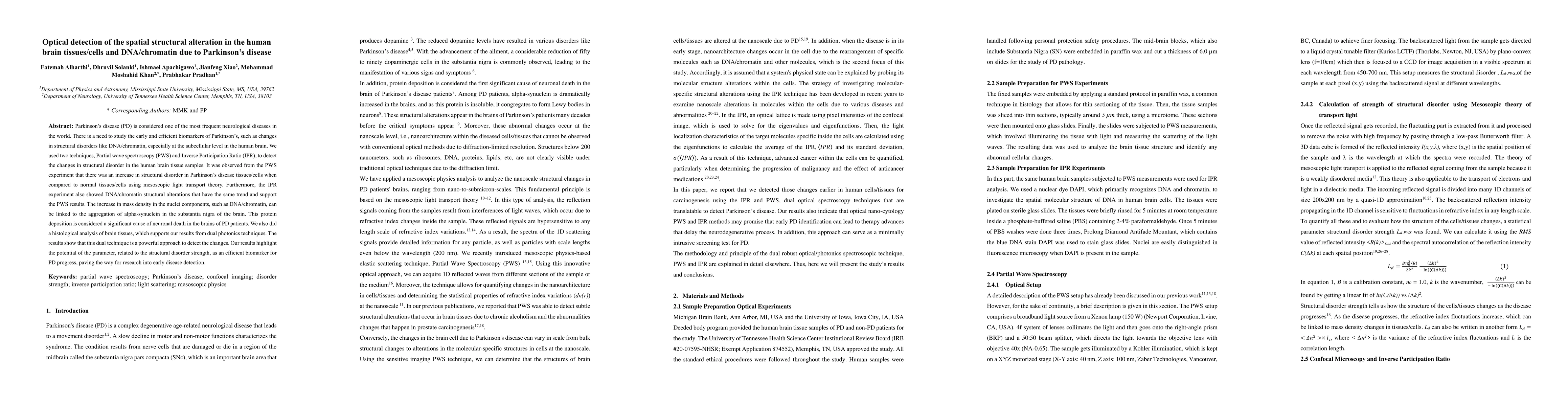
Parkinsons disease (PD) is considered one of the most frequent neurological diseases in the world. There is a need to study the early and efficient biomarkers of Parkinsons, such as changes in structu...

Thumbnail for An AI-directed analytical study on the optical transmission microscopic
  images of Pseudomonas aeruginosa in planktonic and biofilm states

An AI-directed analytical study on the optical transmission microscopic images of Pseudomonas aeruginosa in planktonic and biofilm states

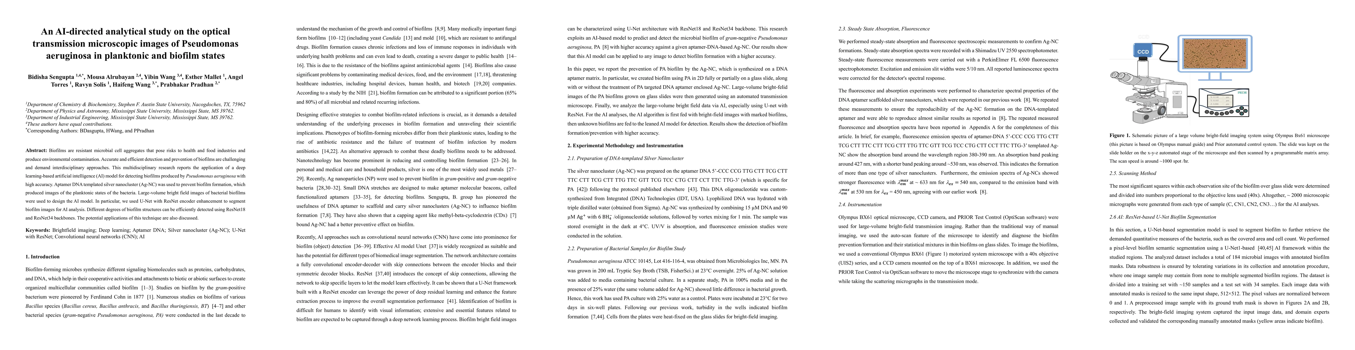
Biofilms are resistant microbial cell aggregates that pose risks to health and food industries and produce environmental contamination. Accurate and efficient detection and prevention of biofilms are ...

Thumbnail for Dual Photonics Probing of Nano- to Submicron-Scale Structural
  Alterations in Human Brain Tissues or Cells and Chromatin or DNA with the
  Progression of Alzheimers Disease

Dual Photonics Probing of Nano- to Submicron-Scale Structural Alterations in Human Brain Tissues or Cells and Chromatin or DNA with the Progression of Alzheimers Disease

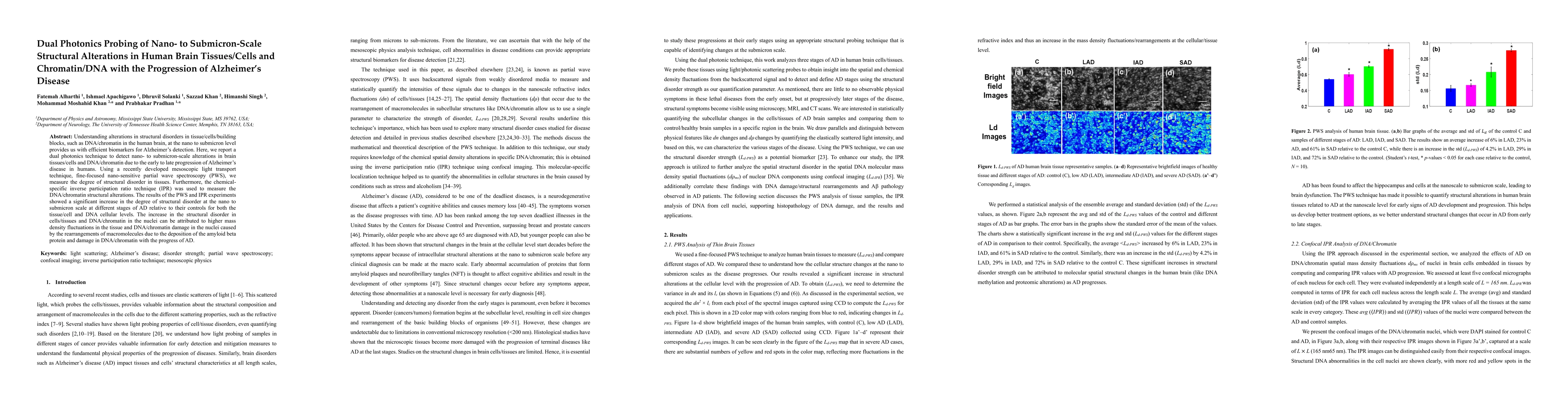
Understanding alterations in structural disorders in tissue or cells or building blocks, such as DNA or chromatin in the human brain, at the nano to submicron level provides us with efficient biomarke...

Thumbnail for Photonics detection of molecular-specific spatial structural alterations
  in cell nuclei due to chronic alcoholism and probiotics treatments on colon
  cancer via a light localization method using confocal imaging

Photonics detection of molecular-specific spatial structural alterations in cell nuclei due to chronic alcoholism and probiotics treatments on colon cancer via a light localization method using confocal imaging

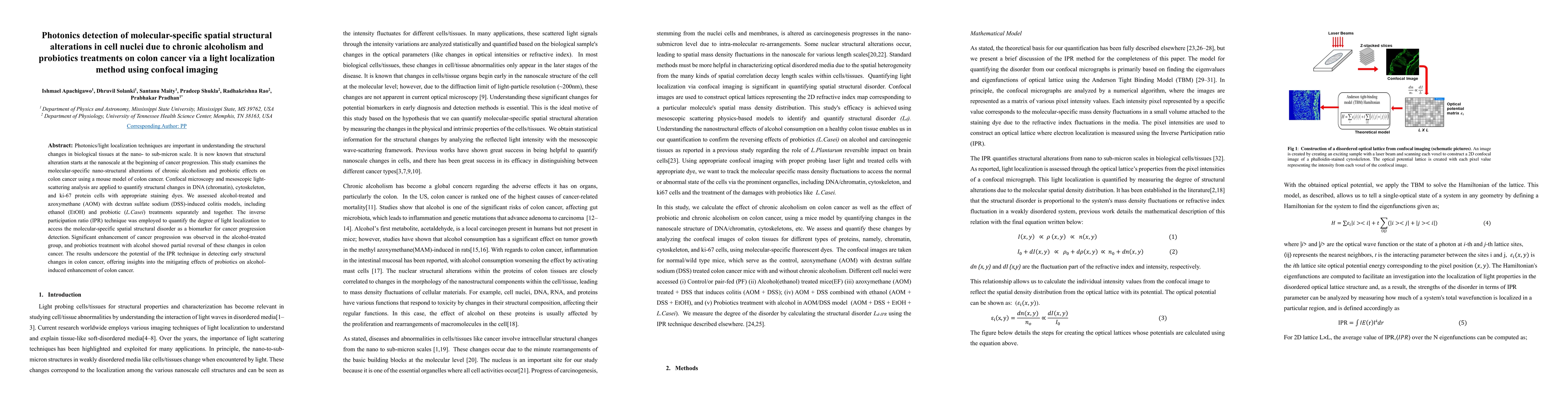
Photonics/light localization techniques are important in understanding the structural changes in biological tissues at the nano- to sub-micron scale. It is now known that structural alteration starts ...

Thumbnail for Decoding Breast Cancer in X-ray Mammograms: A Multi-Parameter Approach
  Using Fractals, Multifractals, and Structural Disorder Analysis

Decoding Breast Cancer in X-ray Mammograms: A Multi-Parameter Approach Using Fractals, Multifractals, and Structural Disorder Analysis

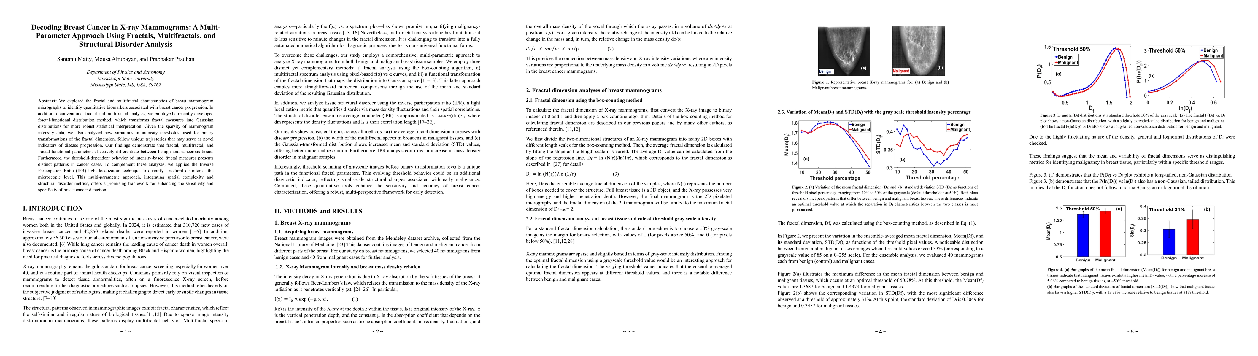
We explored the fractal and multifractal characteristics of breast mammogram micrographs to identify quantitative biomarkers associated with breast cancer progression. In addition to conventional frac...

Thumbnail for Evaluation and optimization of deep learning models for enhanced
  detection of brain cancer using transmission optical microscopy of thin brain
  tissue samples

Evaluation and optimization of deep learning models for enhanced detection of brain cancer using transmission optical microscopy of thin brain tissue samples

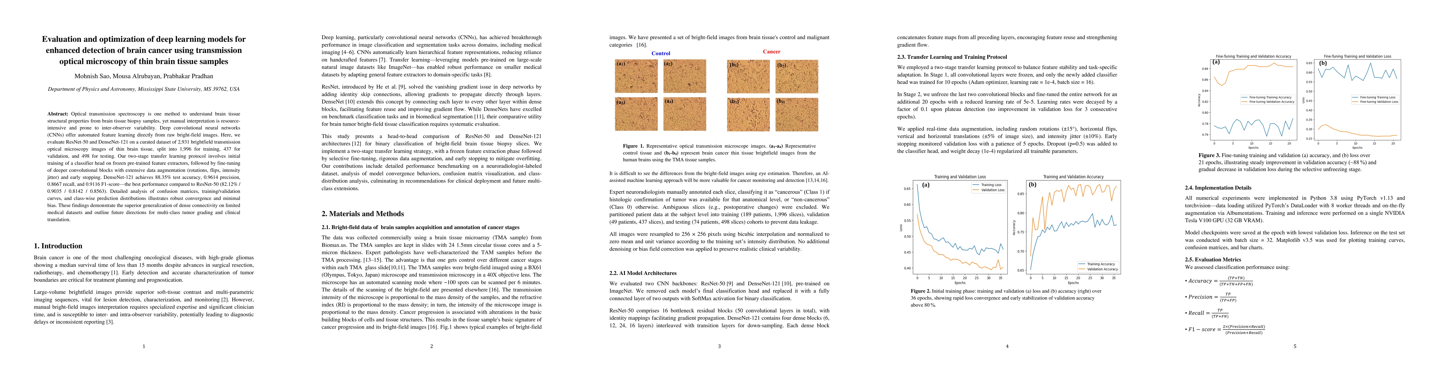
Optical transmission spectroscopy is one method to understand brain tissue structural properties from brain tissue biopsy samples, yet manual interpretation is resource intensive and prone to inter ob...

Thumbnail for Automated grading and staging of ovarian cancer using deep learning on
  the transmission optical microscopy bright-field images of thin biopsy tissue
  samples

Automated grading and staging of ovarian cancer using deep learning on the transmission optical microscopy bright-field images of thin biopsy tissue samples

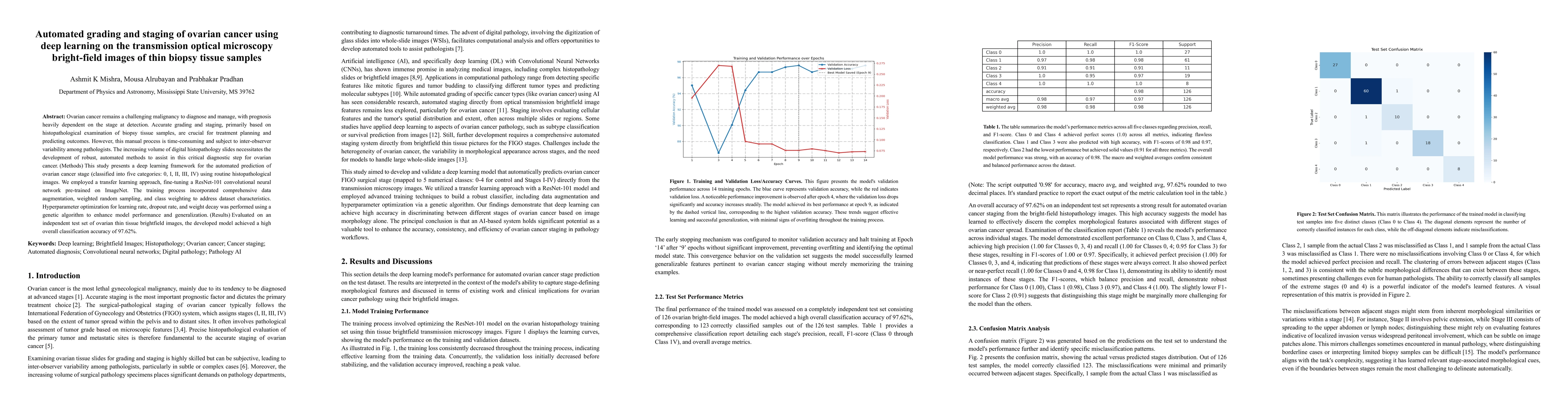
Ovarian cancer remains a challenging malignancy to diagnose and manage, with prognosis heavily dependent on the stage at detection. Accurate grading and staging, primarily based on histopathological e...

Thumbnail for Comprehensive Multimodal and Multiscale Analysis of Alzheimer Disease in 5xFAD Mice: Optical Spectroscopies, TEM, Neuropathological, and Behavioral Investigations

Comprehensive Multimodal and Multiscale Analysis of Alzheimer Disease in 5xFAD Mice: Optical Spectroscopies, TEM, Neuropathological, and Behavioral Investigations

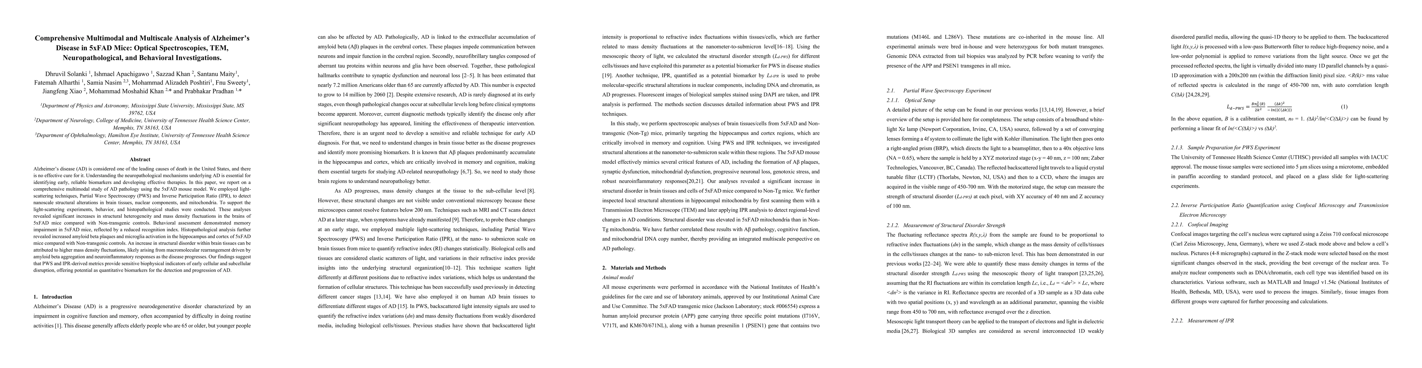
Alzheimer disease (AD) is considered one of the leading causes of death in the United States, and there is no effective cure for it. Understanding the neuropathological mechanisms underlying AD is ess...

Thumbnail for Alterations of brain tissue structural complexity and disorder in Alzheimer's disease (AD): Fractal, multifractal, fractal transformation, and disorder strength analyses

Alterations of brain tissue structural complexity and disorder in Alzheimer's disease (AD): Fractal, multifractal, fractal transformation, and disorder strength analyses

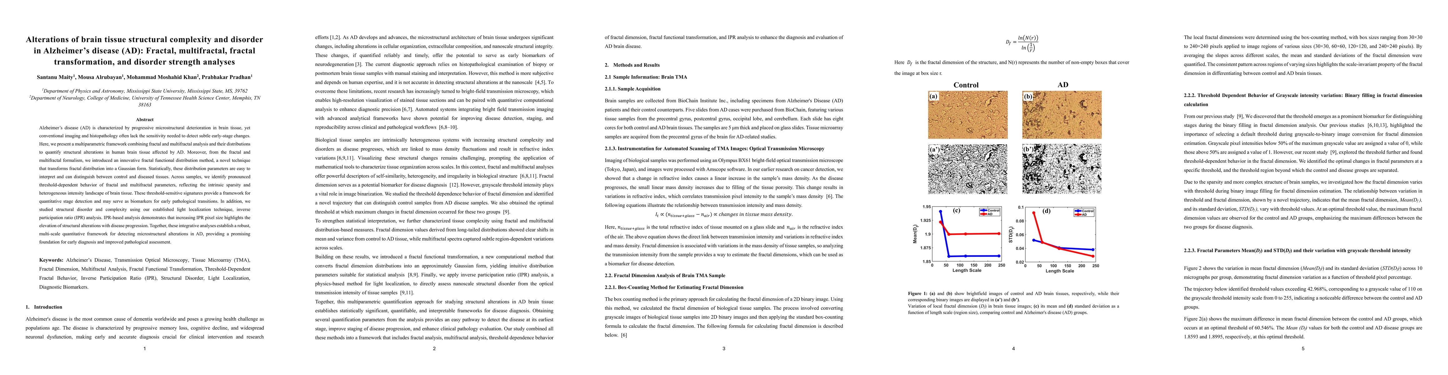
Alzheimer's disease (AD) is characterized by progressive microstructural deterioration in brain tissue, yet conventional imaging and histopathology often lack the sensitivity needed to detect subtle e...

Thumbnail for Alterations of brain tissue structural complexity and disorder in Parkinson disease (PD): Fractal, multifractal, fractal transformation, and disorder strength analyses

Alterations of brain tissue structural complexity and disorder in Parkinson disease (PD): Fractal, multifractal, fractal transformation, and disorder strength analyses

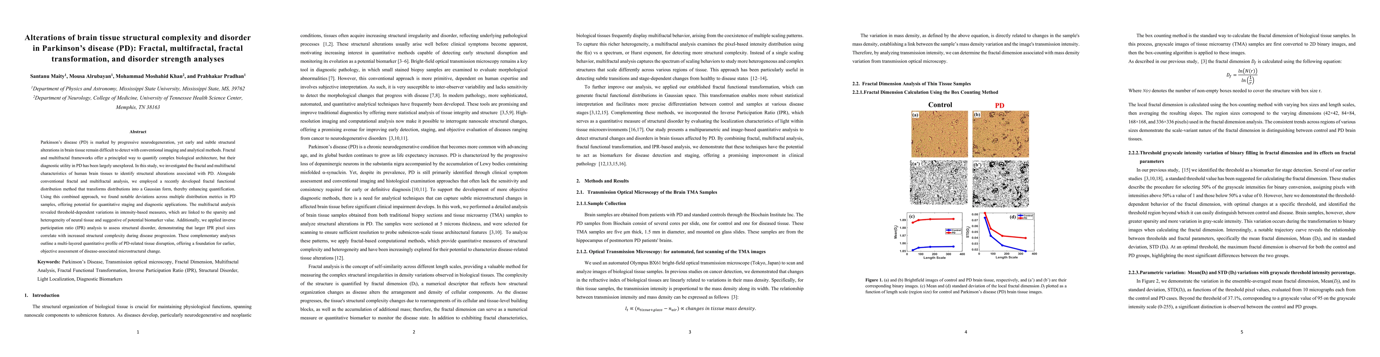
Parkinson disease (PD) is marked by progressive neurodegeneration, yet early and subtle structural alterations in brain tissue remain difficult to detect with conventional imaging and analytical metho...

Thumbnail for Quantitative Characterization of Brain Tissue Alterations in Brain Cancer Using Fractal, Multifractal, and IPR Metrics

Quantitative Characterization of Brain Tissue Alterations in Brain Cancer Using Fractal, Multifractal, and IPR Metrics

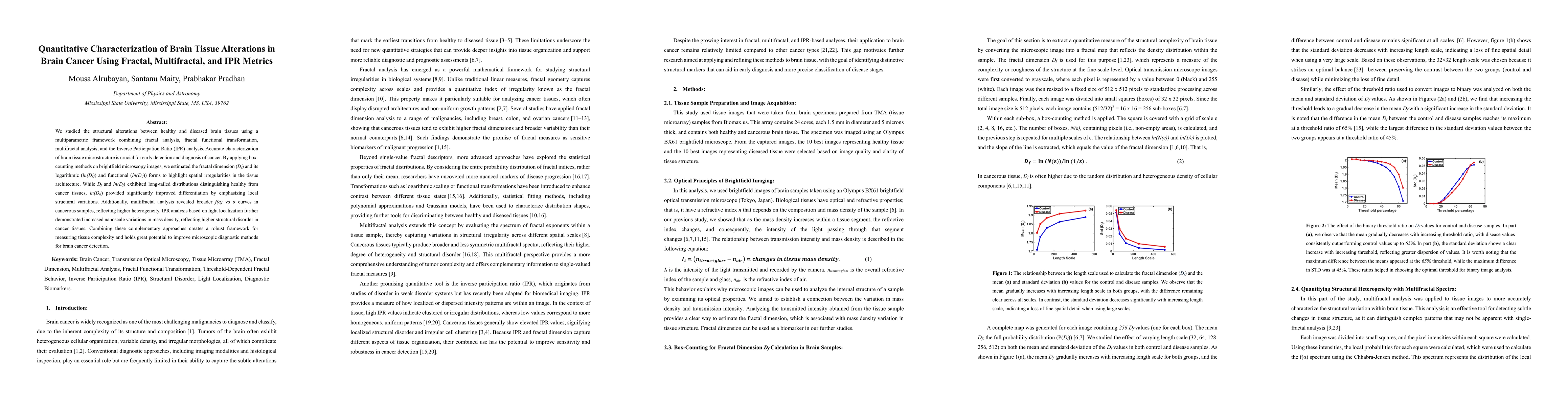
We studied the structural alterations between healthy and diseased brain tissues using a multiparametric framework combining fractal analysis, fractal functional transformation, multifractal analysis,...

This website uses cookies to ensure you get the best experience. Learn more