Summary
Ovarian cancer remains a challenging malignancy to diagnose and manage, with prognosis heavily dependent on the stage at detection. Accurate grading and staging, primarily based on histopathological examination of biopsy tissue samples, are crucial for treatment planning and predicting outcomes. However, this manual process is time-consuming and subject to inter-observer variability among pathologists. The increasing volume of digital histopathology slides necessitates the development of robust, automated methods to assist in this critical diagnostic step for ovarian cancer. (Methods) This study presents a deep learning framework for the automated prediction of ovarian cancer stage (classified into five categories: 0, I, II, III, IV) using routine histopathological images. We employed a transfer learning approach, fine-tuning a ResNet-101 convolutional neural network pre-trained on ImageNet. The training process incorporated comprehensive data augmentation, weighted random sampling, and class weighting to address dataset characteristics. Hyperparameter optimization for learning rate, dropout rate, and weight decay was performed using a genetic algorithm to enhance model performance and generalization. (Results) Evaluated on an independent test set of ovarian thin tissue brightfield images, the developed model achieved a high overall classification accuracy of 97.62%.
AI Key Findings
Generated Jun 08, 2025
Methodology
The research employs a deep learning framework using a fine-tuned ResNet-101 CNN pre-trained on ImageNet. It incorporates transfer learning, data augmentation, weighted random sampling, class weighting, and hyperparameter optimization via a genetic algorithm for improved performance and generalization.
Key Results
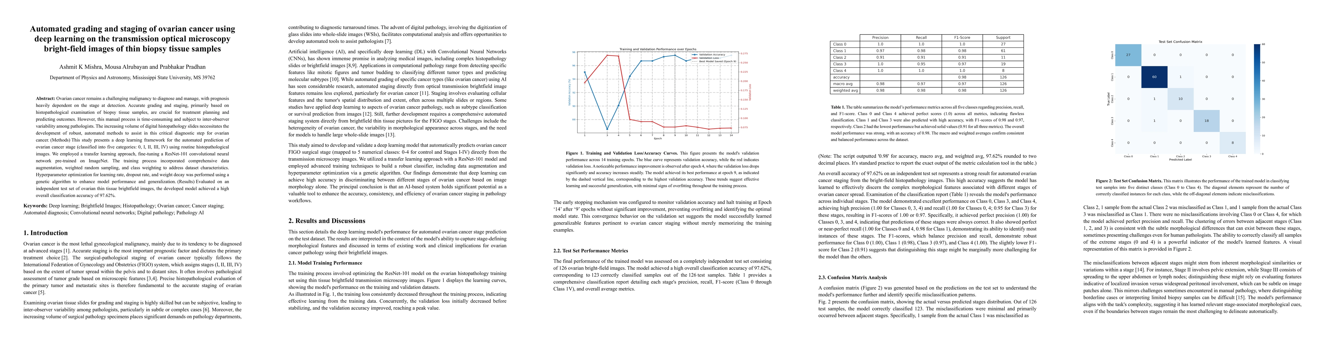
- The model achieved an overall classification accuracy of 97.62% on an independent test set of ovarian thin tissue brightfield images.
- The model predicts ovarian cancer stage (classified into five categories: 0, I, II, III, IV) from routine histopathological images.
Significance
This research is important as it addresses the time-consuming and variable nature of manual ovarian cancer grading and staging, which is critical for treatment planning and outcome prediction. The automated method can potentially improve diagnostic consistency and efficiency in handling the increasing volume of digital histopathology slides.
Technical Contribution
The development of a deep learning framework for automated ovarian cancer staging using transfer learning and optimized hyperparameters, demonstrating high accuracy on independent test data.
Novelty
This work stands out by applying transfer learning to a ResNet-101 CNN for ovarian cancer staging, addressing a critical diagnostic challenge with high accuracy and potentially improving diagnostic consistency.
Limitations
- The study was limited to thin tissue brightfield images; performance with other imaging modalities or thicker samples remains unexplored.
- Generalizability to diverse pathologist practices and institutions wasn't assessed.
Future Work
- Investigate model performance with various imaging modalities and tissue sample types.
- Evaluate the model's applicability across different pathologist practices and institutions.
Paper Details
PDF Preview
Citation Network
Current paper (gray), citations (green), references (blue)
Display is limited for performance on very large graphs.
Similar Papers
Found 4 papersEvaluation and optimization of deep learning models for enhanced detection of brain cancer using transmission optical microscopy of thin brain tissue samples
Mousa Alrubayan, Prabhakar Pradhan, Mohnish Sao
Lymphocyte Classification in Hyperspectral Images of Ovarian Cancer Tissue Biopsy Samples
John Cisler, Benjamin Paulson, Andrew Crisler et al.
Digital Volumetric Biopsy Cores Improve Gleason Grading of Prostate Cancer Using Deep Learning
Zichen Wang, William Speier, Corey W. Arnold et al.
No citations found for this paper.
Comments (0)