ArxivLens
P
Researcher

Priscille de Dumast

9 papers on arXiv

9 arXiv Papers
22 Total Publications
Profile

Academic Profile

Name P. D. Dumast
Total Papers 22
Scholar Profile View on Semantic Scholar
Metrics

Statistics

9 arXiv Papers
22 Total Publications
Publications

Papers on arXiv

Thumbnail for Multi-Center Fetal Brain Tissue Annotation (FeTA) Challenge 2022 Results
arXiv
Multi-Center Fetal Brain Tissue Annotation (FeTA) Challenge 2022 Results

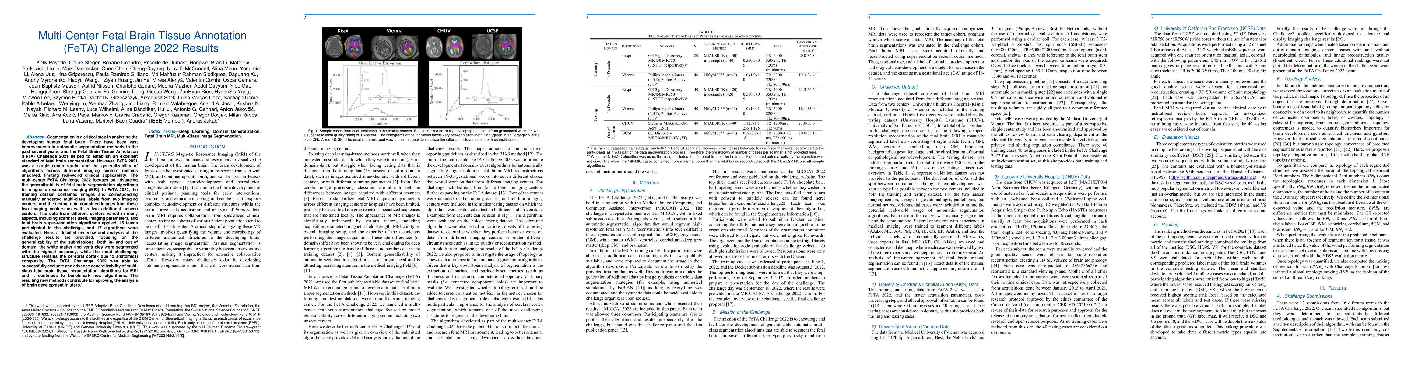
Segmentation is a critical step in analyzing the developing human fetal brain. There have been vast improvements in automatic segmentation methods in the past several years, and the Fetal Brain Tiss...

Thumbnail for Domain generalization in fetal brain MRI segmentation \\with
  multi-reconstruction augmentation
arXiv
Domain generalization in fetal brain MRI segmentation \\with multi-reconstruction augmentation

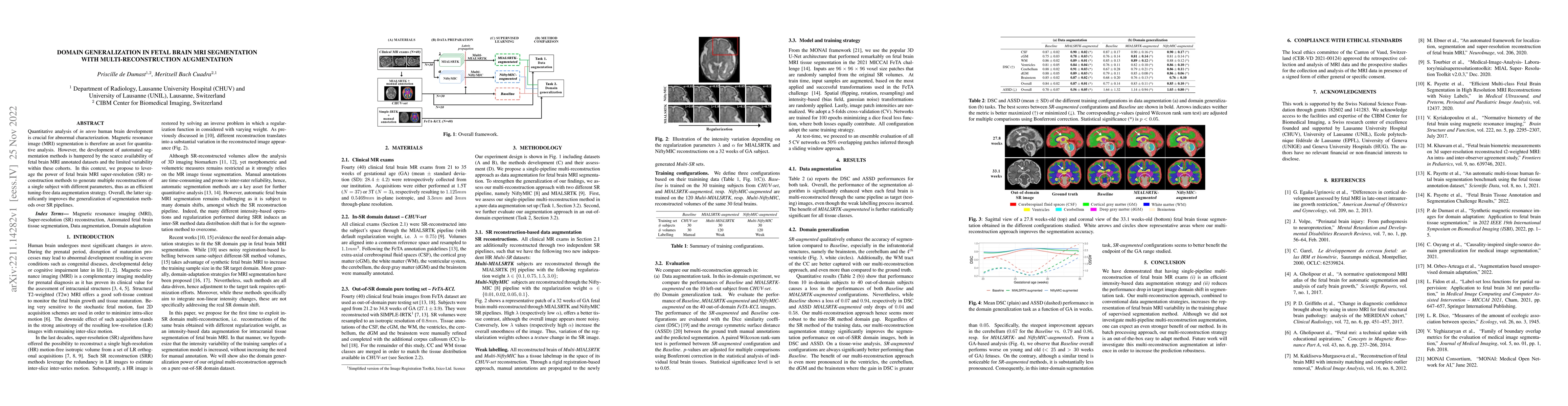
Quantitative analysis of in utero human brain development is crucial for abnormal characterization. Magnetic resonance image (MRI) segmentation is therefore an asset for quantitative analysis. Howev...

Thumbnail for Simulation-based parameter optimization for fetal brain MRI
  super-resolution reconstruction
arXiv
Simulation-based parameter optimization for fetal brain MRI super-resolution reconstruction

Tuning the regularization hyperparameter $\alpha$ in inverse problems has been a longstanding problem. This is particularly true in the case of fetal brain magnetic resonance imaging, where an isotr...

Thumbnail for Multi-dimensional topological loss for cortical plate segmentation in
  fetal brain MRI
arXiv
Multi-dimensional topological loss for cortical plate segmentation in fetal brain MRI

The fetal cortical plate (CP) undergoes drastic morphological changes during the in utero development. Therefore, CP growth and folding patterns are key indicator in the assessment of the brain deve...

Thumbnail for Fetal Brain Tissue Annotation and Segmentation Challenge Results
arXiv
Fetal Brain Tissue Annotation and Segmentation Challenge Results

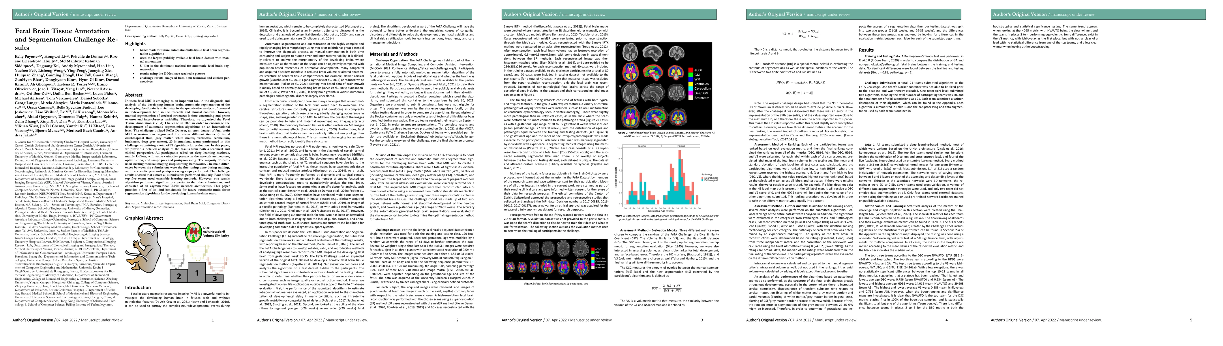
In-utero fetal MRI is emerging as an important tool in the diagnosis and analysis of the developing human brain. Automatic segmentation of the developing fetal brain is a vital step in the quantitat...

Thumbnail for 4D iterative reconstruction of brain fMRI in the moving fetus
arXiv
4D iterative reconstruction of brain fMRI in the moving fetus

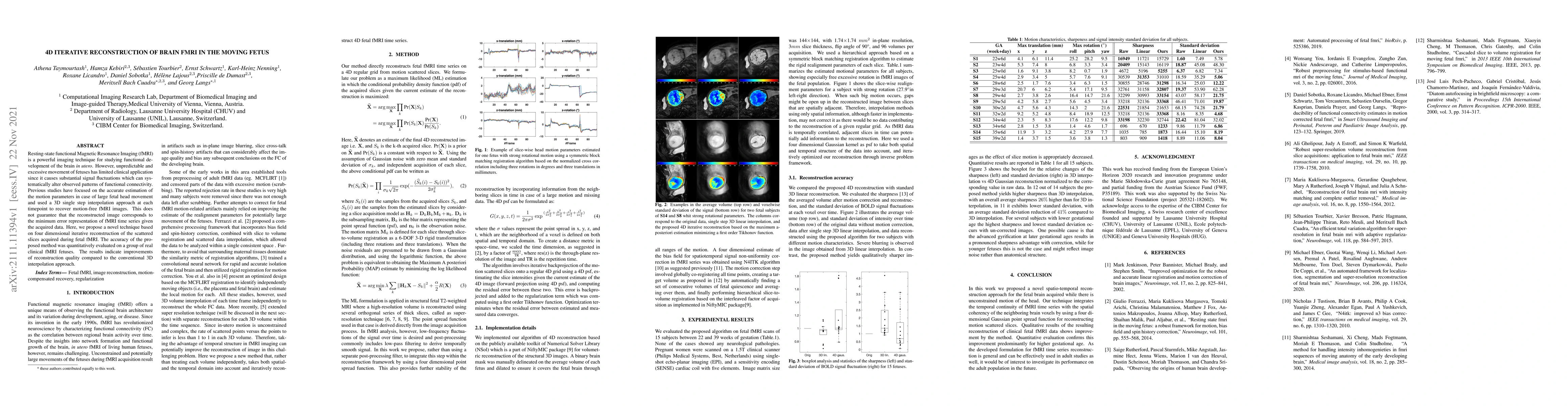
Resting-state functional Magnetic Resonance Imaging (fMRI) is a powerful imaging technique for studying functional development of the brain in utero. However, unpredictable and excessive movement of...

Thumbnail for Synthetic magnetic resonance images for domain adaptation: Application
  to fetal brain tissue segmentation
arXiv
Synthetic magnetic resonance images for domain adaptation: Application to fetal brain tissue segmentation

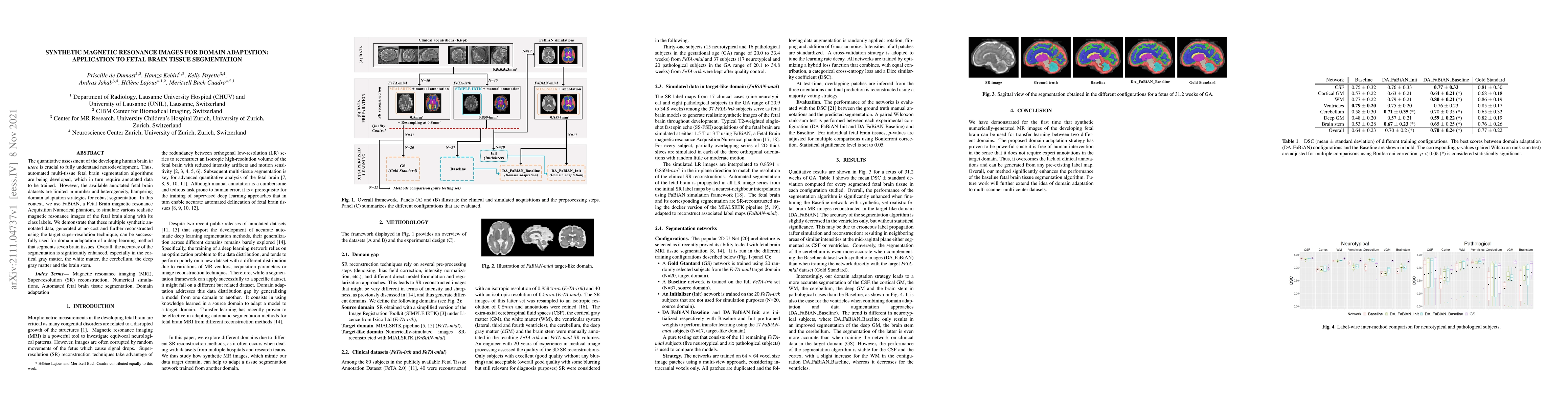
The quantitative assessment of the developing human brain in utero is crucial to fully understand neurodevelopment. Thus, automated multi-tissue fetal brain segmentation algorithms are being develop...

Thumbnail for FaBiAN: A Fetal Brain magnetic resonance Acquisition Numerical phantom
arXiv
FaBiAN: A Fetal Brain magnetic resonance Acquisition Numerical phantom

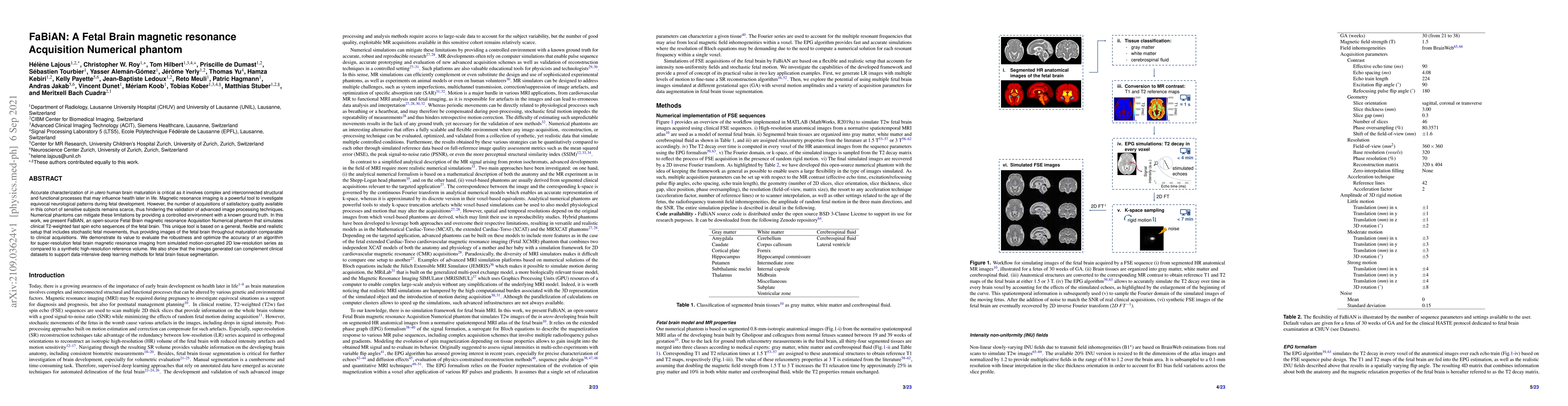
Accurate characterization of in utero human brain maturation is critical as it involves complex and interconnected structural and functional processes that may influence health later in life. Magnet...

Thumbnail for T2 Mapping from Super-Resolution-Reconstructed Clinical Fast Spin Echo
  Magnetic Resonance Acquisitions
arXiv
T2 Mapping from Super-Resolution-Reconstructed Clinical Fast Spin Echo Magnetic Resonance Acquisitions

Relaxometry studies in preterm and at-term newborns have provided insight into brain microstructure, thus opening new avenues for studying normal brain development and supporting diagnosis in equivo...

This website uses cookies to ensure you get the best experience. Learn more