Academic Profile
Statistics
Similar Authors
Papers on arXiv
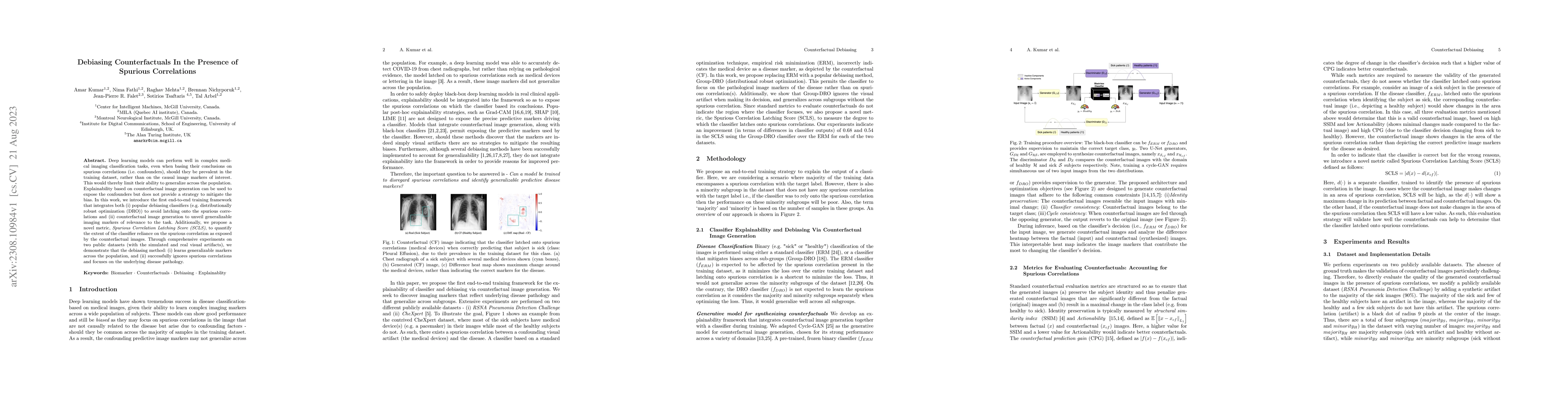
Deep learning models can perform well in complex medical imaging classification tasks, even when basing their conclusions on spurious correlations (i.e. confounders), should they be prevalent in the...
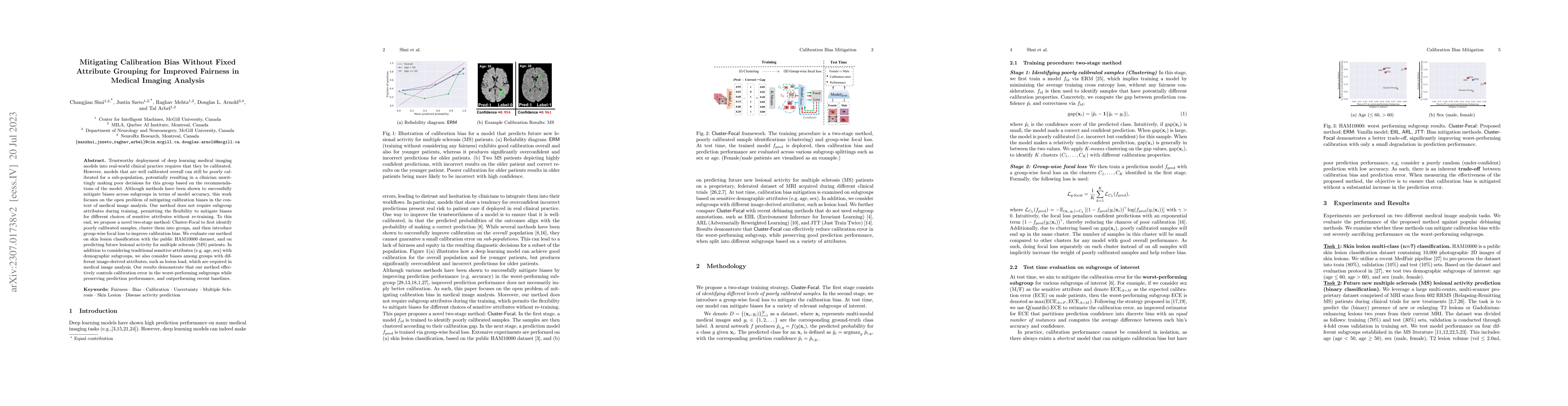
Trustworthy deployment of deep learning medical imaging models into real-world clinical practice requires that they be calibrated. However, models that are well calibrated overall can still be poorl...
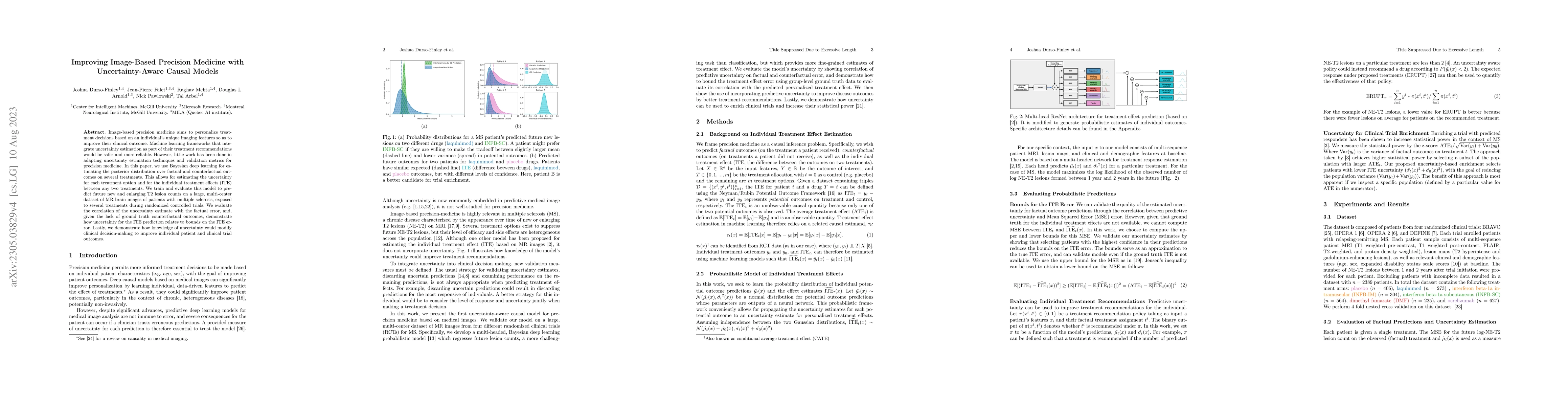
Image-based precision medicine aims to personalize treatment decisions based on an individual's unique imaging features so as to improve their clinical outcome. Machine learning frameworks that inte...
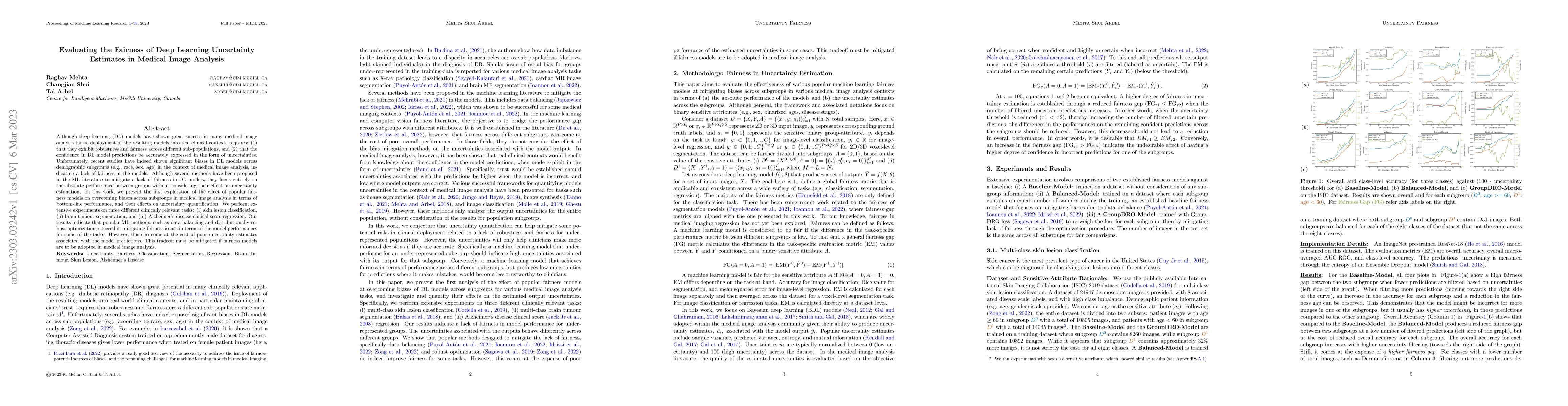
Although deep learning (DL) models have shown great success in many medical image analysis tasks, deployment of the resulting models into real clinical contexts requires: (1) that they exhibit robus...
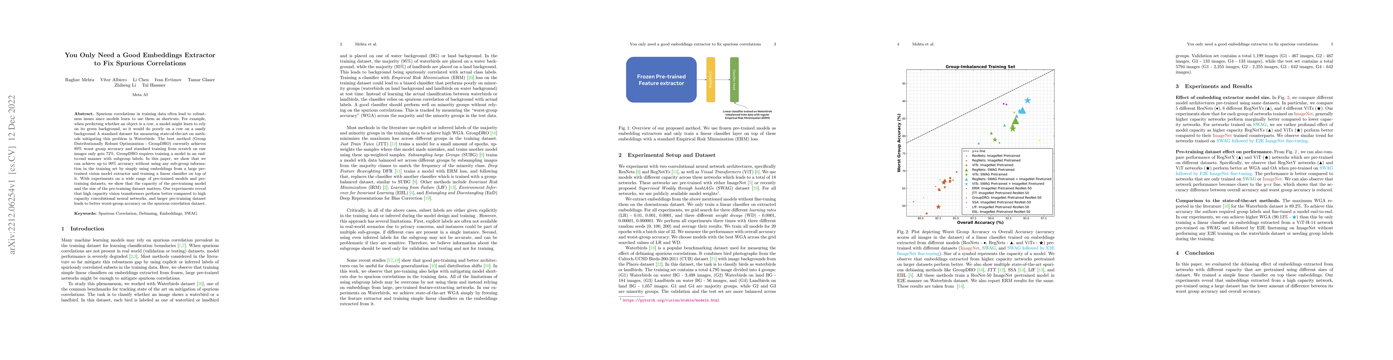
Spurious correlations in training data often lead to robustness issues since models learn to use them as shortcuts. For example, when predicting whether an object is a cow, a model might learn to re...
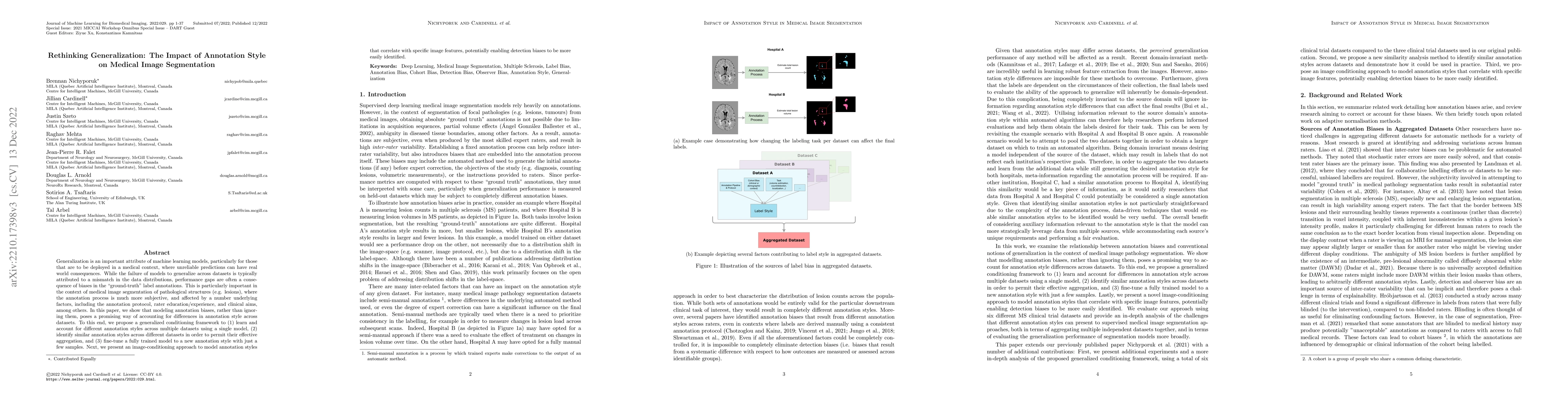
Generalization is an important attribute of machine learning models, particularly for those that are to be deployed in a medical context, where unreliable predictions can have real world consequence...
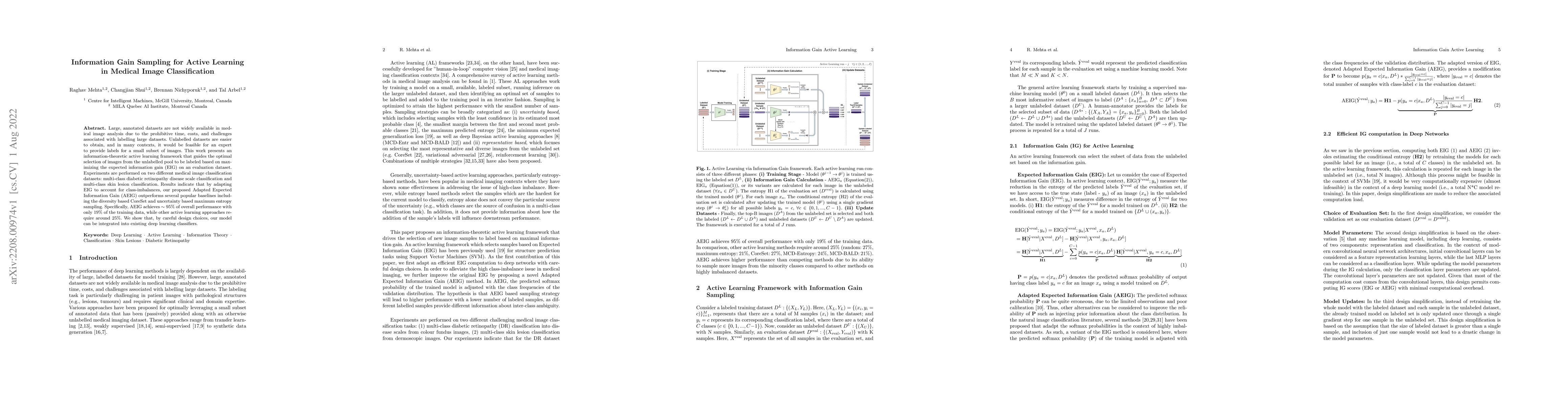
Large, annotated datasets are not widely available in medical image analysis due to the prohibitive time, costs, and challenges associated with labelling large datasets. Unlabelled datasets are easi...
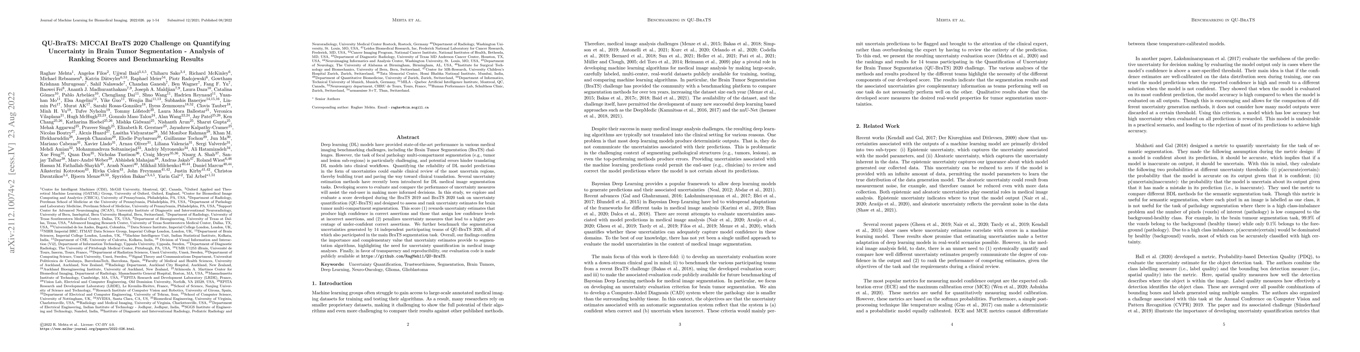
Deep learning (DL) models have provided state-of-the-art performance in various medical imaging benchmarking challenges, including the Brain Tumor Segmentation (BraTS) challenges. However, the task ...
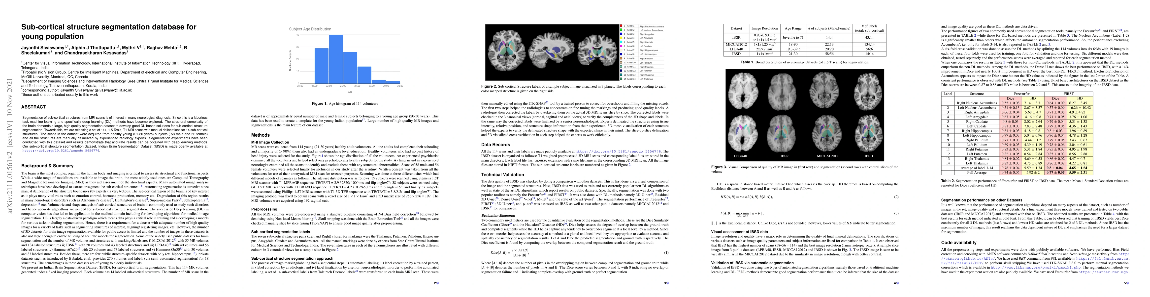
Segmentation of sub-cortical structures from MRI scans is of interest in many neurological diagnosis. Since this is a laborious task machine learning and specifically deep learning (DL) methods have...
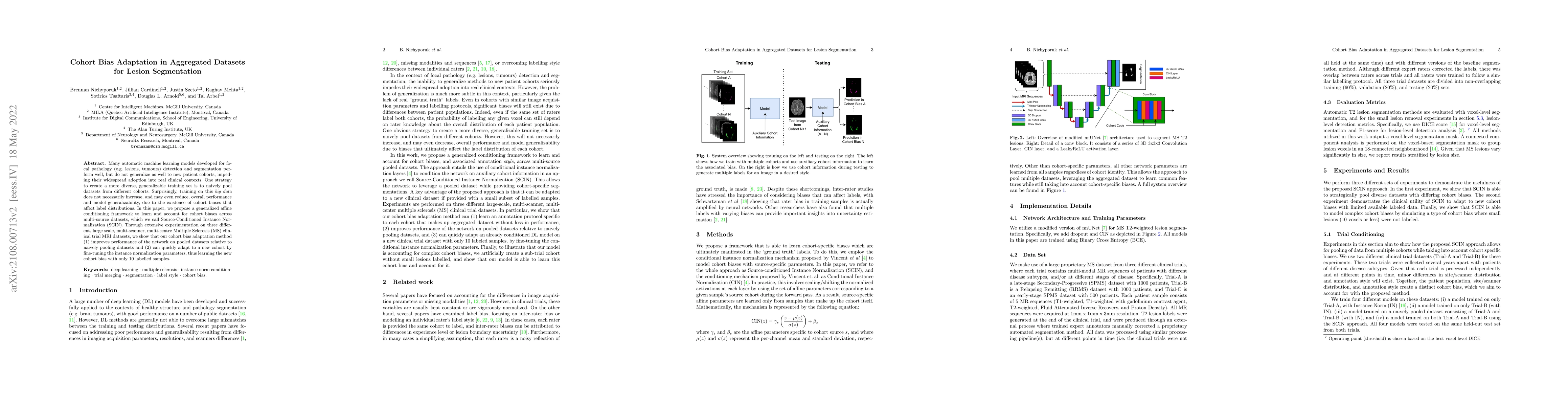
Many automatic machine learning models developed for focal pathology (e.g. lesions, tumours) detection and segmentation perform well, but do not generalize as well to new patient cohorts, impeding t...
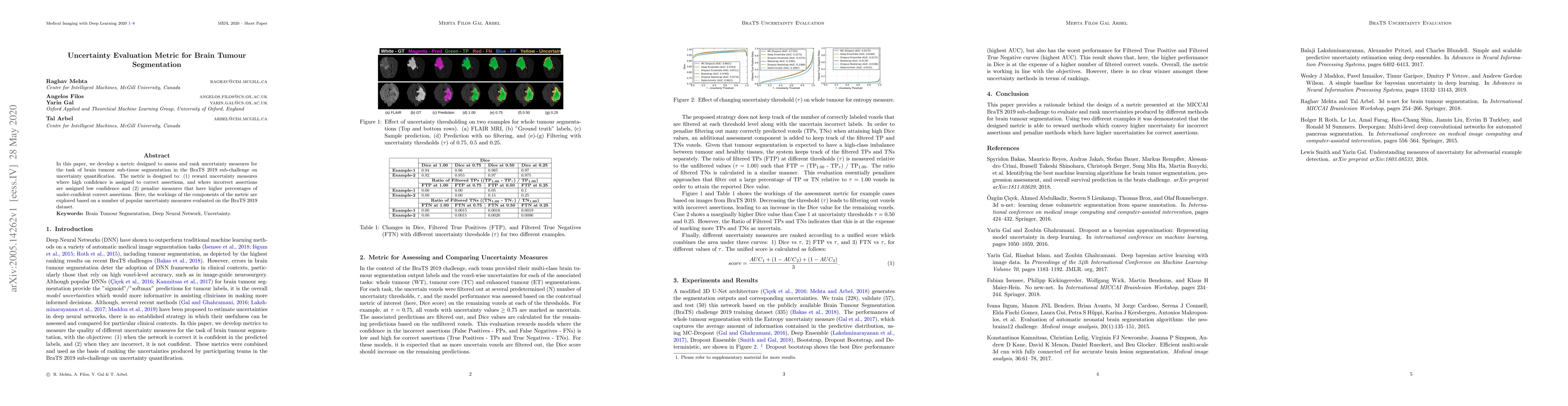
In this paper, we develop a metric designed to assess and rank uncertainty measures for the task of brain tumour sub-tissue segmentation in the BraTS 2019 sub-challenge on uncertainty quantification...
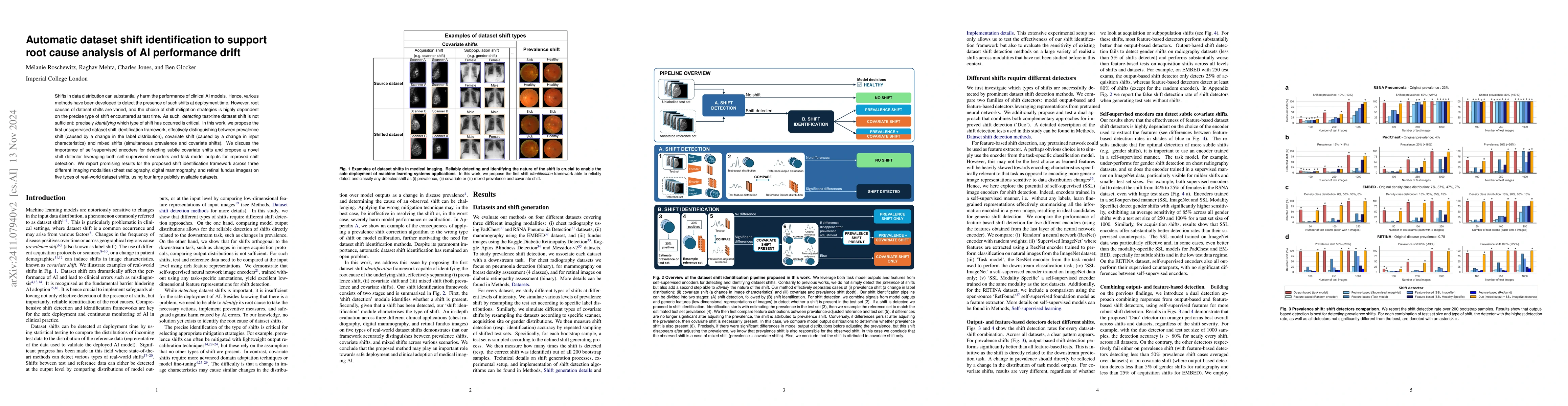
Shifts in data distribution can substantially harm the performance of clinical AI models. Hence, various methods have been developed to detect the presence of such shifts at deployment time. However, ...
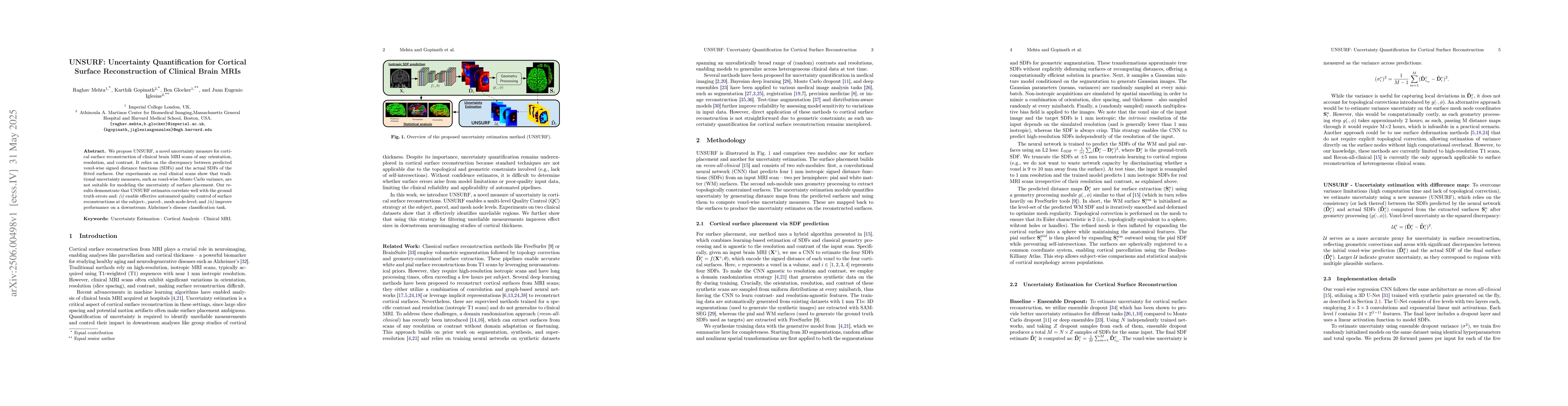
We propose UNSURF, a novel uncertainty measure for cortical surface reconstruction of clinical brain MRI scans of any orientation, resolution, and contrast. It relies on the discrepancy between predic...
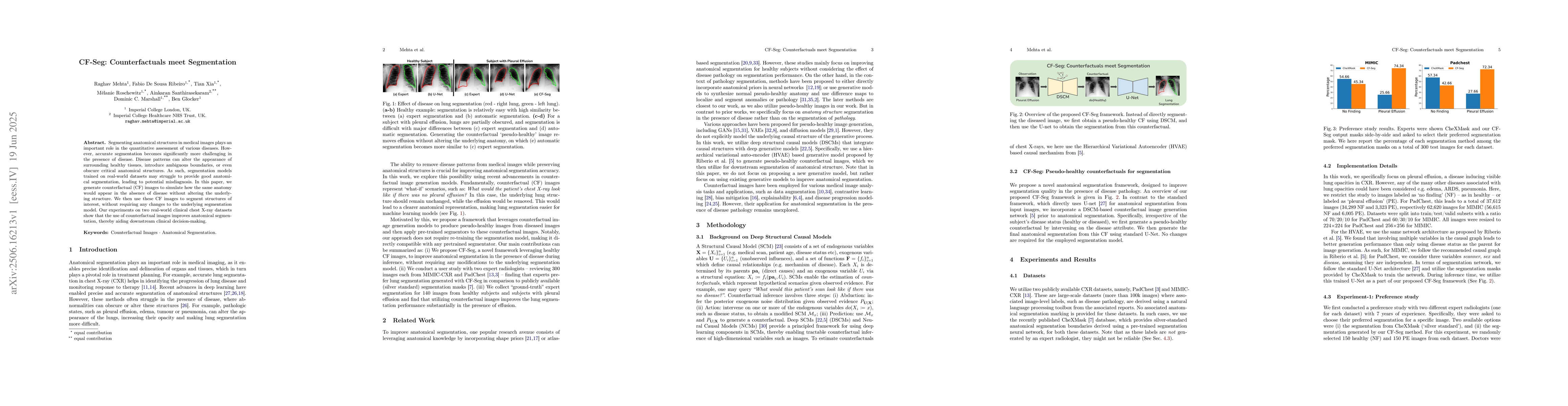
Segmenting anatomical structures in medical images plays an important role in the quantitative assessment of various diseases. However, accurate segmentation becomes significantly more challenging in ...
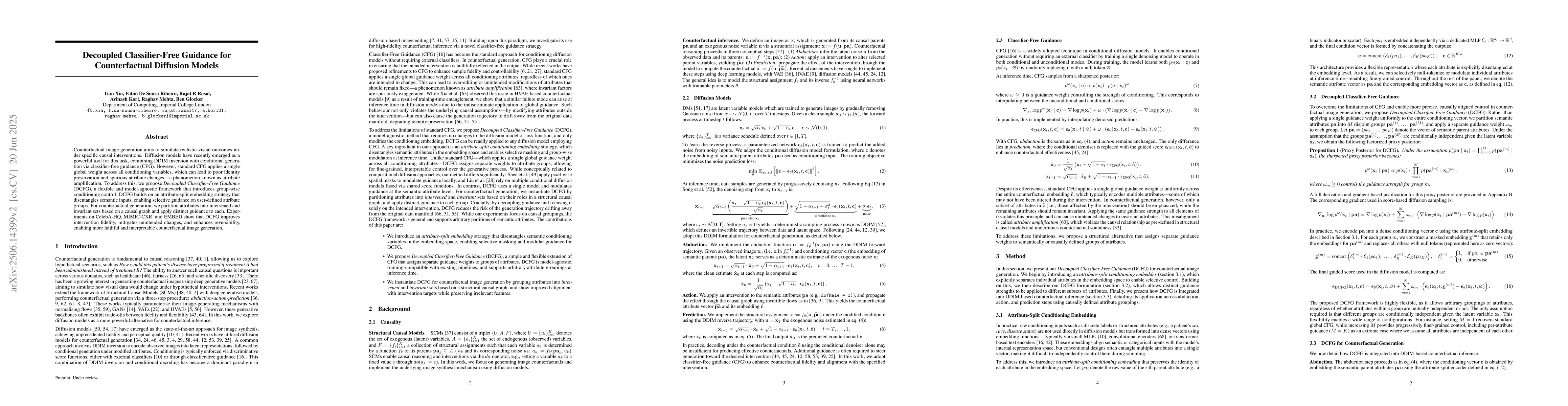
Counterfactual image generation aims to simulate realistic visual outcomes under specific causal interventions. Diffusion models have recently emerged as a powerful tool for this task, combining DDIM ...
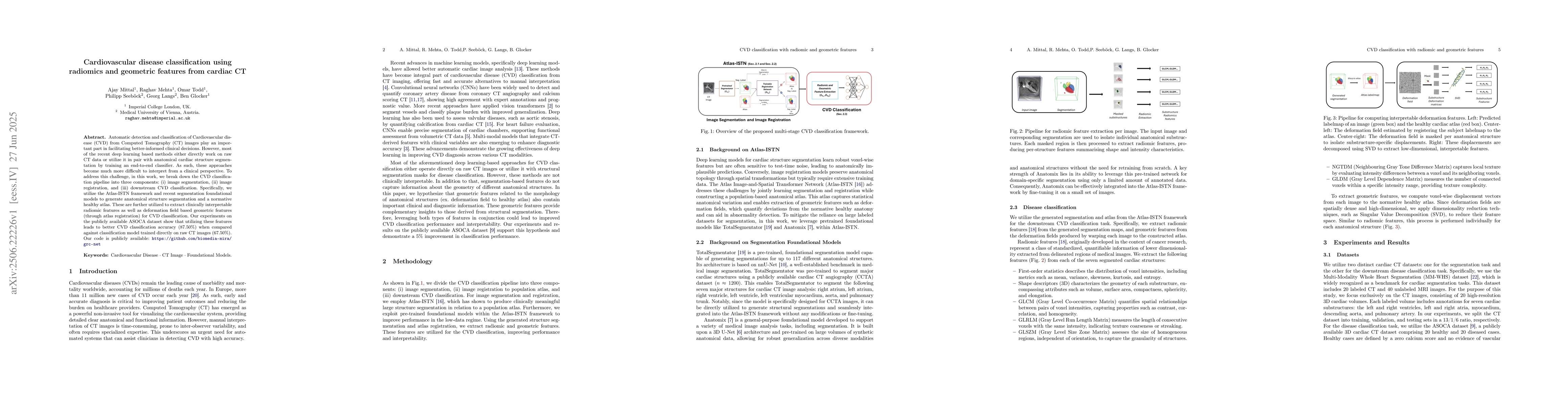
Automatic detection and classification of Cardiovascular disease (CVD) from Computed Tomography (CT) images play an important part in facilitating better-informed clinical decisions. However, most of ...
We conduct an extensive study on the state of calibration under real-world dataset shift for image classification. Our work provides important insights on the choice of post-hoc and in-training calibr...
Systematic mislabelling affecting specific subgroups (i.e., label bias) in medical imaging datasets represents an understudied issue concerning the fairness of medical AI systems. In this work, we inv...
We introduce the Flow Stochastic Segmentation Network (Flow-SSN), a generative segmentation model family featuring discrete-time autoregressive and modern continuous-time flow variants. We prove funda...
Counterfactual image generation is a powerful tool for augmenting training data, de-biasing datasets, and modeling disease. Current approaches rely on external classifiers or regressors to increase th...
Image segmentation relies on large annotated datasets, which are expensive and slow to produce. Silver-standard (AI-generated) labels are easier to obtain, but they risk introducing bias. Self-supervi...
Counterfactual image generation enables controlled data augmentation, bias mitigation, and disease modeling. However, existing methods guided by external classifiers or regressors are limited to subje...
Deep learning models in medical imaging often fail when deployed in new clinical environments due to distribution shifts in demographics, scanner hardware, or acquisition protocols. A central challeng...
Foundation models are increasingly used as image feature extractors for mammography, but their robustness under external domain shift remains unclear. We benchmark 15 foundation-model backbones across...
The automatic detection and classification of cardiovascular disease (CVD) from computed tomography (CT) images plays an important role in clinical practice. Recently, a hybrid pipeline (GRC-Net) for ...