Cardiovascular disease classification using radiomics and geometric features from cardiac CT
Publication
Metrics
AI Quick Summary
This research introduces a novel CVD classification method using radiomics and geometric features from cardiac CT scans, breaking down the pipeline into image segmentation, registration, and classification. The approach, tested on the ASOCA dataset, outperforms direct CT image classification with 87.50% accuracy, offering improved interpretability for clinical use.
Paper Preview
Abstract
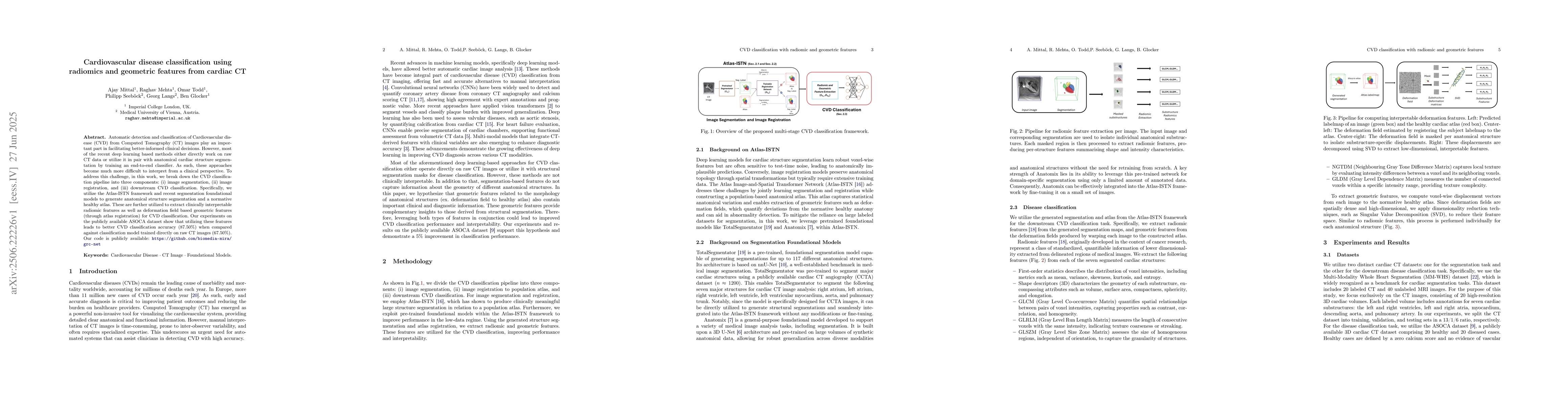
Automatic detection and classification of Cardiovascular disease (CVD) from Computed Tomography (CT) images play an important part in facilitating better-informed clinical decisions. However, most of the recent deep learning based methods either directly work on raw CT data or utilize it in pair with anatomical cardiac structure segmentation by training an end-to-end classifier. As such, these approaches become much more difficult to interpret from a clinical perspective. To address this challenge, in this work, we break down the CVD classification pipeline into three components: (i) image segmentation, (ii) image registration, and (iii) downstream CVD classification. Specifically, we utilize the Atlas-ISTN framework and recent segmentation foundational models to generate anatomical structure segmentation and a normative healthy atlas. These are further utilized to extract clinically interpretable radiomic features as well as deformation field based geometric features (through atlas registration) for CVD classification. Our experiments on the publicly available ASOCA dataset show that utilizing these features leads to better CVD classification accuracy (87.50\%) when compared against classification model trained directly on raw CT images (67.50\%). Our code is publicly available: https://github.com/biomedia-mira/grc-net
AI Key Findings
Get AI-generated insights about this paper's methodology, results, significance, and more — seven facets brought into focus.
Impact
Paper Details
Authors
PDF Preview
Citation Network
Current paper (gray), citations (green), references (blue)
Display is limited for performance on very large graphs.
Discussion 0