Academic Profile
Statistics
Similar Authors
Papers on arXiv
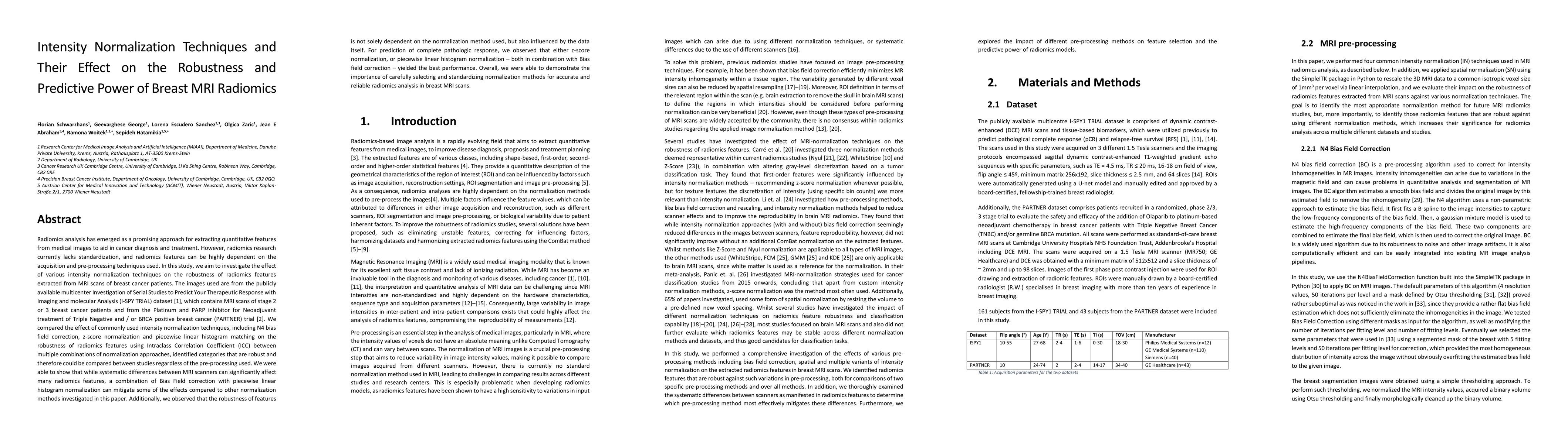
Radiomics analysis has emerged as a promising approach for extracting quantitative features from medical images to aid in cancer diagnosis and treatment. However, radiomics research currently lacks ...
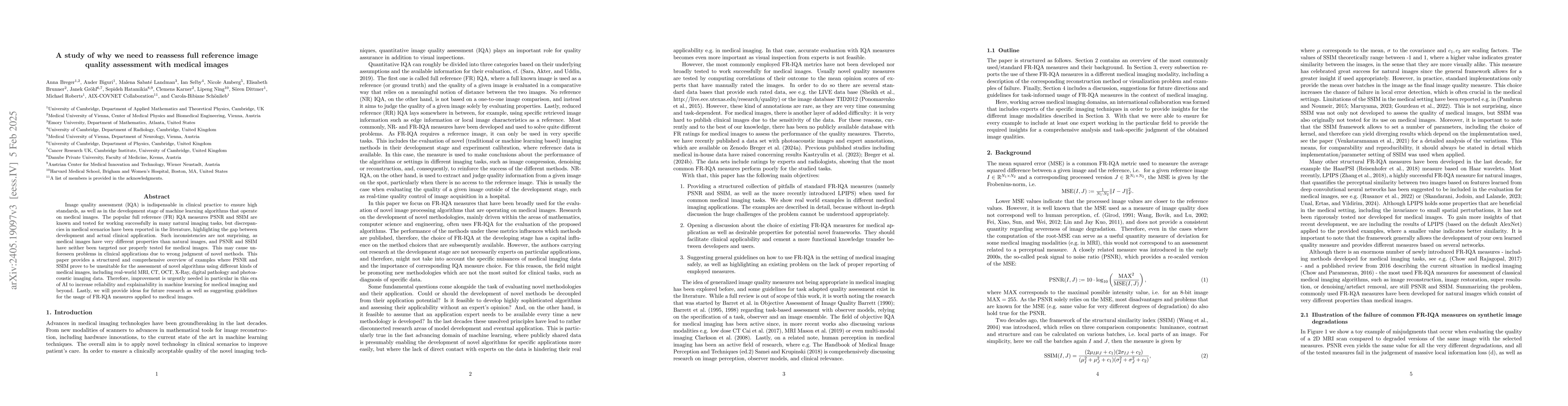
Image quality assessment (IQA) is not just indispensable in clinical practice to ensure high standards, but also in the development stage of novel algorithms that operate on medical images with refe...
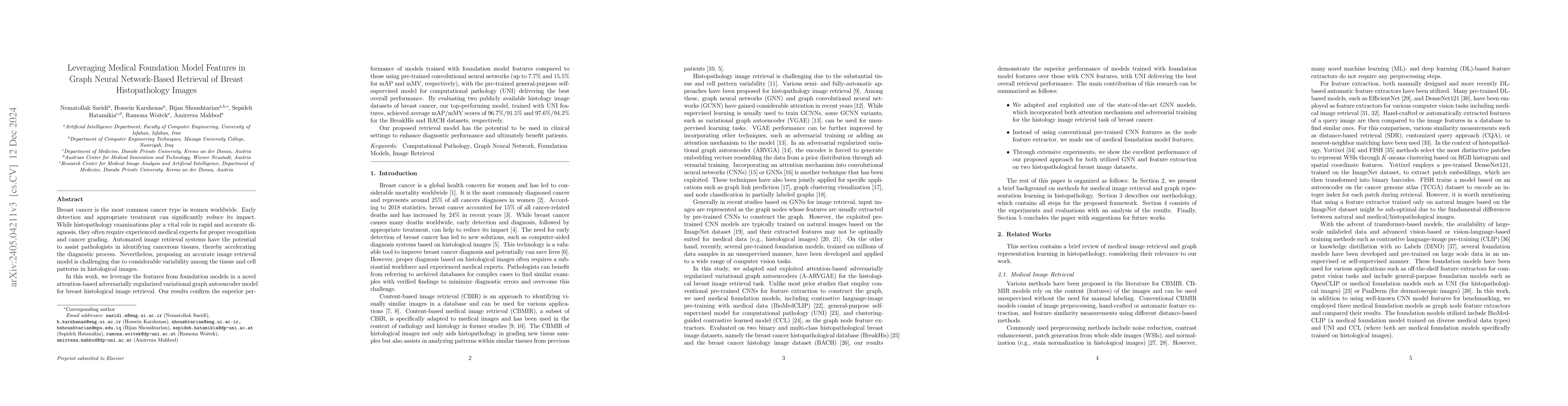
Breast cancer is the most common cancer type in women worldwide. Early detection and appropriate treatment can significantly reduce its impact. While histopathology examinations play a vital role in...
With the advent of digital pathology and microscopic systems that can scan and save whole slide histological images automatically, there is a growing trend to use computerized methods to analyze acq...
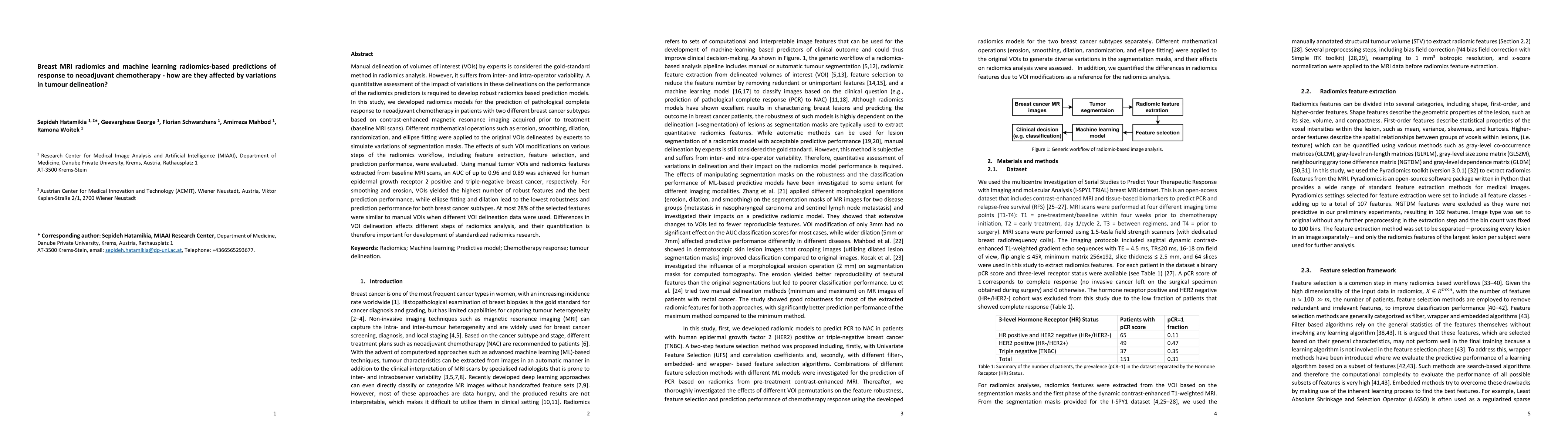
Manual delineation of volumes of interest (VOIs) by experts is considered the gold-standard method in radiomics analysis. However, it suffers from inter- and intra-operator variability. A quantitati...
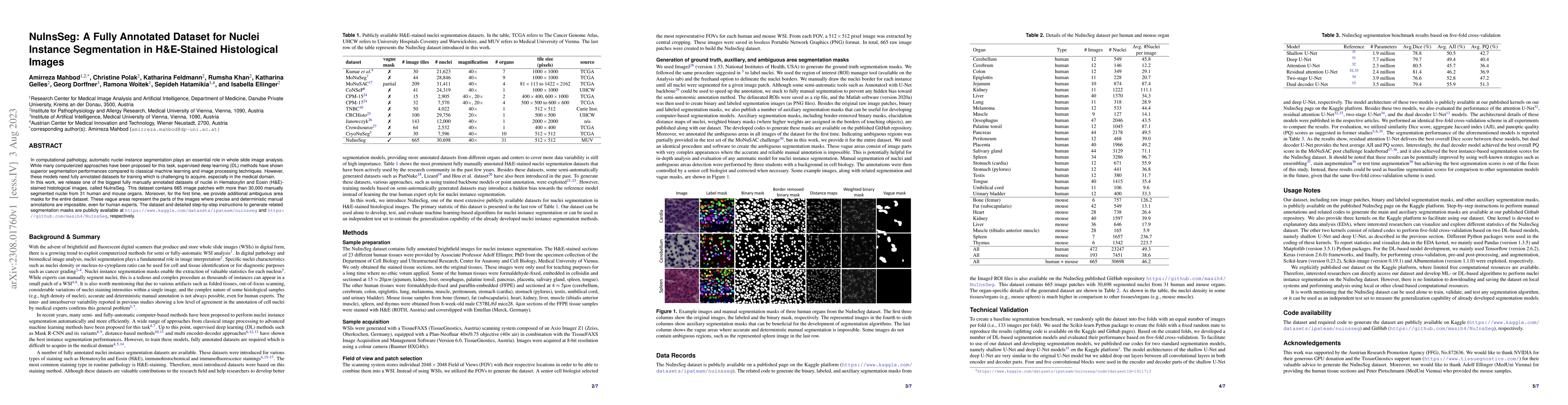
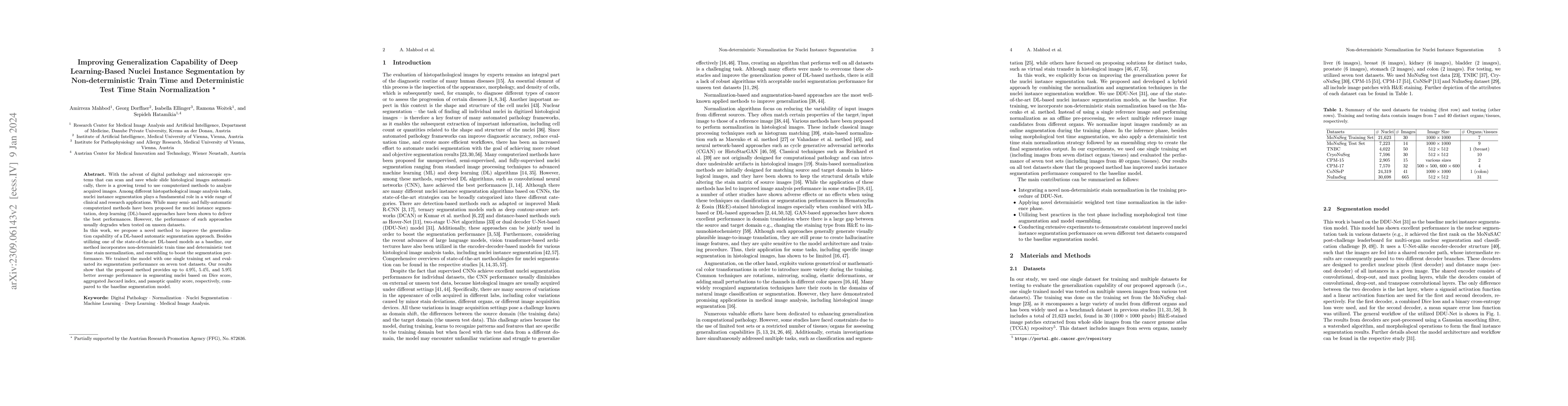
In computational pathology, automatic nuclei instance segmentation plays an essential role in whole slide image analysis. While many computerized approaches have been proposed for this task, supervi...
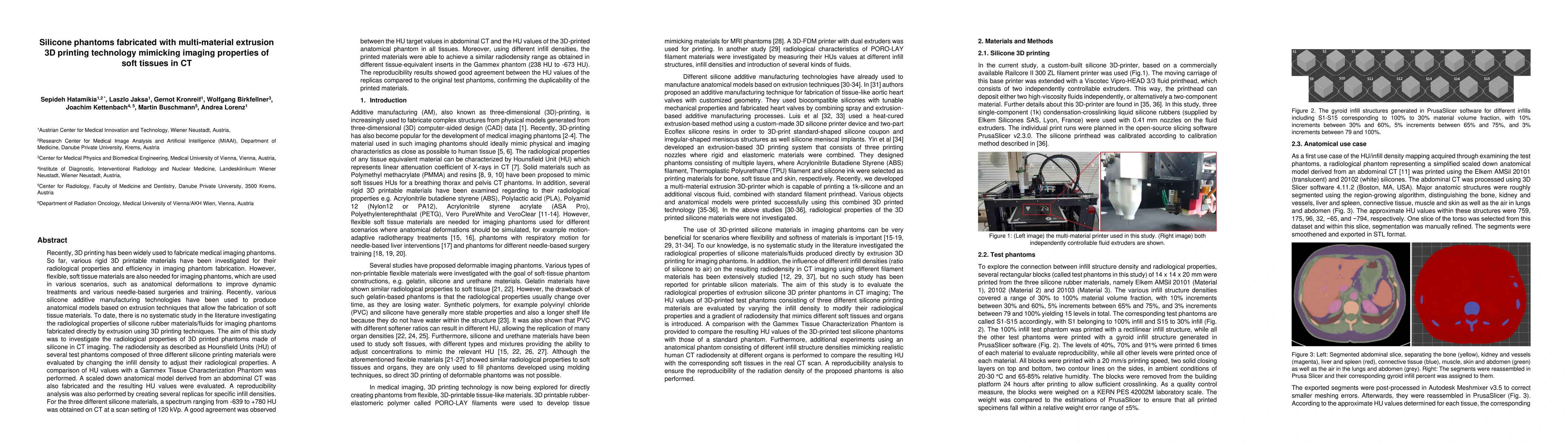
Recently, 3D printing has been widely used to fabricate medical imaging phantoms. So far, various rigid 3D printable materials have been investigated for their radiological properties and efficiency...
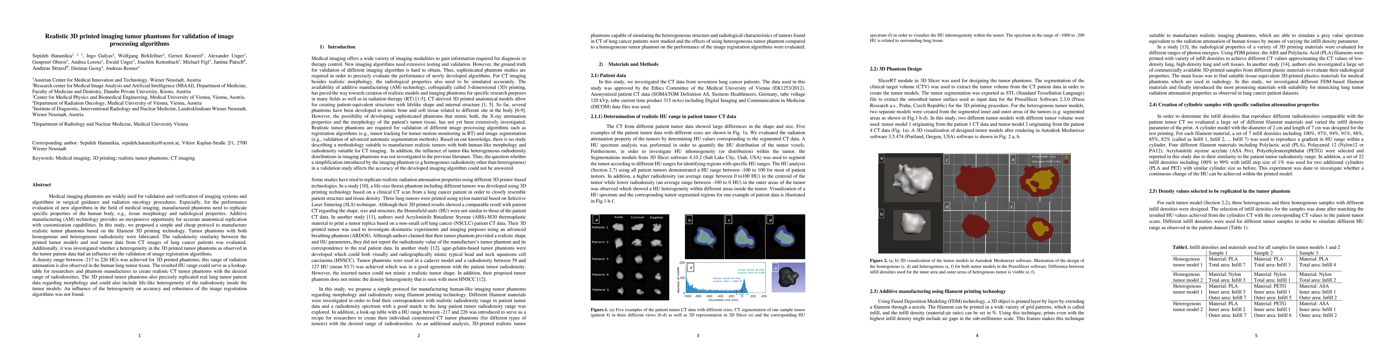
Medical imaging phantoms are widely used for validation and verification of imaging systems and algorithms in surgical guidance and radiation oncology procedures. Especially, for the performance eva...
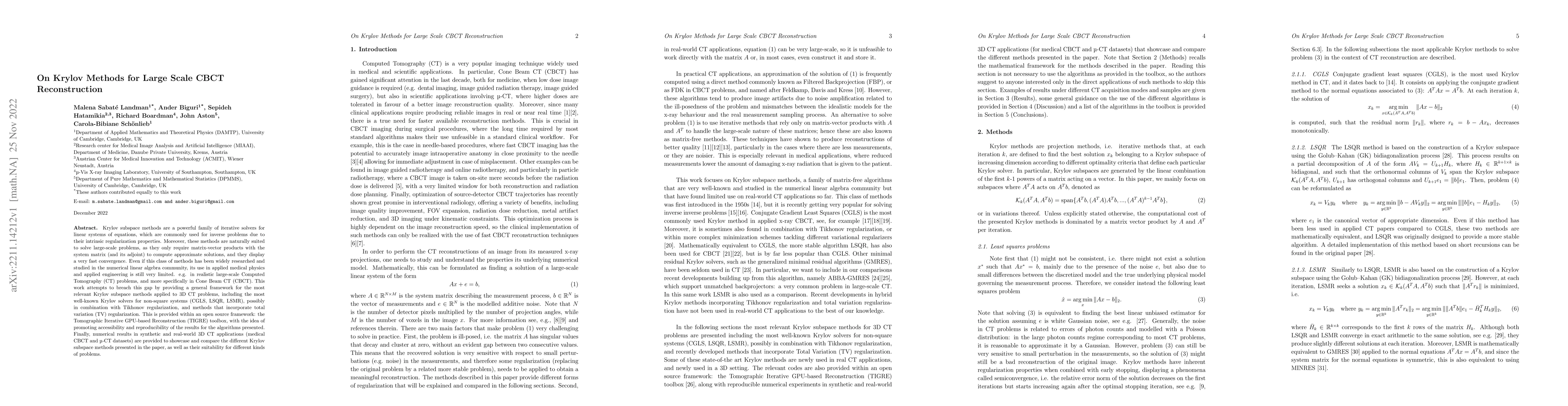
Krylov subspace methods are a powerful family of iterative solvers for linear systems of equations, which are commonly used for inverse problems due to their intrinsic regularization properties. Mor...
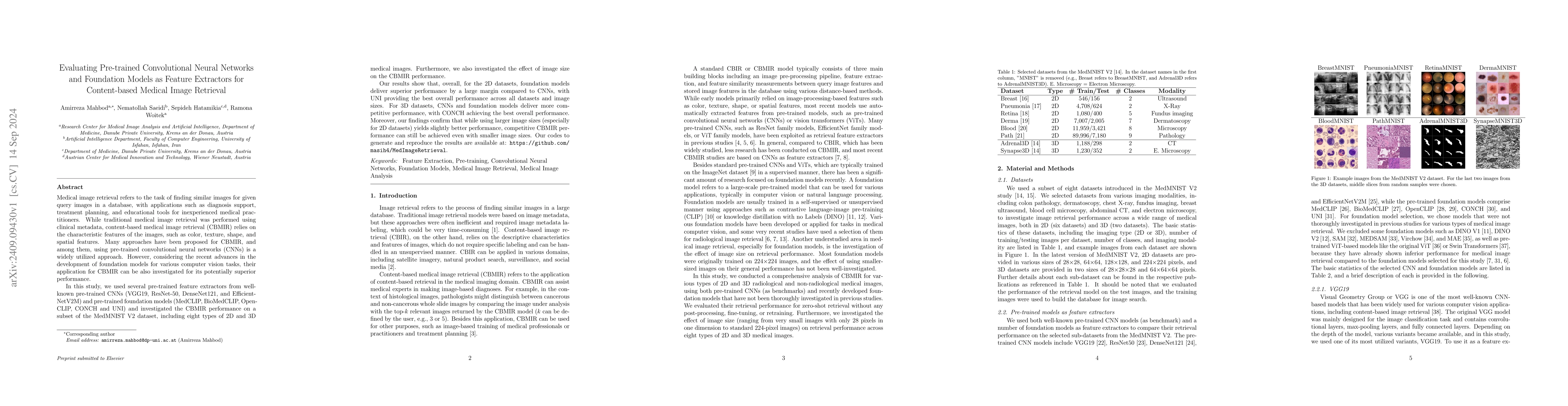
Medical image retrieval refers to the task of finding similar images for given query images in a database, with applications such as diagnosis support, treatment planning, and educational tools for in...
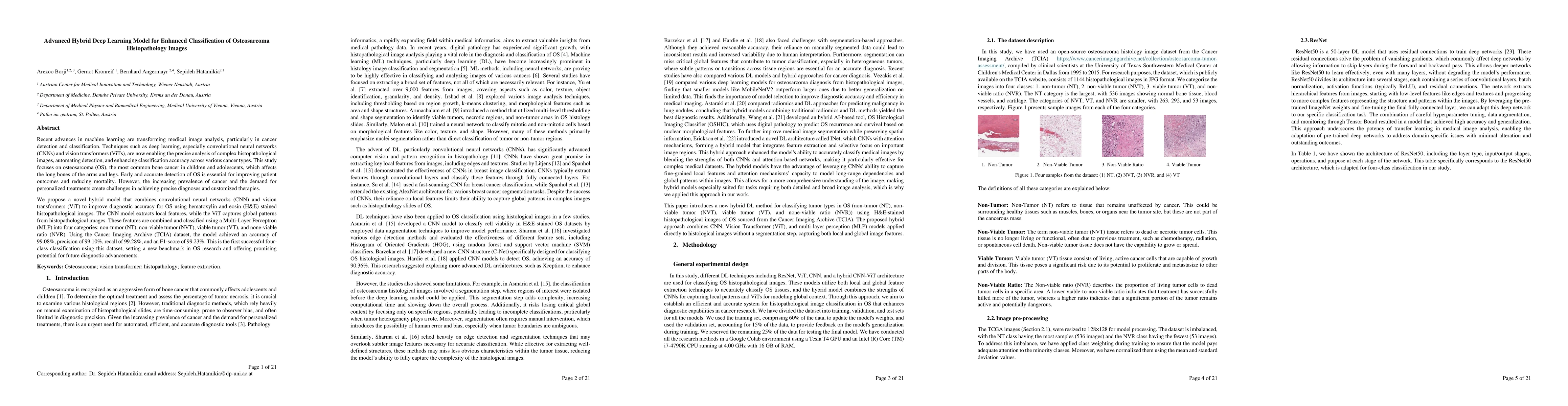
Recent advances in machine learning are transforming medical image analysis, particularly in cancer detection and classification. Techniques such as deep learning, especially convolutional neural netw...
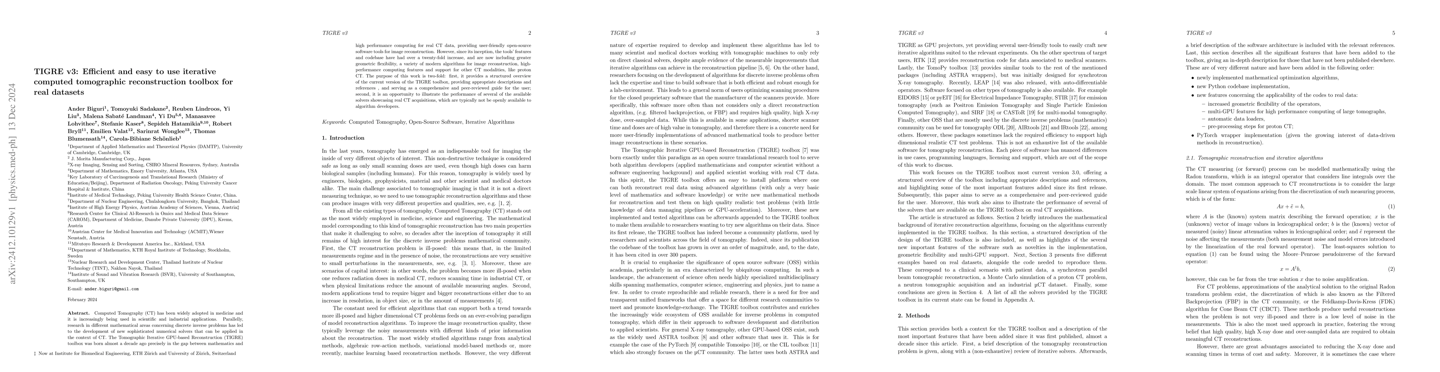
Computed Tomography (CT) has been widely adopted in medicine and it is increasingly being used in scientific and industrial applications. Parallelly, research in different mathematical areas concernin...
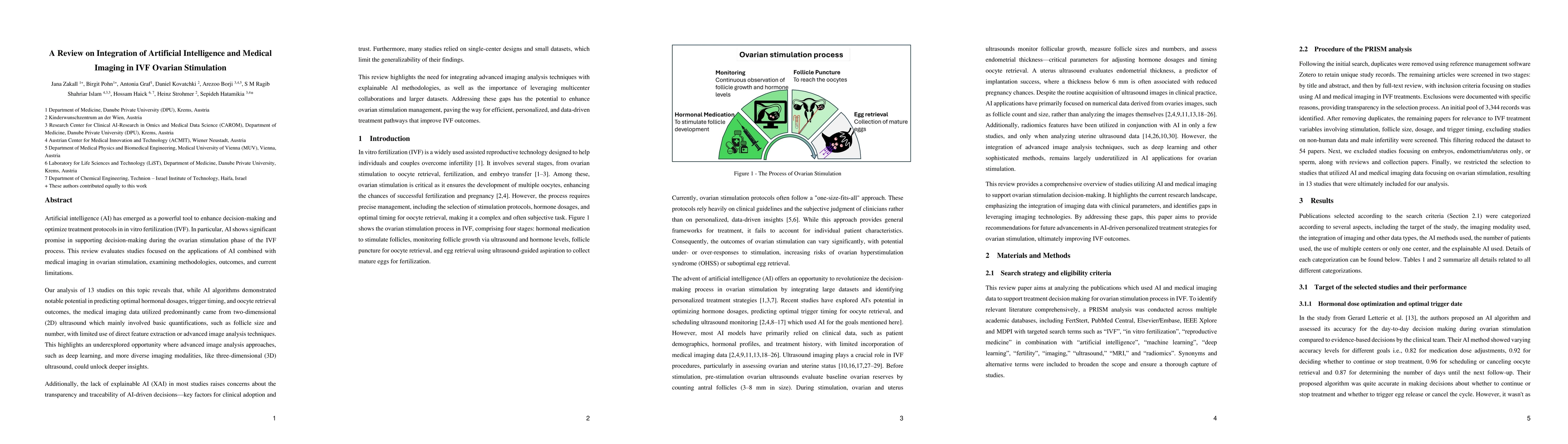
Artificial intelligence (AI) has emerged as a powerful tool to enhance decision-making and optimize treatment protocols in in vitro fertilization (IVF). In particular, AI shows significant promise in ...
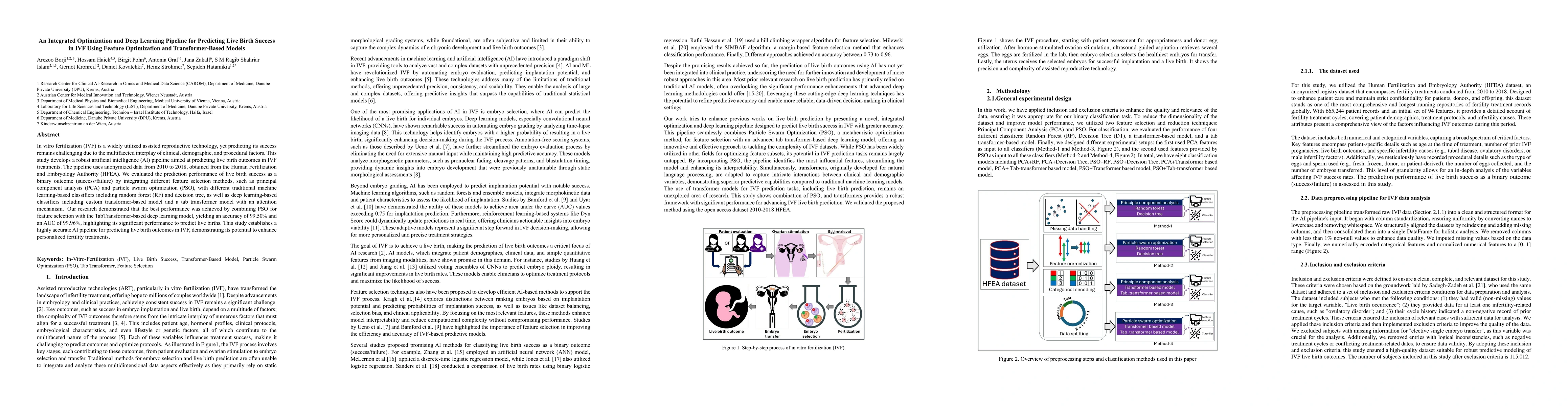
In vitro fertilization (IVF) is a widely utilized assisted reproductive technology, yet predicting its success remains challenging due to the multifaceted interplay of clinical, demographic, and proce...
To develop and externally validate integrated ultrasound nomograms combining BIRADS features and quantitative morphometric characteristics, and to compare their performance with expert radiologists an...
Melanoma is the most lethal form of skin cancer, with an increasing incidence rate worldwide. Analyzing histological images of melanoma by localizing and classifying tissues and cell nuclei is conside...
This work introduces a new efficient iterative solver for the reconstruction of real-time cone-beam computed tomography (CBCT), which is based on the Prior Image Constrained Compressed Sensing (PICCS)...
Background: The 2022 update of the Ovarian-Adnexal Reporting and Data System (O-RADS) ultrasound classification refines risk stratification for adnexal lesions, yet human interpretation remains subjec...
Objectives: High-grade serous ovarian carcinoma (HGSOC) is typically diagnosed at an advanced stage with extensive peritoneal metastases, making treatment challenging. Neoadjuvant chemotherapy (NACT) ...
Nuclei instance segmentation in hematoxylin and eosin (H&E)-stained images plays an important role in automated histological image analysis, with various applications in downstream tasks. While severa...
Autism spectrum disorder (ASD) is a complex neurodevelopmental condition characterized by atypical functional brain connectivity and subtle structural alterations. rs-fMRI has been widely used to iden...
Breast cancer is a highly heterogeneous disease with diverse molecular profiles. The PAM50 gene signature is widely recognized as a standard for classifying breast cancer into intrinsic subtypes, enab...
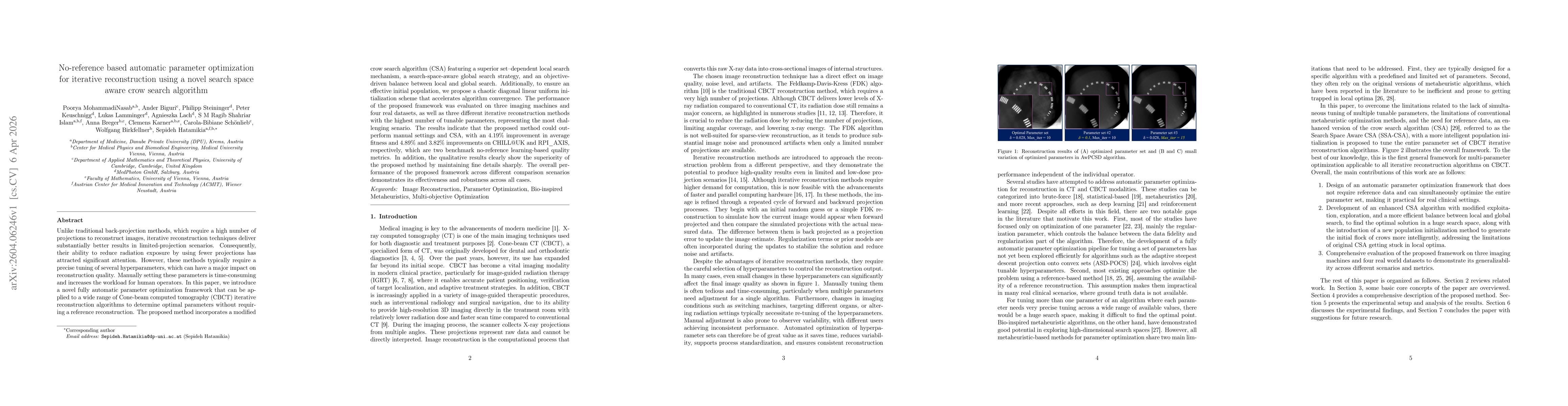
Iterative reconstruction technique's ability to reduce radiation exposure by using fewer projections has attracted significant attention. However, these methods typically require a precise tuning of s...
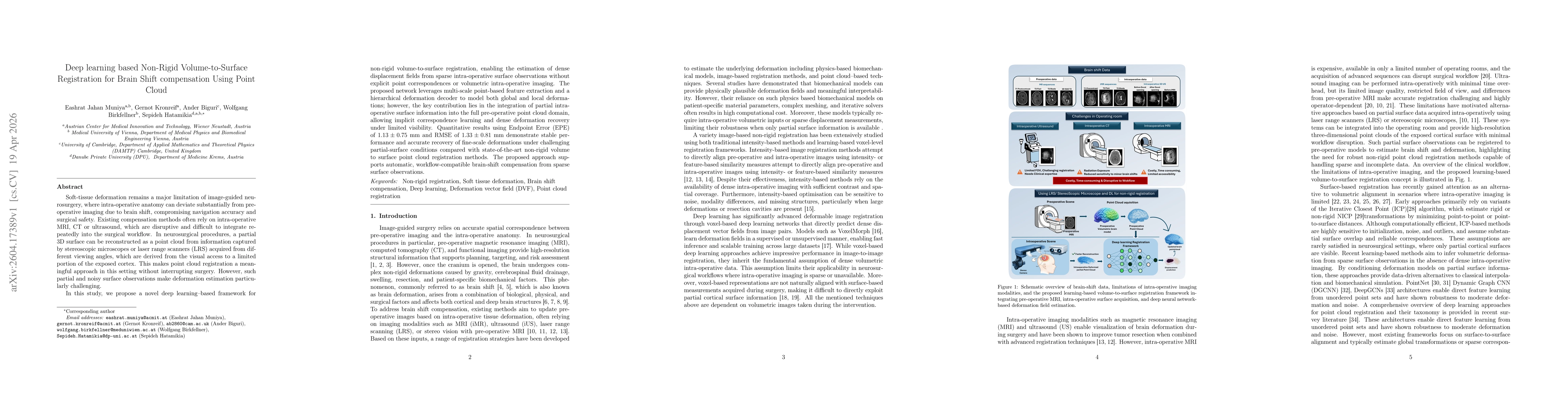
Soft-tissue deformation remains a major limitation in image-guided neurosurgery, where intra-operative anatomy can deviate substantially from pre-operative imaging due to brain shift, compromising nav...