The research presents NuInsSeg, a fully annotated dataset for nuclei instance segmentation in H&E-stained histological images, containing 665 image patches with over 30,000 manually segmented nuclei from 31 human and mouse organs. The dataset also includes ambiguous area masks for parts of images where precise annotations are impossible.
NuInsSeg: A Fully Annotated Dataset for Nuclei Instance Segmentation in H&E-Stained Histological Images
Publication
Metrics
AI Quick Summary
The paper introduces NuInsSeg, a large, fully annotated dataset for nuclei instance segmentation in H&E-stained histological images, comprising 665 patches with over 30,000 manually segmented nuclei from 31 organs. It also includes ambiguous area masks to indicate regions where precise annotations are challenging, aiming to enhance supervised deep learning methods in computational pathology.
Quick Answers
What is "NuInsSeg: A Fully Annotated Dataset for Nuclei Instance Segmentation in H&E-Stained Histological Images" about?
The paper introduces NuInsSeg, a large, fully annotated dataset for nuclei instance segmentation in H&E-stained histological images, comprising 665 patches with over 30,000 manually segmented nuclei from 31 organs. It also includes ambiguous area masks to indicate regions where precise annotations are challenging, aiming to enhance supervised deep learning methods in computational pathology.
What methodology did the authors use?
The research presents NuInsSeg, a fully annotated dataset for nuclei instance segmentation in H&E-stained histological images, containing 665 image patches with over 30,000 manually segmented nuclei from 31 human and mouse organs. The dataset also includes ambiguous area masks for parts of images where precise annotations are impossible. More in Methodology →
What are the key results?
NuInsSeg is one of the largest publicly available datasets for nuclei segmentation in H&E-stained histological images. — The dataset provides additional ambiguous area masks, which are crucial for in-depth analysis and evaluation of automatic nuclei instance segmentation models. More in Key Results →
Why is this work significant?
This dataset is significant for developing, testing, and evaluating machine learning-based algorithms for nuclei instance segmentation, as well as for estimating the generalization capability of existing methods. More in Significance →
What are the main limitations?
The dataset is limited to H&E-stained histological images from specific human and mouse organs. — The manual annotation process might introduce inter- and intra-observer variability. More in Limitations →
Abstract
In computational pathology, automatic nuclei instance segmentation plays an essential role in whole slide image analysis. While many computerized approaches have been proposed for this task, supervised deep learning (DL) methods have shown superior segmentation performances compared to classical machine learning and image processing techniques. However, these models need fully annotated datasets for training which is challenging to acquire, especially in the medical domain. In this work, we release one of the biggest fully manually annotated datasets of nuclei in Hematoxylin and Eosin (H&E)-stained histological images, called NuInsSeg. This dataset contains 665 image patches with more than 30,000 manually segmented nuclei from 31 human and mouse organs. Moreover, for the first time, we provide additional ambiguous area masks for the entire dataset. These vague areas represent the parts of the images where precise and deterministic manual annotations are impossible, even for human experts. The dataset and detailed step-by-step instructions to generate related segmentation masks are publicly available at https://www.kaggle.com/datasets/ipateam/nuinsseg and https://github.com/masih4/NuInsSeg, respectively.
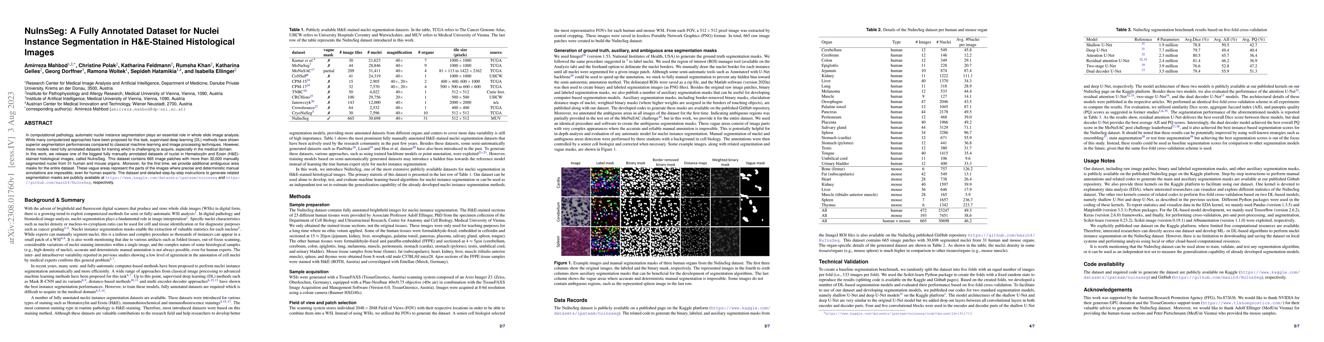
Key Findings, in focus
Seven facets of this paper, analysed and brought into focus by AI.
This dataset is significant for developing, testing, and evaluating machine learning-based algorithms for nuclei instance segmentation, as well as for estimating the generalization capability of existing methods.
- NuInsSeg is one of the largest publicly available datasets for nuclei segmentation in H&E-stained histological images.
- The dataset provides additional ambiguous area masks, which are crucial for in-depth analysis and evaluation of automatic nuclei instance segmentation models.
This dataset is significant for developing, testing, and evaluating machine learning-based algorithms for nuclei instance segmentation, as well as for estimating the generalization capability of existing methods.
The primary technical contribution is the creation of a large, fully annotated dataset with ambiguous area masks for nuclei instance segmentation in H&E-stained histological images.
NuInsSeg stands out by providing not only fully annotated image patches but also ambiguous area masks, which are essential for comprehensive analysis and evaluation of nuclei instance segmentation models.
- The dataset is limited to H&E-stained histological images from specific human and mouse organs.
- The manual annotation process might introduce inter- and intra-observer variability.
- The dataset can be expanded to include more organ types and staining methods.
- Further research can explore the application of semi-automatic or automated annotation methods to increase efficiency and reduce human bias.
Impact
Paper Details
Authors
PDF Preview
Key Terms
Citation Network
Current paper (gray), citations (green), references (blue)
Display is limited for performance on very large graphs.
Discussion 0