ArxivLens
A
Researcher

Amirreza Mahbod

21 papers on arXiv

21 arXiv Papers
32 Total Publications
Profile

Academic Profile

Name A. Mahbod
Total Papers 32
Scholar Profile View on Semantic Scholar
Metrics

Statistics

21 arXiv Papers
32 Total Publications
Publications

Papers on arXiv

Thumbnail for Breast Histopathology Image Retrieval by Attention-based Adversarially
  Regularized Variational Graph Autoencoder with Contrastive Learning-Based
  Feature Extraction
arXiv
Breast Histopathology Image Retrieval by Attention-based Adversarially Regularized Variational Graph Autoencoder with Contrastive Learning-Based Feature Extraction

Breast cancer is the most common cancer type in women worldwide. Early detection and appropriate treatment can significantly reduce its impact. While histopathology examinations play a vital role in...

Thumbnail for Improving Generalization Capability of Deep Learning-Based Nuclei
  Instance Segmentation by Non-deterministic Train Time and Deterministic Test
  Time Stain Normalization
arXiv
Improving Generalization Capability of Deep Learning-Based Nuclei Instance Segmentation by Non-deterministic Train Time and Deterministic Test Time Stain Normalization

With the advent of digital pathology and microscopic systems that can scan and save whole slide histological images automatically, there is a growing trend to use computerized methods to analyze acq...

Thumbnail for Breast MRI radiomics and machine learning radiomics-based predictions of
  response to neoadjuvant chemotherapy -- how are they affected by variations
  in tumour delineation?
arXiv
Breast MRI radiomics and machine learning radiomics-based predictions of response to neoadjuvant chemotherapy -- how are they affected by variations in tumour delineation?

Manual delineation of volumes of interest (VOIs) by experts is considered the gold-standard method in radiomics analysis. However, it suffers from inter- and intra-operator variability. A quantitati...

Thumbnail for NuInsSeg: A Fully Annotated Dataset for Nuclei Instance Segmentation in
  H&E-Stained Histological Images
arXiv
NuInsSeg: A Fully Annotated Dataset for Nuclei Instance Segmentation in H&E-Stained Histological Images

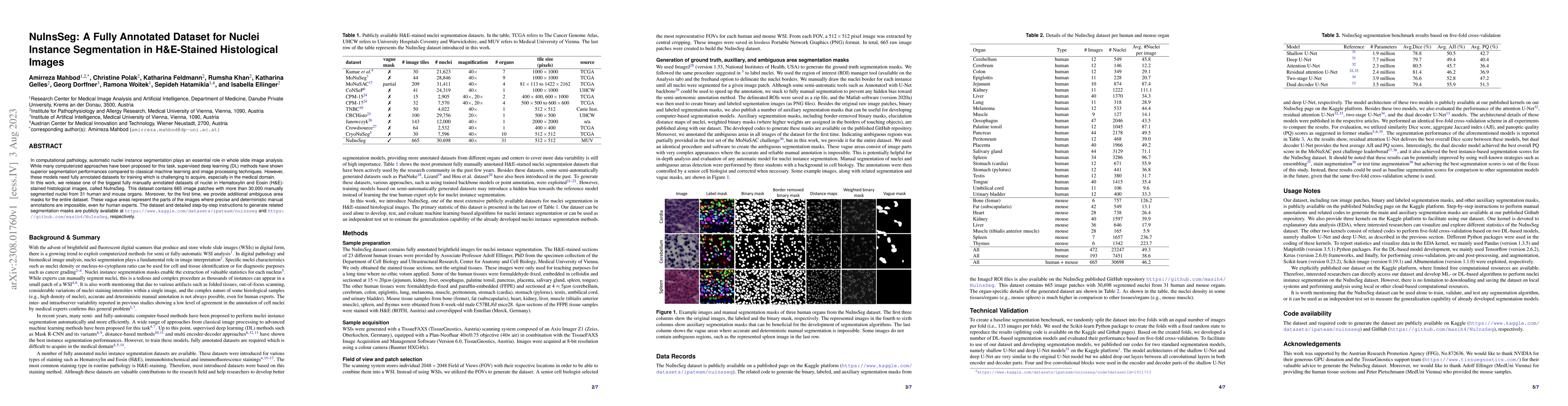
In computational pathology, automatic nuclei instance segmentation plays an essential role in whole slide image analysis. While many computerized approaches have been proposed for this task, supervi...

Thumbnail for Deep Neural Network Pruning for Nuclei Instance Segmentation in
  Hematoxylin & Eosin-Stained Histological Images
arXiv
Deep Neural Network Pruning for Nuclei Instance Segmentation in Hematoxylin & Eosin-Stained Histological Images

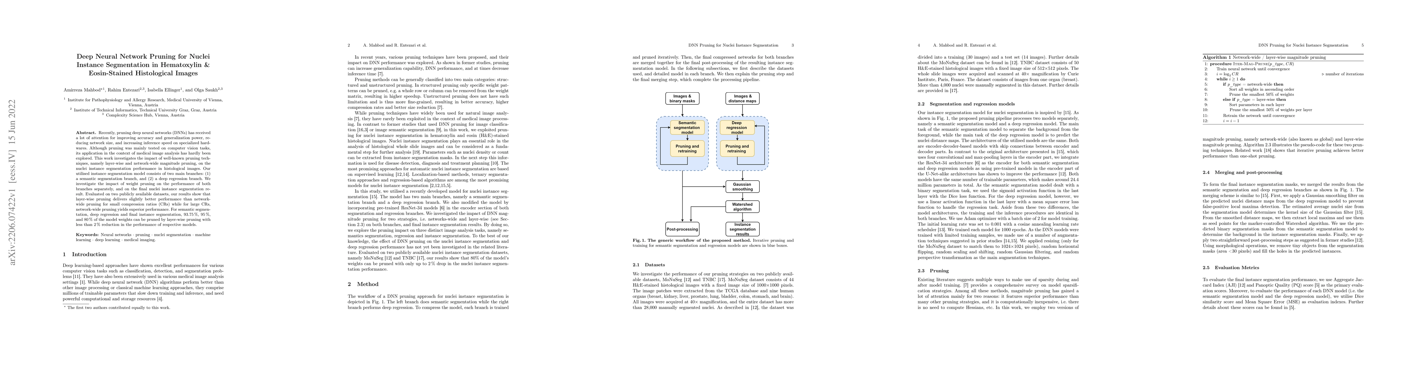
Recently, pruning deep neural networks (DNNs) has received a lot of attention for improving accuracy and generalization power, reducing network size, and increasing inference speed on specialized ha...

Thumbnail for FUSeg: The Foot Ulcer Segmentation Challenge
arXiv
FUSeg: The Foot Ulcer Segmentation Challenge

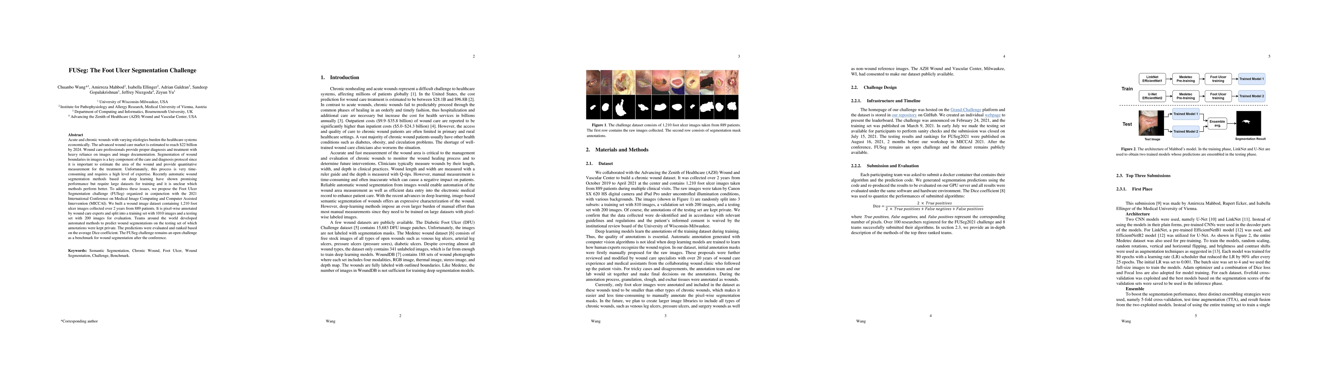
Acute and chronic wounds with varying etiologies burden the healthcare systems economically. The advanced wound care market is estimated to reach $22 billion by 2024. Wound care professionals provid...

Thumbnail for Automatic Foot Ulcer Segmentation Using an Ensemble of Convolutional
  Neural Networks
arXiv
Automatic Foot Ulcer Segmentation Using an Ensemble of Convolutional Neural Networks

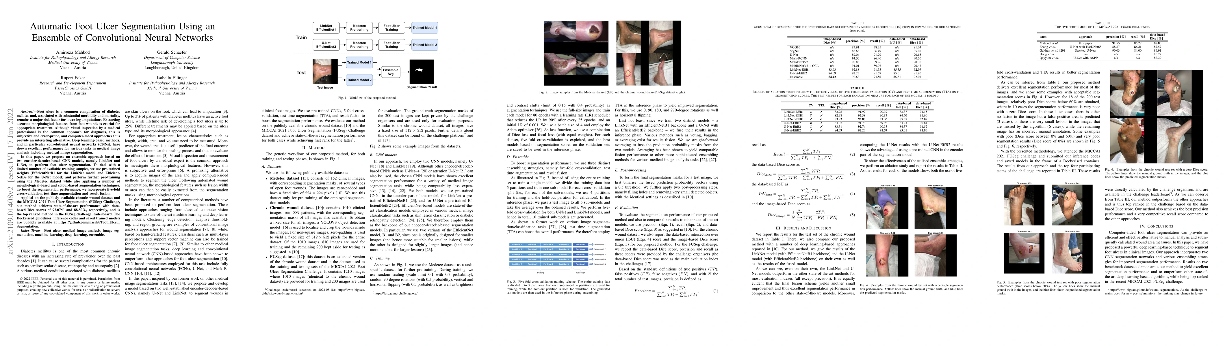
Foot ulcer is a common complication of diabetes mellitus and, associated with substantial morbidity and mortality, remains a major risk factor for lower leg amputations. Extracting accurate morpholo...

Thumbnail for CryoNuSeg: A Dataset for Nuclei Instance Segmentation of Cryosectioned
  H&E-Stained Histological Images
arXiv
CryoNuSeg: A Dataset for Nuclei Instance Segmentation of Cryosectioned H&E-Stained Histological Images

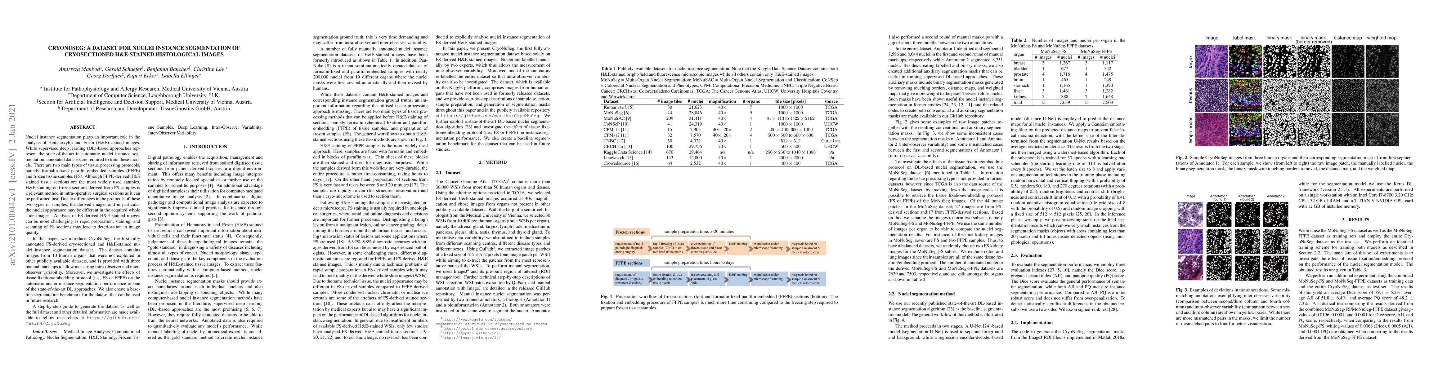
Nuclei instance segmentation plays an important role in the analysis of Hematoxylin and Eosin (H&E)-stained images. While supervised deep learning (DL)-based approaches represent the state-of-the-ar...

Thumbnail for Pollen Grain Microscopic Image Classification Using an Ensemble of
  Fine-Tuned Deep Convolutional Neural Networks
arXiv
Pollen Grain Microscopic Image Classification Using an Ensemble of Fine-Tuned Deep Convolutional Neural Networks

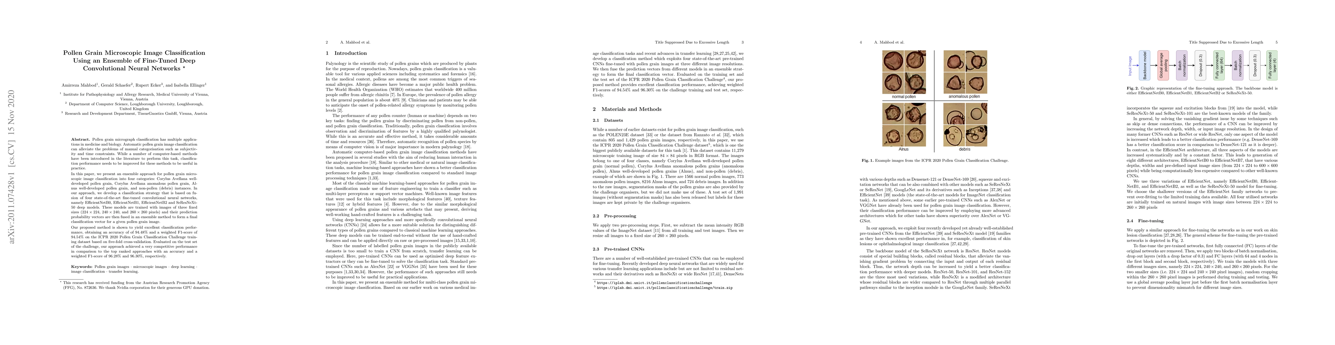
Pollen grain micrograph classification has multiple applications in medicine and biology. Automatic pollen grain image classification can alleviate the problems of manual categorisation such as subj...

Thumbnail for The Effects of Skin Lesion Segmentation on the Performance of
  Dermatoscopic Image Classification
arXiv
The Effects of Skin Lesion Segmentation on the Performance of Dermatoscopic Image Classification

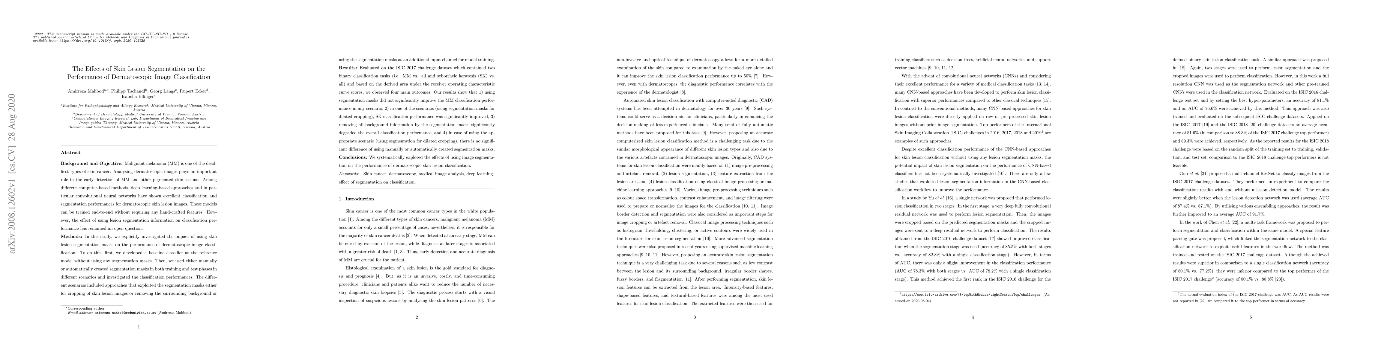
Malignant melanoma (MM) is one of the deadliest types of skin cancer. Analysing dermatoscopic images plays an important role in the early detection of MM and other pigmented skin lesions. Among diff...

Thumbnail for Investigating and Exploiting Image Resolution for Transfer
  Learning-based Skin Lesion Classification
arXiv
Investigating and Exploiting Image Resolution for Transfer Learning-based Skin Lesion Classification

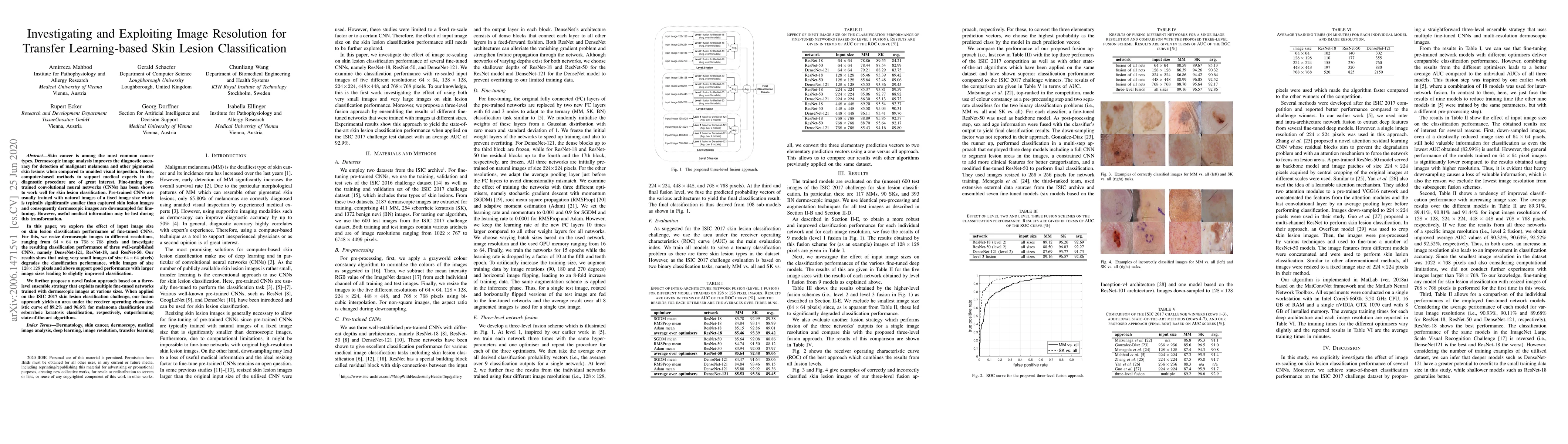
Skin cancer is among the most common cancer types. Dermoscopic image analysis improves the diagnostic accuracy for detection of malignant melanoma and other pigmented skin lesions when compared to u...

Thumbnail for WCEbleedGen: A wireless capsule endoscopy dataset and its benchmarking
  for automatic bleeding classification, detection, and segmentation
arXiv
WCEbleedGen: A wireless capsule endoscopy dataset and its benchmarking for automatic bleeding classification, detection, and segmentation

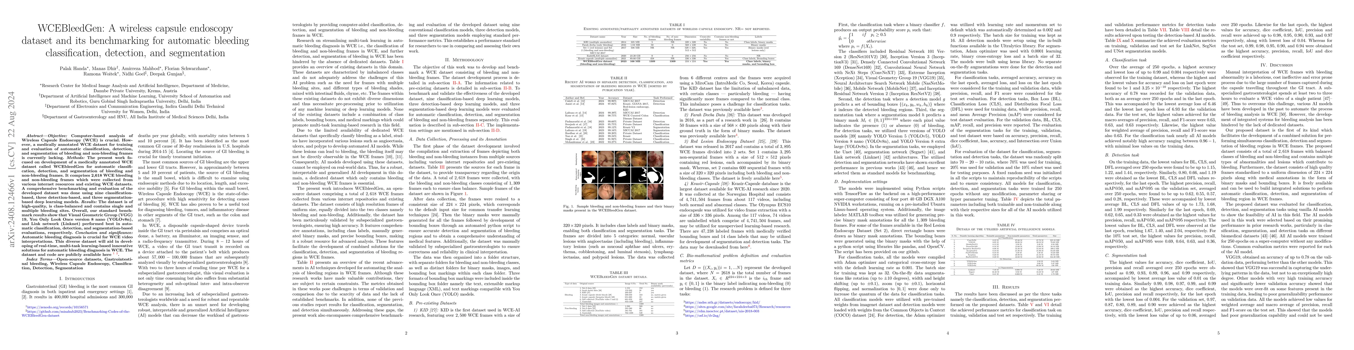
Computer-based analysis of Wireless Capsule Endoscopy (WCE) is crucial. However, a medically annotated WCE dataset for training and evaluation of automatic classification, detection, and segmentation ...

Thumbnail for Capsule Vision 2024 Challenge: Multi-Class Abnormality Classification
  for Video Capsule Endoscopy
arXiv
Capsule Vision 2024 Challenge: Multi-Class Abnormality Classification for Video Capsule Endoscopy

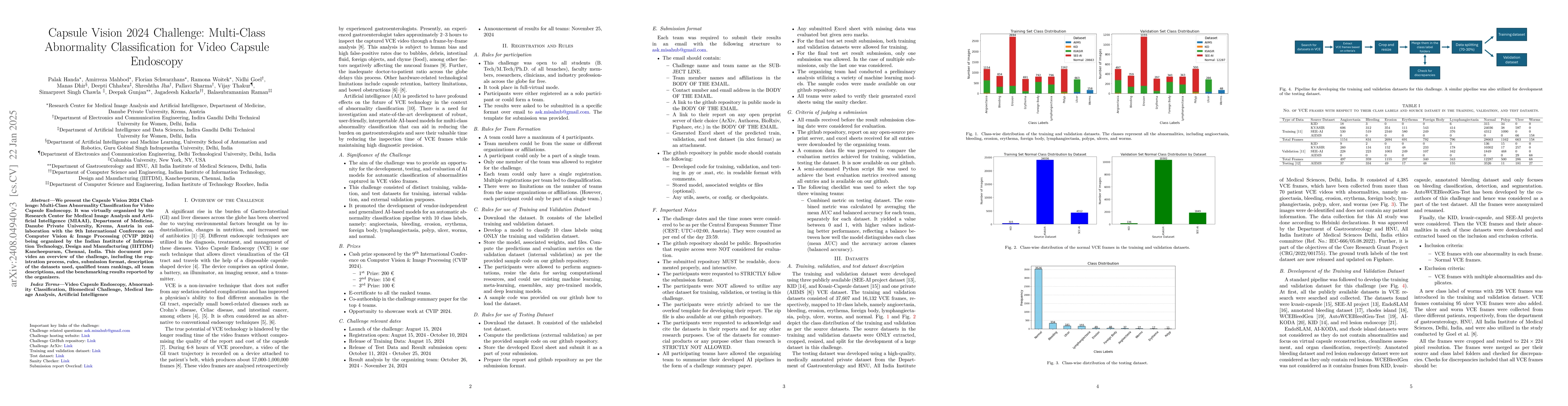
We present the Capsule Vision 2024 Challenge: Multi-Class Abnormality Classification for Video Capsule Endoscopy. It is being virtually organized by the Research Center for Medical Image Analysis and ...

Thumbnail for Evaluating Pre-trained Convolutional Neural Networks and Foundation
  Models as Feature Extractors for Content-based Medical Image Retrieval
arXiv
Evaluating Pre-trained Convolutional Neural Networks and Foundation Models as Feature Extractors for Content-based Medical Image Retrieval

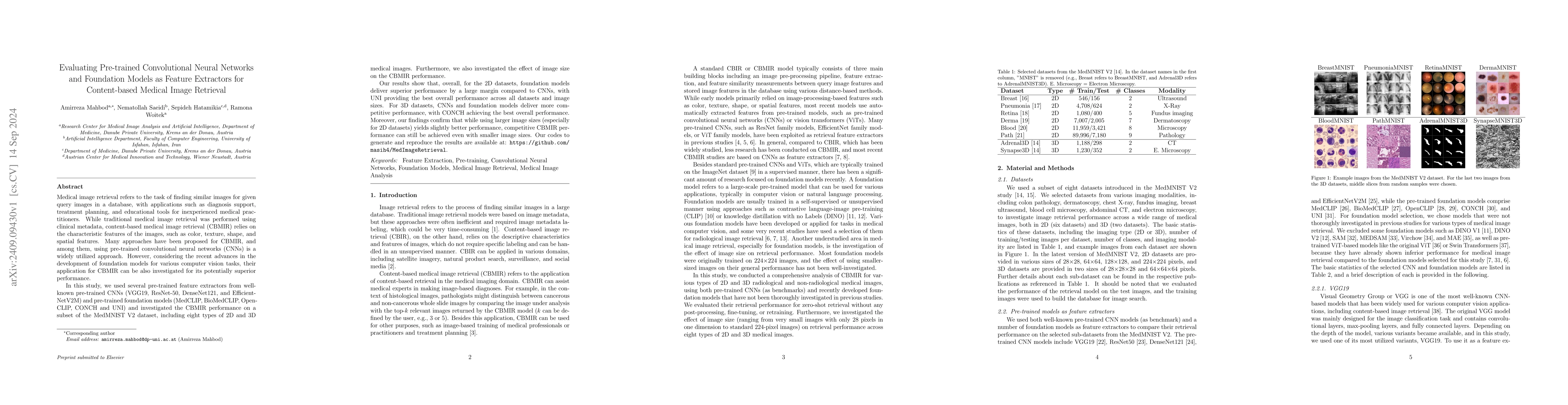
Medical image retrieval refers to the task of finding similar images for given query images in a database, with applications such as diagnosis support, treatment planning, and educational tools for in...

Thumbnail for Fusion of Foundation and Vision Transformer Model Features for
  Dermatoscopic Image Classification
arXiv
Fusion of Foundation and Vision Transformer Model Features for Dermatoscopic Image Classification

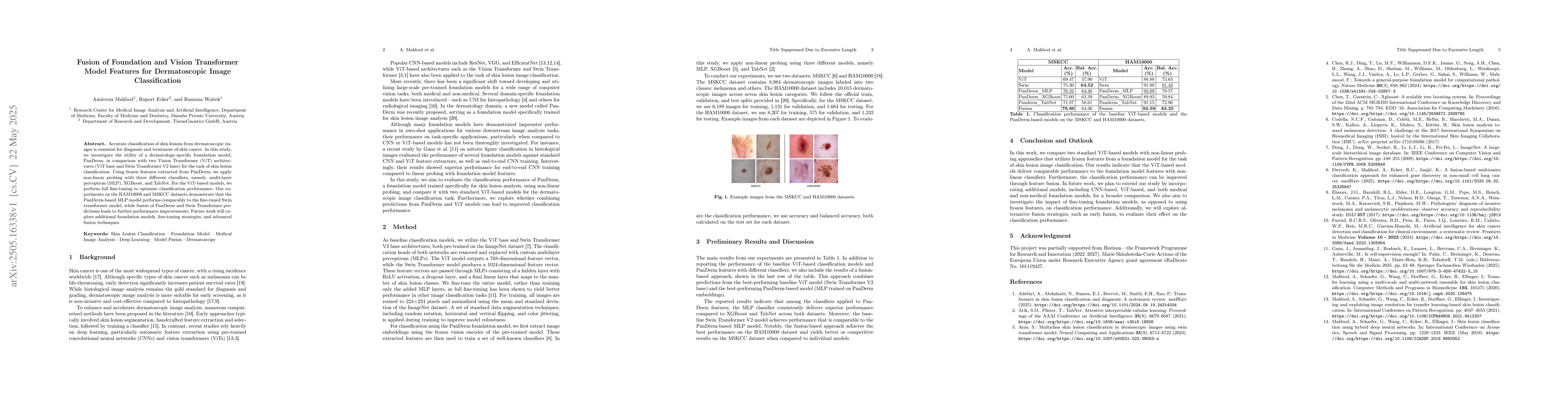
Accurate classification of skin lesions from dermatoscopic images is essential for diagnosis and treatment of skin cancer. In this study, we investigate the utility of a dermatology-specific foundatio...

Thumbnail for A Multi-Stage Auto-Context Deep Learning Framework for Tissue and Nuclei
  Segmentation and Classification in H&E-Stained Histological Images of
  Advanced Melanoma
arXiv
A Multi-Stage Auto-Context Deep Learning Framework for Tissue and Nuclei Segmentation and Classification in H&E-Stained Histological Images of Advanced Melanoma

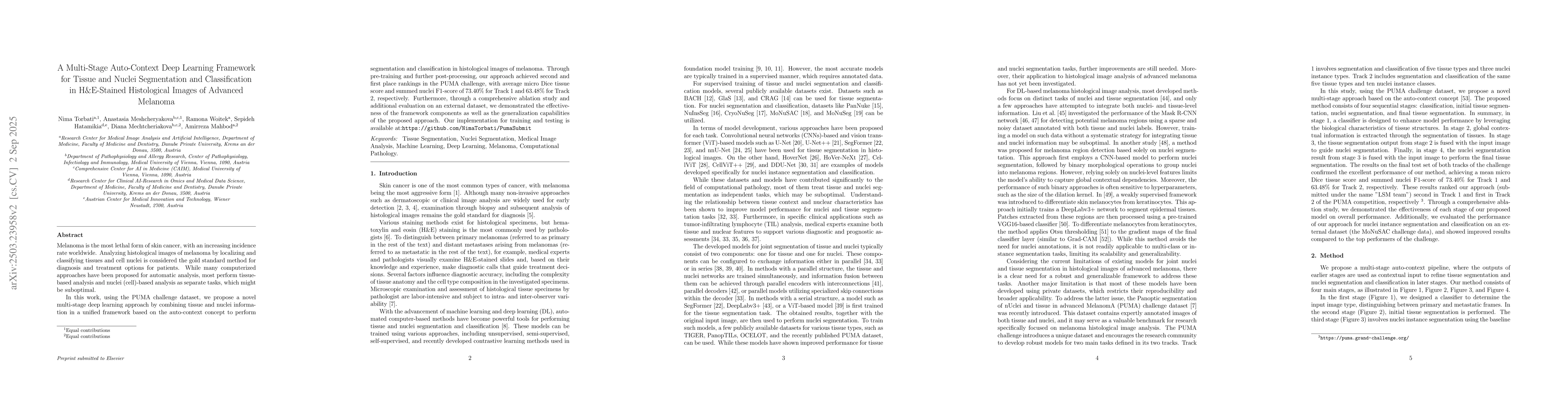
Melanoma is the most lethal form of skin cancer, with an increasing incidence rate worldwide. Analyzing histological images of melanoma by localizing and classifying tissues and cell nuclei is conside...

Thumbnail for ACS-SegNet: An Attention-Based CNN-SegFormer Segmentation Network for
  Tissue Segmentation in Histopathology
arXiv
ACS-SegNet: An Attention-Based CNN-SegFormer Segmentation Network for Tissue Segmentation in Histopathology

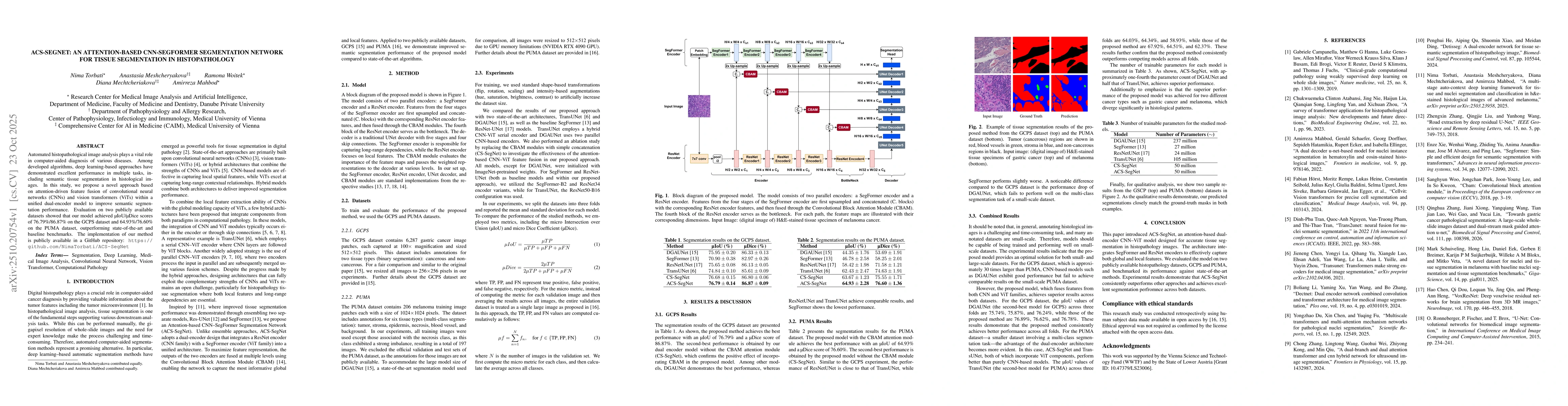
Automated histopathological image analysis plays a vital role in computer-aided diagnosis of various diseases. Among developed algorithms, deep learning-based approaches have demonstrated excellent pe...

Thumbnail for Developing Predictive and Robust Radiomics Models for Chemotherapy Response in High-Grade Serous Ovarian Carcinoma
arXiv
Developing Predictive and Robust Radiomics Models for Chemotherapy Response in High-Grade Serous Ovarian Carcinoma

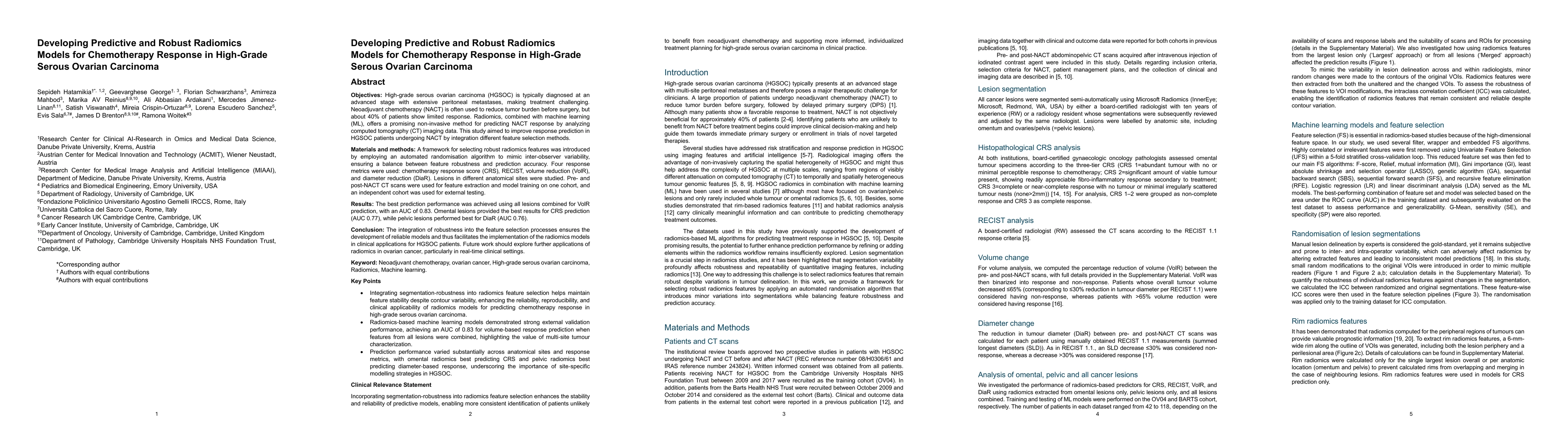
Objectives: High-grade serous ovarian carcinoma (HGSOC) is typically diagnosed at an advanced stage with extensive peritoneal metastases, making treatment challenging. Neoadjuvant chemotherapy (NACT) ...

Thumbnail for NucFuseRank: Dataset Fusion and Performance Ranking for Nuclei Instance Segmentation
arXiv
NucFuseRank: Dataset Fusion and Performance Ranking for Nuclei Instance Segmentation

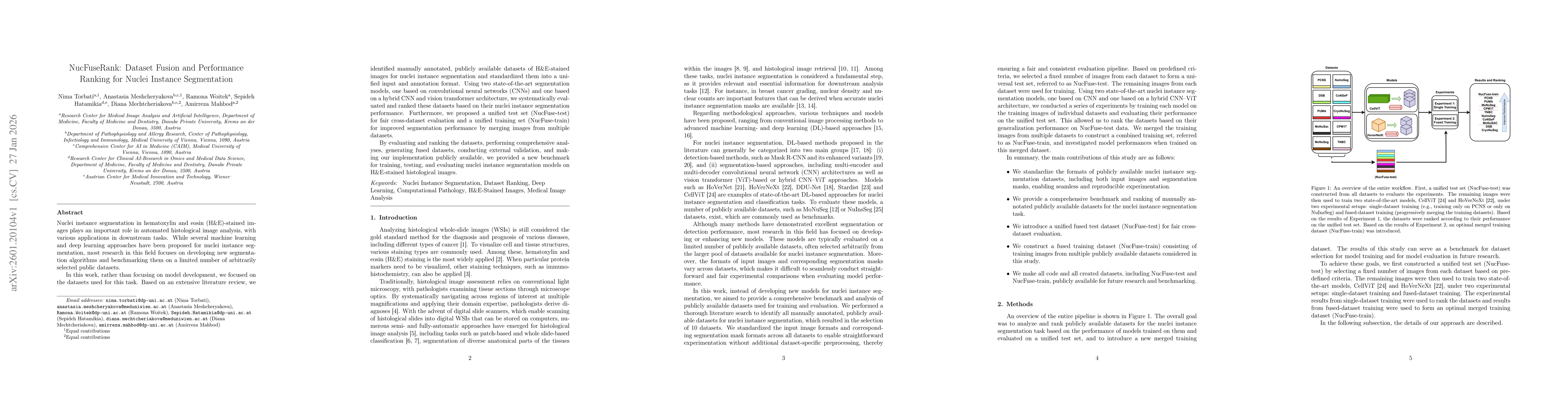
Nuclei instance segmentation in hematoxylin and eosin (H&E)-stained images plays an important role in automated histological image analysis, with various applications in downstream tasks. While severa...

Thumbnail for Effect of Patch Size on Fine-Tuning Vision Transformers in Two-Dimensional and Three-Dimensional Medical Image Classification
arXiv
Effect of Patch Size on Fine-Tuning Vision Transformers in Two-Dimensional and Three-Dimensional Medical Image Classification

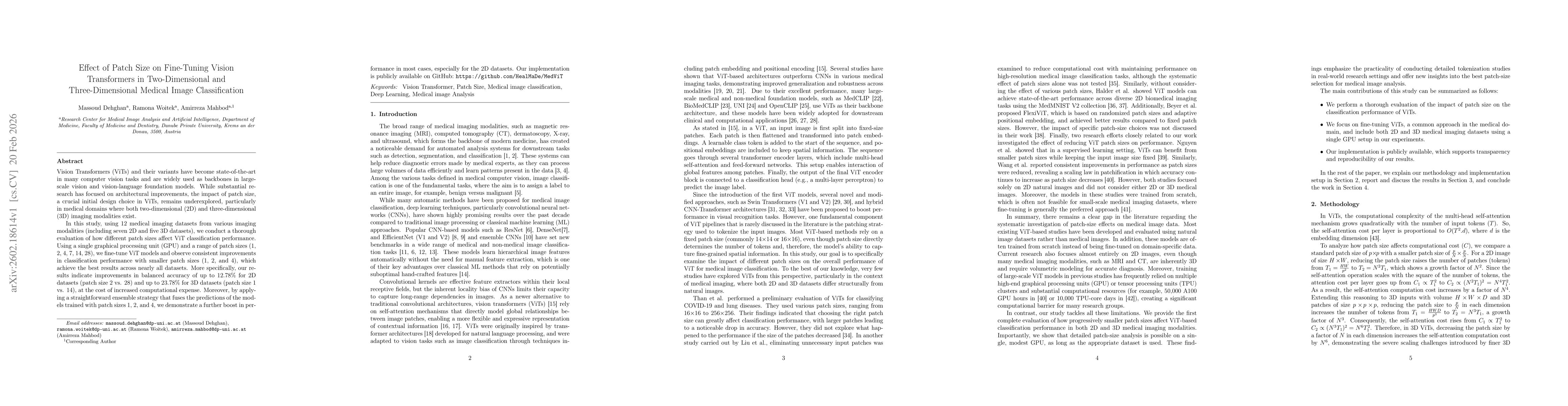
Vision Transformers (ViTs) and their variants have become state-of-the-art in many computer vision tasks and are widely used as backbones in large-scale vision and vision-language foundation models. W...

Thumbnail for NucEval: A Robust Evaluation Framework for Nuclear Instance Segmentation
arXiv
NucEval: A Robust Evaluation Framework for Nuclear Instance Segmentation

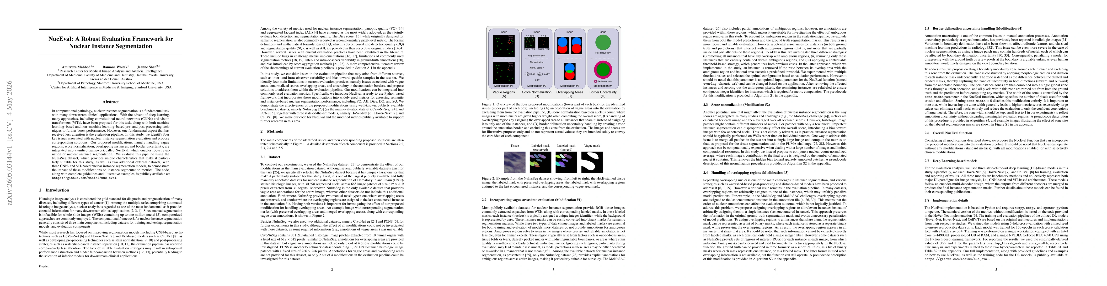
In computational pathology, nuclear instance segmentation is a fundamental task with many downstream clinical applications. With the advent of deep learning, many approaches, including convolutional n...

This website uses cookies to ensure you get the best experience. Learn more