The paper proposes ACS-SegNet, a dual-encoder model combining SegFormer (vision transformer) and ResNet (CNN) with CBAM attention modules for histopathological tissue segmentation. The model was evaluated on GCPS and PUMA datasets using µIoU and µDice metrics.
ACS-SegNet: An Attention-Based CNN-SegFormer Segmentation Network for Tissue Segmentation in Histopathology
Publication
Metrics
Quick Answers
What methodology did the authors use?
The paper proposes ACS-SegNet, a dual-encoder model combining SegFormer (vision transformer) and ResNet (CNN) with CBAM attention modules for histopathological tissue segmentation. The model was evaluated on GCPS and PUMA datasets using µIoU and µDice metrics. More in Methodology →
What are the key results?
ACS-SegNet achieved µIoU/µDice scores of 76.79%/86.87% on GCPS and 64.93%/76.60% on PUMA, outperforming state-of-the-art benchmarks. — The dual-encoder architecture with CBAM attention modules significantly improved segmentation performance compared to single-encoder models like DGAUNet and TransUNet. More in Key Results →
Why is this work significant?
This research advances automated histopathological analysis by providing a robust segmentation solution for tissue types, aiding in computer-aided diagnosis and improving accuracy in cancer detection. More in Significance →
What are the main limitations?
The model's performance may degrade on extremely imbalanced datasets, as seen with the PUMA dataset's necrosis class. — The method requires significant computational resources due to the combination of CNN and ViT components. More in Limitations →
Abstract
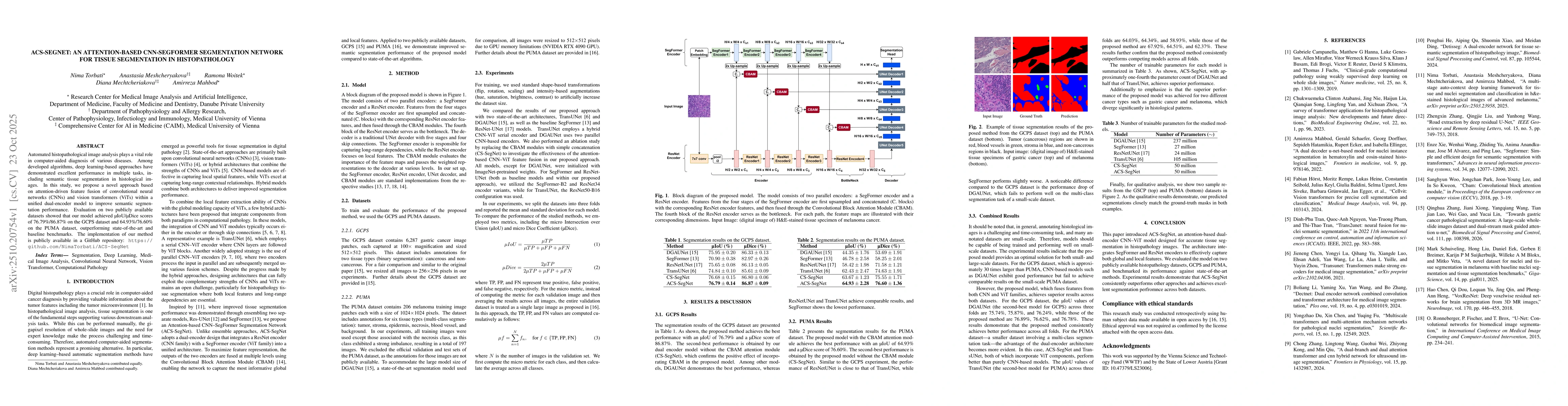
Automated histopathological image analysis plays a vital role in computer-aided diagnosis of various diseases. Among developed algorithms, deep learning-based approaches have demonstrated excellent performance in multiple tasks, including semantic tissue segmentation in histological images. In this study, we propose a novel approach based on attention-driven feature fusion of convolutional neural networks (CNNs) and vision transformers (ViTs) within a unified dual-encoder model to improve semantic segmentation performance. Evaluation on two publicly available datasets showed that our model achieved {\mu}IoU/{\mu}Dice scores of 76.79%/86.87% on the GCPS dataset and 64.93%/76.60% on the PUMA dataset, outperforming state-of-the-art and baseline benchmarks. The implementation of our method is publicly available in a GitHub repository: https://github.com/NimaTorbati/ACS-SegNet
Key Findings, in focus
Seven facets of this paper, analysed and brought into focus by AI.
This research advances automated histopathological analysis by providing a robust segmentation solution for tissue types, aiding in computer-aided diagnosis and improving accuracy in cancer detection.
- ACS-SegNet achieved µIoU/µDice scores of 76.79%/86.87% on GCPS and 64.93%/76.60% on PUMA, outperforming state-of-the-art benchmarks.
- The dual-encoder architecture with CBAM attention modules significantly improved segmentation performance compared to single-encoder models like DGAUNet and TransUNet.
- The model demonstrated consistent performance across different dataset sizes and cancer types (gastric cancer and melanoma).
This research advances automated histopathological analysis by providing a robust segmentation solution for tissue types, aiding in computer-aided diagnosis and improving accuracy in cancer detection.
Integration of SegFormer (ViT) and ResNet (CNN) encoders with CBAM attention modules in a unified dual-encoder architecture for enhanced feature fusion.
The novel combination of CNN and ViT-based encoders with attention mechanisms provides superior performance for histopathological segmentation compared to existing methods.
- The model's performance may degrade on extremely imbalanced datasets, as seen with the PUMA dataset's necrosis class.
- The method requires significant computational resources due to the combination of CNN and ViT components.
- Exploring more efficient attention mechanisms to reduce computational overhead.
- Investigating the application of this framework to other histopathological tasks beyond tissue segmentation.
Discussion 0