Academic Profile
Statistics
Similar Authors
Papers on arXiv
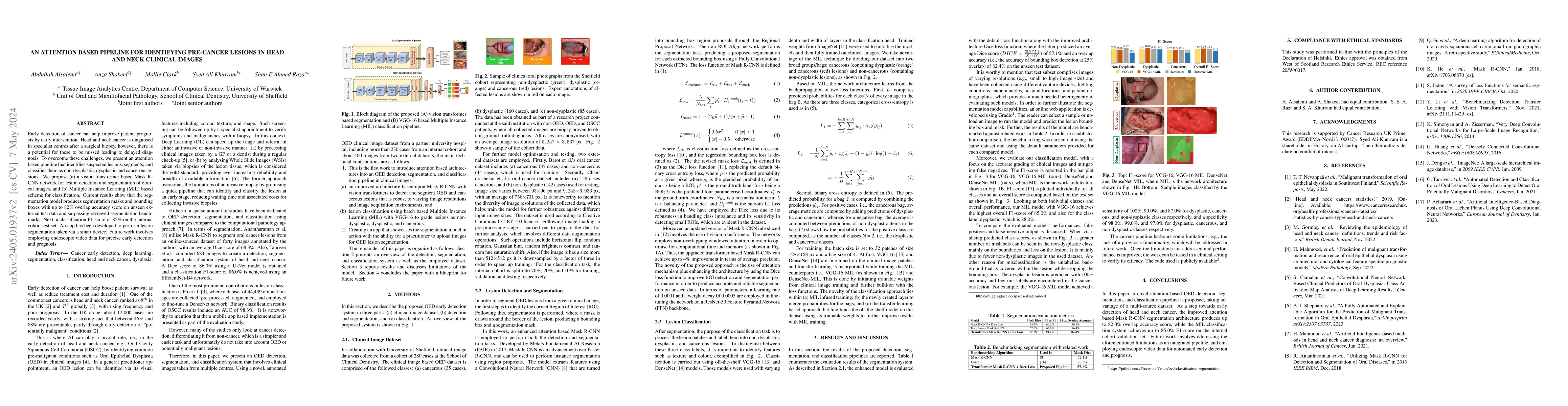
Early detection of cancer can help improve patient prognosis by early intervention. Head and neck cancer is diagnosed in specialist centres after a surgical biopsy, however, there is a potential for...
Whole Slide Images (WSIs) provide exceptional detail for studying tissue architecture at the cell level. To study tumour microenvironment (TME) with the context of various protein biomarkers and cel...
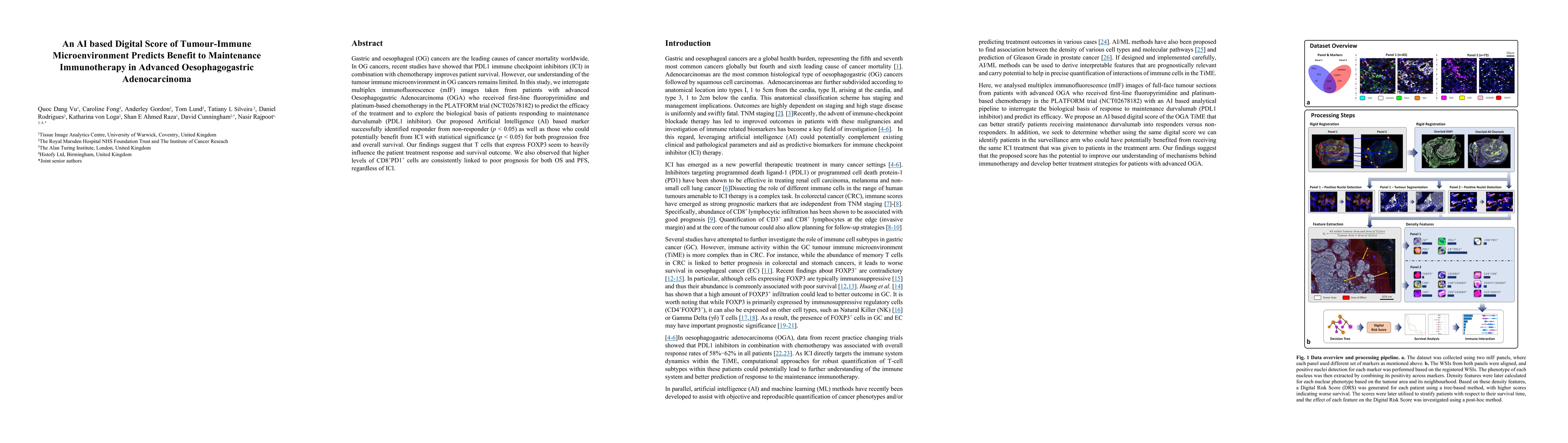
Gastric and oesophageal (OG) cancers are the leading causes of cancer mortality worldwide. In OG cancers, recent studies have showed that PDL1 immune checkpoint inhibitors (ICI) in combination with ...
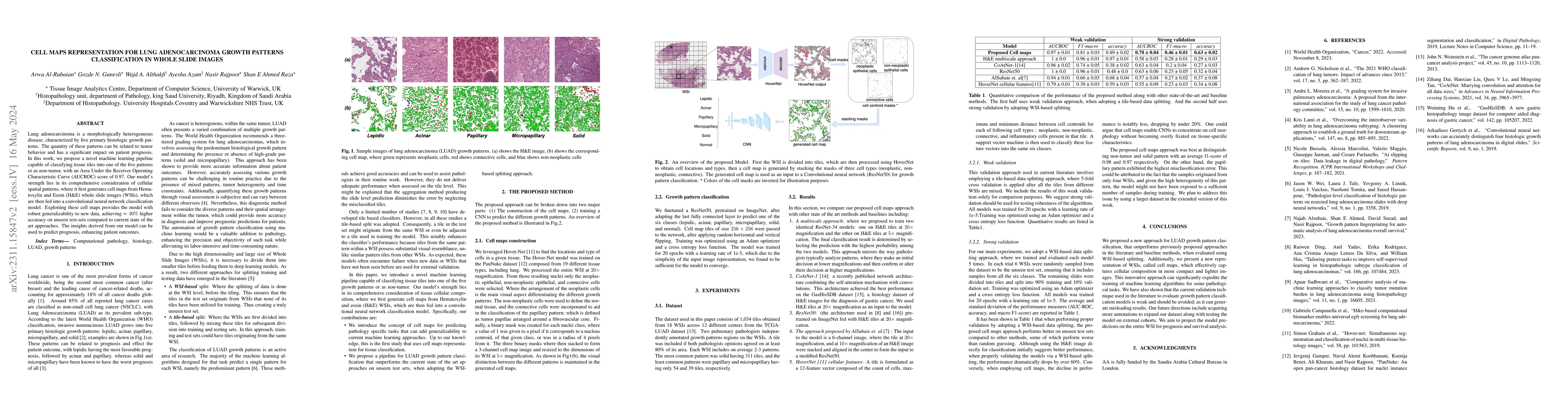
Lung adenocarcinoma is a morphologically heterogeneous disease, characterized by five primary histologic growth patterns. The quantity of these patterns can be related to tumor behavior and has a si...
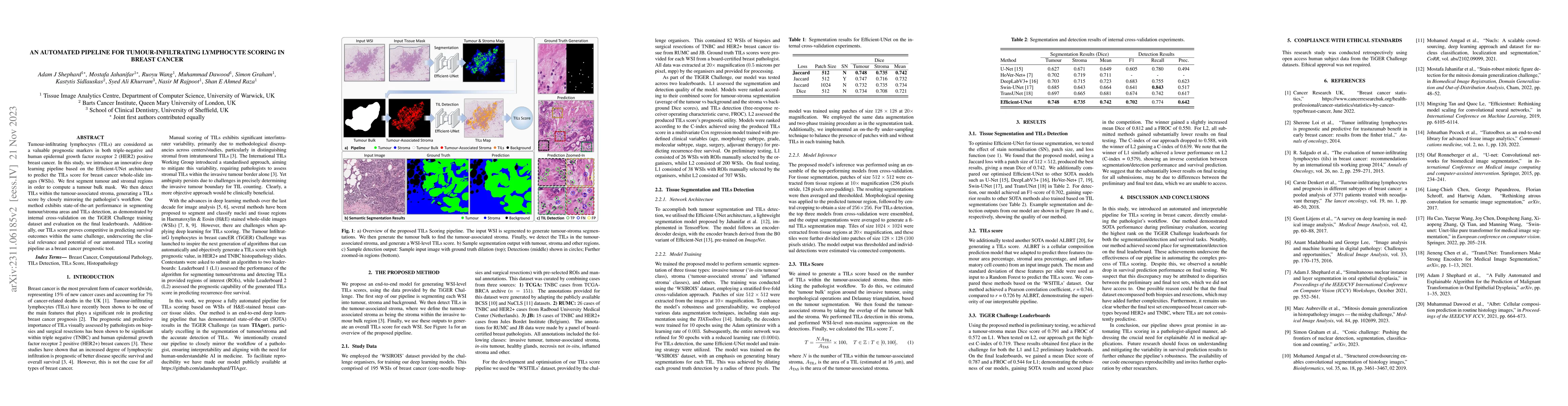
Tumour-infiltrating lymphocytes (TILs) are considered as a valuable prognostic markers in both triple-negative and human epidermal growth factor receptor 2 (HER2) positive breast cancer. In this stu...
Oral epithelial dysplasia (OED) is a premalignant histopathological diagnosis given to lesions of the oral cavity. OED grading is subject to large inter/intra-rater variability, resulting in the und...

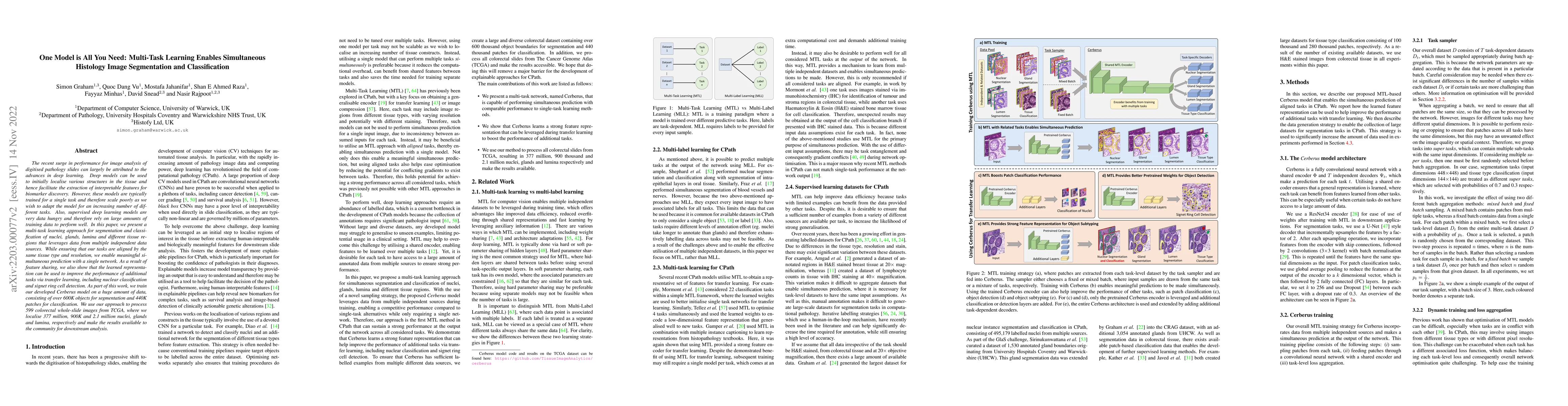
Deep learning models have exhibited exceptional effectiveness in Computational Pathology (CPath) by tackling intricate tasks across an array of histology image analysis applications. Nevertheless, t...
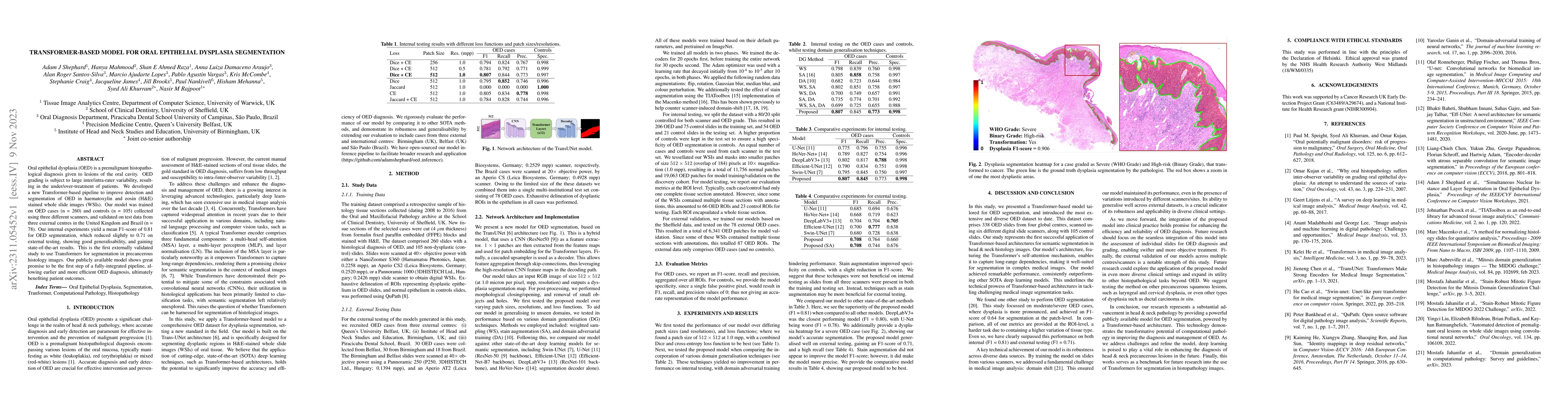
Oral epithelial dysplasia (OED) is a premalignant histopathological diagnosis given to lesions of the oral cavity. Its grading suffers from significant inter-/intra- observer variability, and does n...
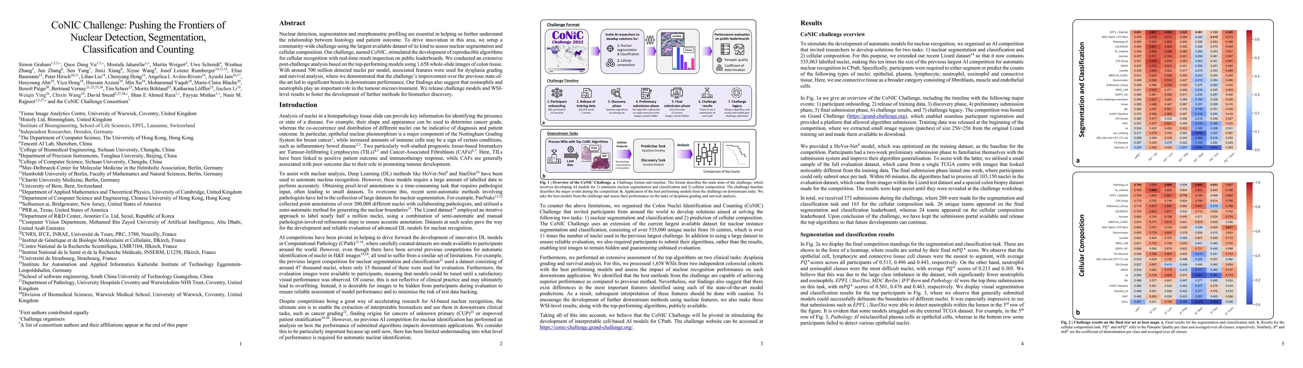
Nuclear detection, segmentation and morphometric profiling are essential in helping us further understand the relationship between histology and patient outcome. To drive innovation in this area, we...
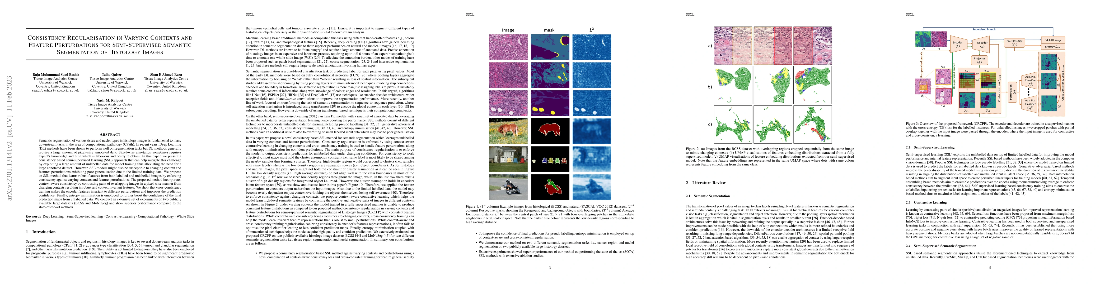
Semantic segmentation of various tissue and nuclei types in histology images is fundamental to many downstream tasks in the area of computational pathology (CPath). In recent years, Deep Learning (D...
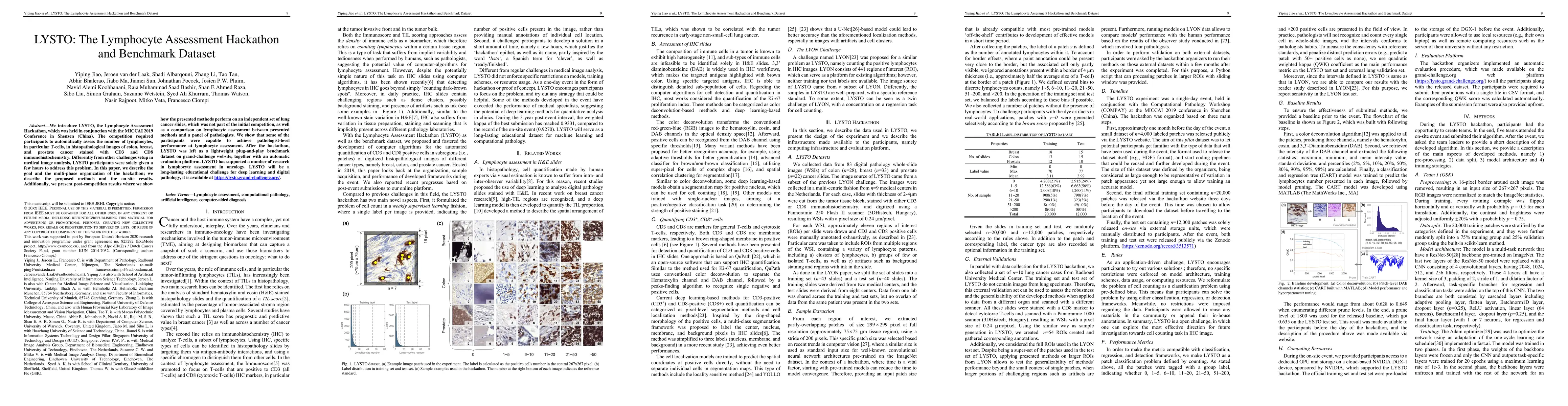
We introduce LYSTO, the Lymphocyte Assessment Hackathon, which was held in conjunction with the MICCAI 2019 Conference in Shenzen (China). The competition required participants to automatically asse...
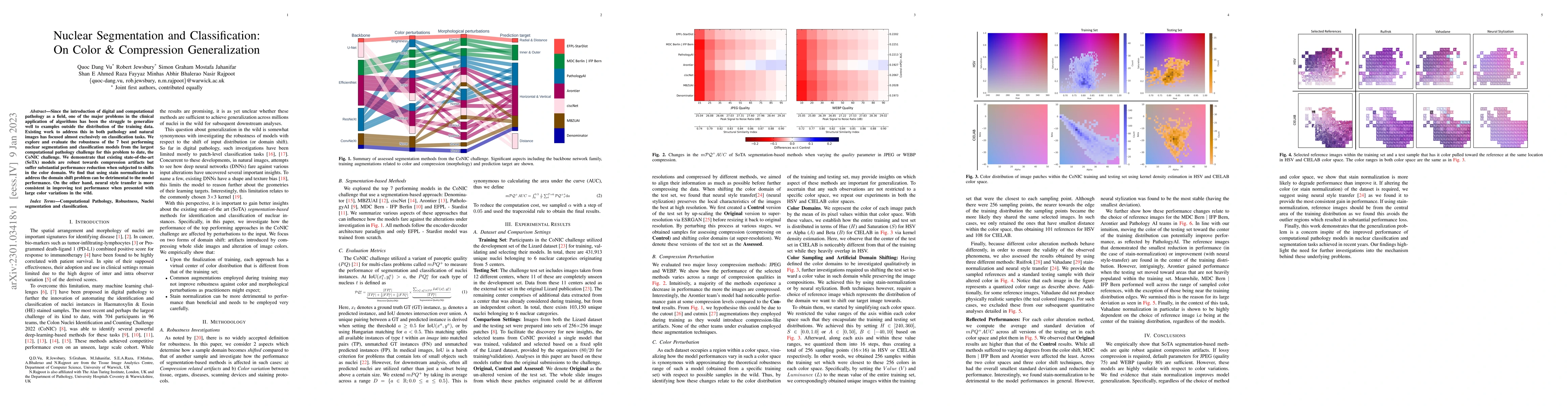
Since the introduction of digital and computational pathology as a field, one of the major problems in the clinical application of algorithms has been the struggle to generalize well to examples out...
Counting of mitotic figures is a fundamental step in grading and prognostication of several cancers. However, manual mitosis counting is tedious and time-consuming. In addition, variation in the app...
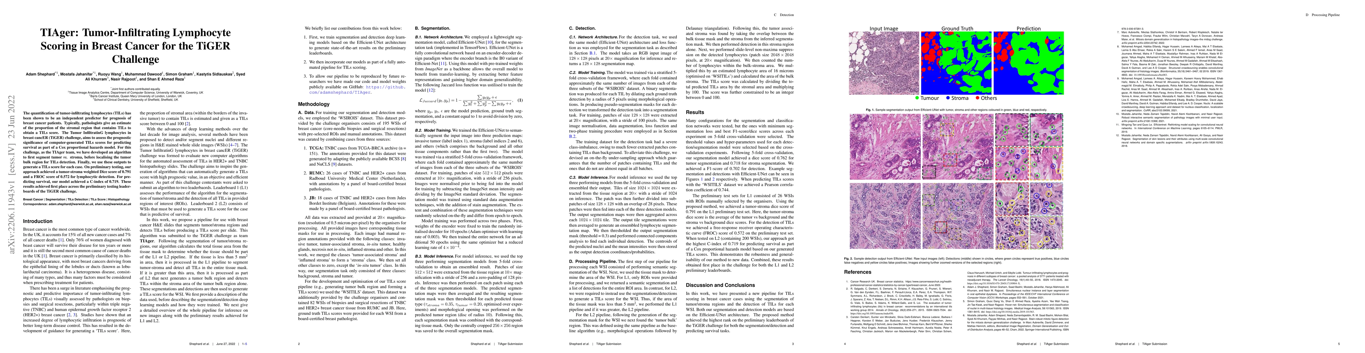
The quantification of tumor-infiltrating lymphocytes (TILs) has been shown to be an independent predictor for prognosis of breast cancer patients. Typically, pathologists give an estimate of the pro...
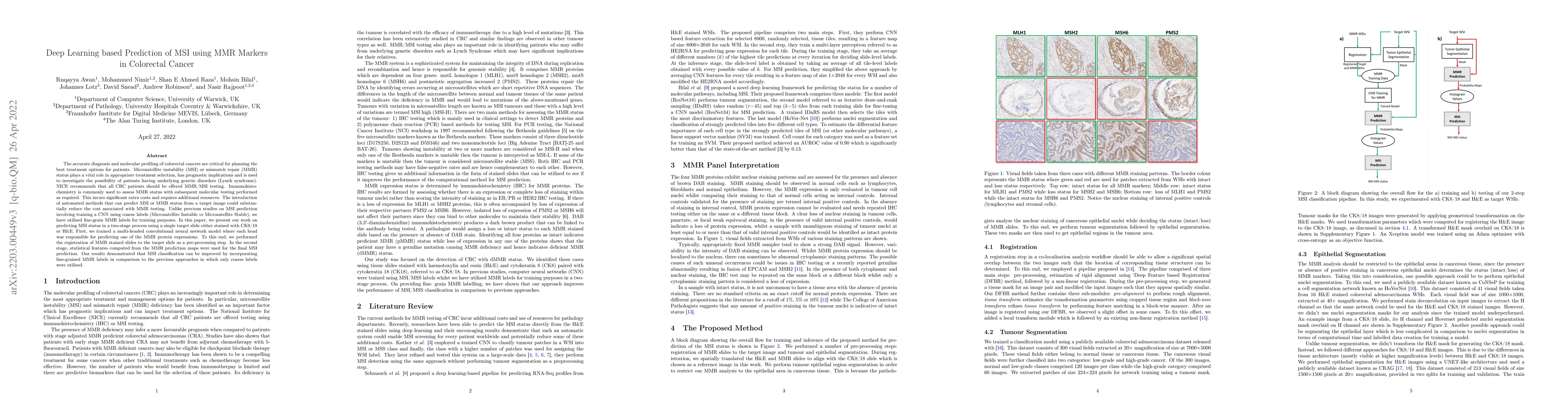
The accurate diagnosis and molecular profiling of colorectal cancers are critical for planning the best treatment options for patients. Microsatellite instability (MSI) or mismatch repair (MMR) stat...
The recent surge in performance for image analysis of digitised pathology slides can largely be attributed to the advances in deep learning. Deep models can be used to initially localise various str...

Cross-slide image analysis provides additional information by analysing the expression of different biomarkers as compared to a single slide analysis. These biomarker stained slides are analysed sid...
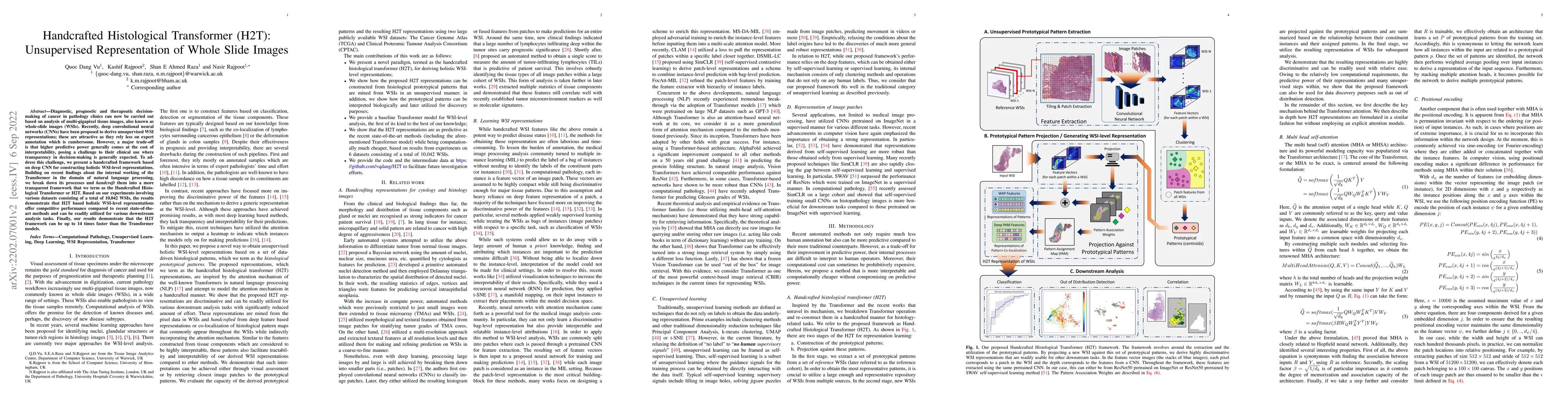
Diagnostic, prognostic and therapeutic decision-making of cancer in pathology clinics can now be carried out based on analysis of multi-gigapixel tissue images, also known as whole-slide images (WSI...
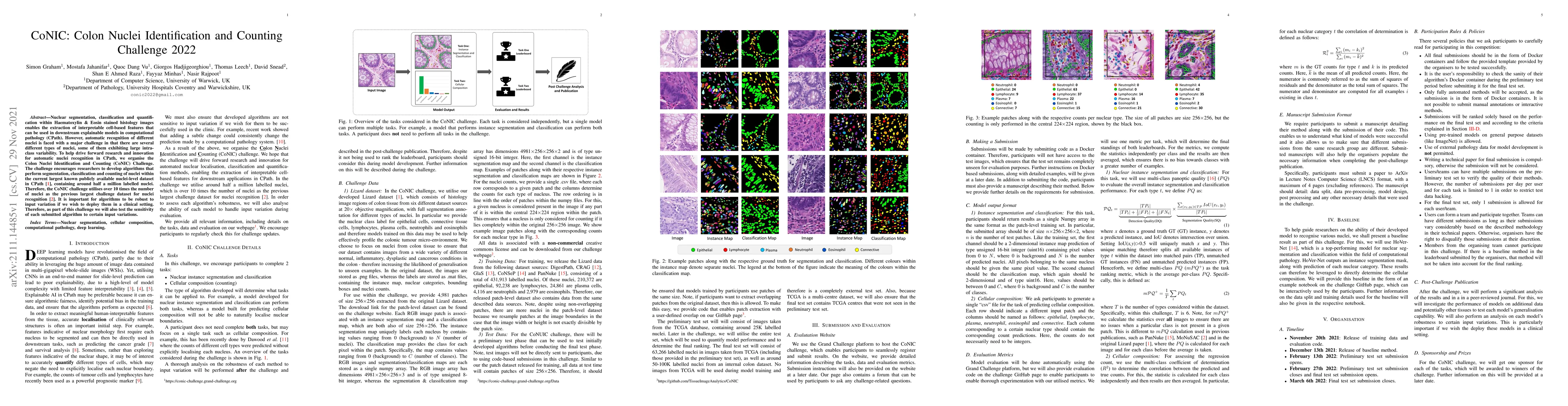
Nuclear segmentation, classification and quantification within Haematoxylin & Eosin stained histology images enables the extraction of interpretable cell-based features that can be used in downstrea...
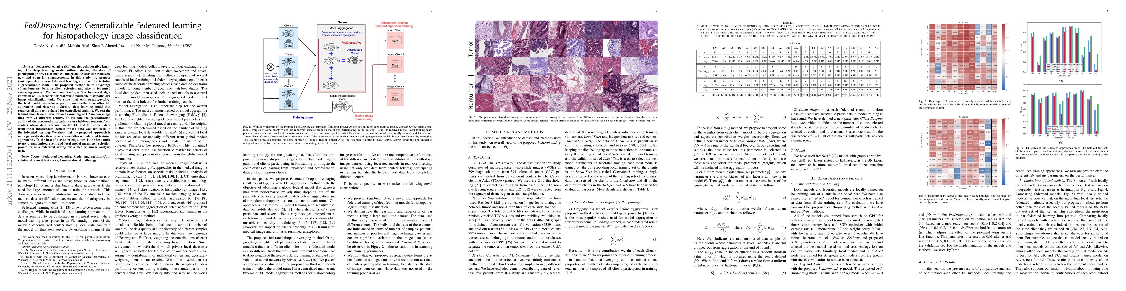
Federated learning (FL) enables collaborative learning of a deep learning model without sharing the data of participating sites. FL in medical image analysis tasks is relatively new and open for enh...
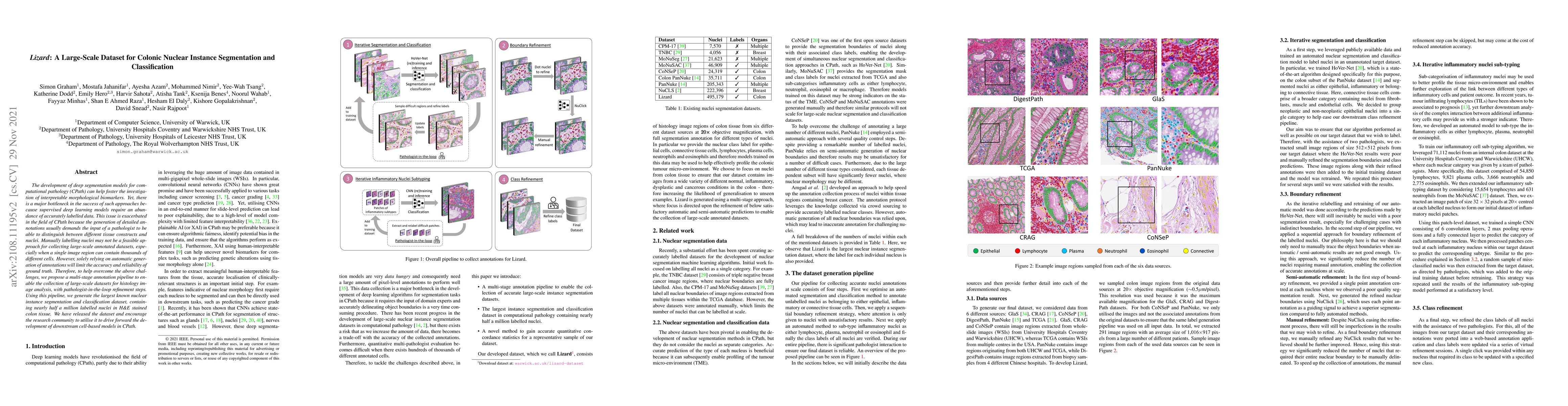
The development of deep segmentation models for computational pathology (CPath) can help foster the investigation of interpretable morphological biomarkers. Yet, there is a major bottleneck in the s...

Recent advances in whole slide imaging (WSI) technology have led to the development of a myriad of computer vision and artificial intelligence (AI) based diagnostic, prognostic, and predictive algor...
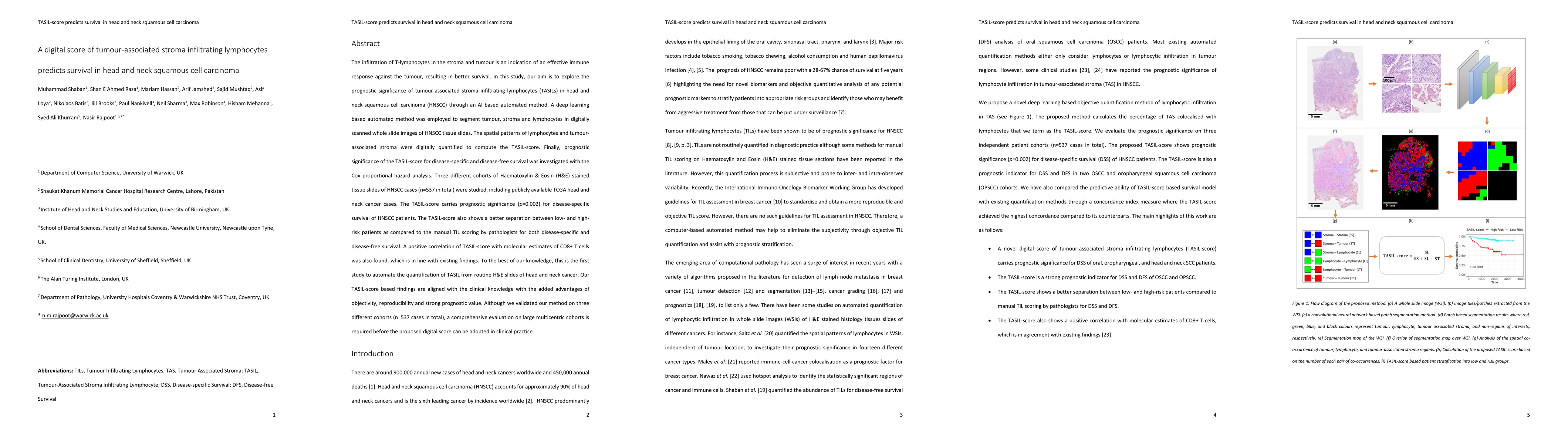
The infiltration of T-lymphocytes in the stroma and tumour is an indication of an effective immune response against the tumour, resulting in better survival. In this study, our aim is to explore the...
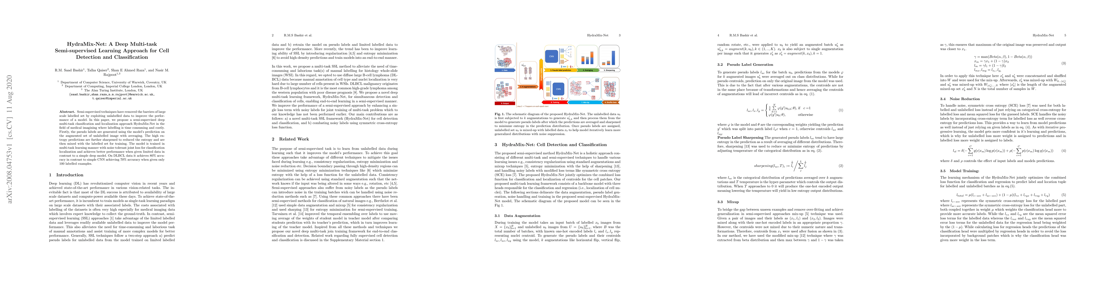
Semi-supervised techniques have removed the barriers of large scale labelled set by exploiting unlabelled data to improve the performance of a model. In this paper, we propose a semi-supervised deep...
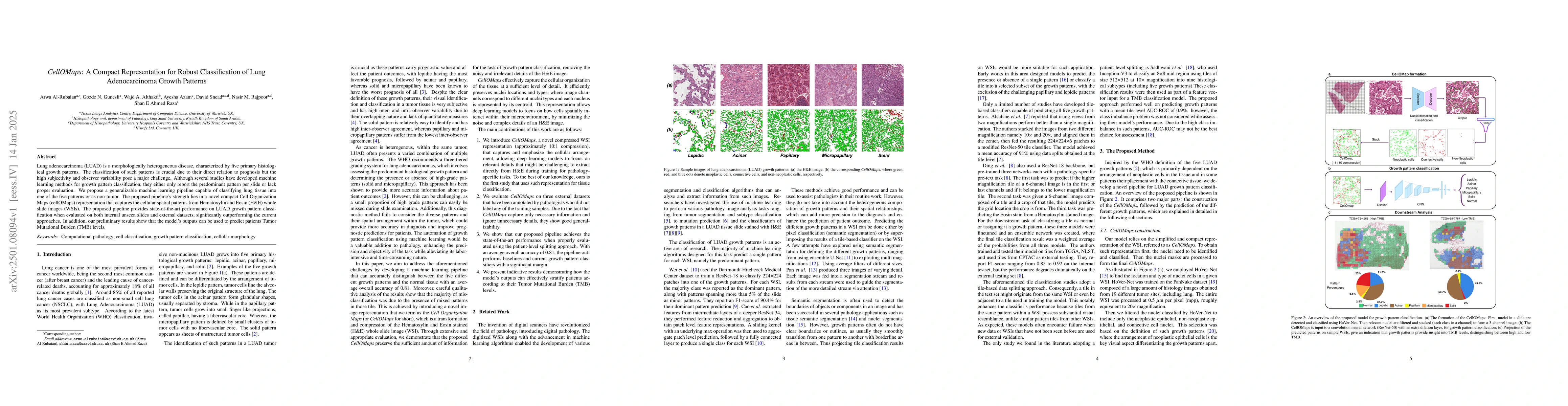
Lung adenocarcinoma (LUAD) is a morphologically heterogeneous disease, characterized by five primary histological growth patterns. The classification of such patterns is crucial due to their direct re...
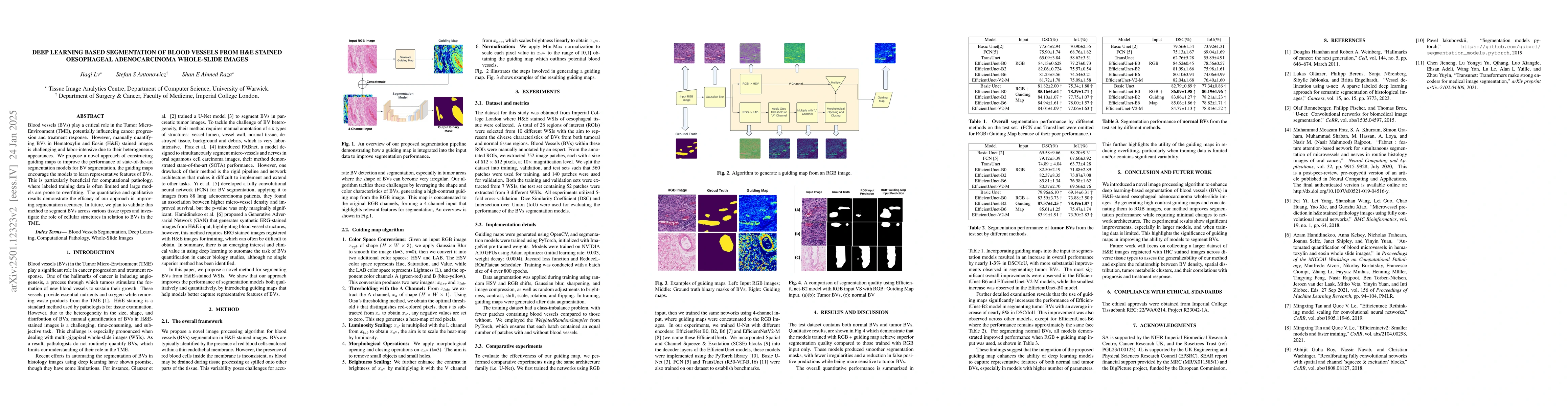
Blood vessels (BVs) play a critical role in the Tumor Micro-Environment (TME), potentially influencing cancer progression and treatment response. However, manually quantifying BVs in Hematoxylin and E...
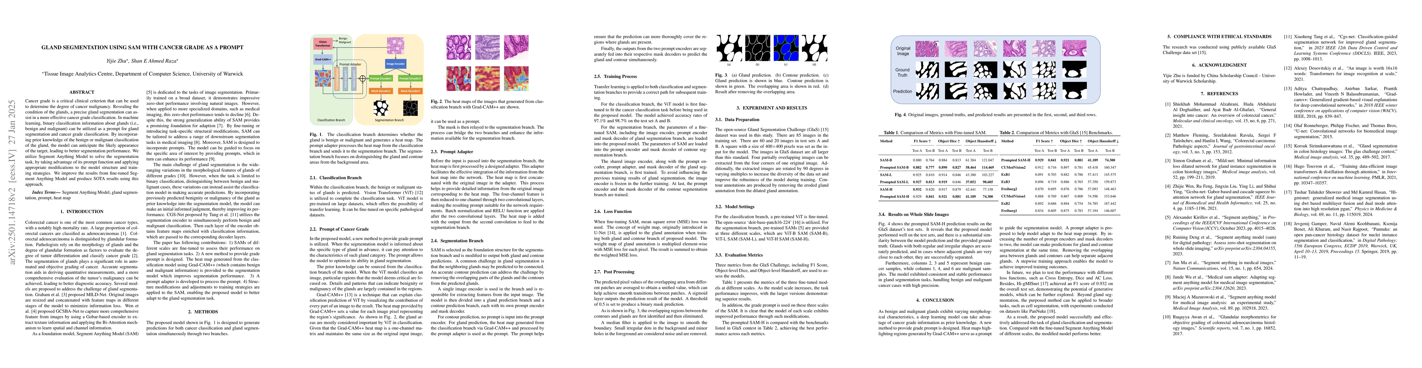
Cancer grade is a critical clinical criterion that can be used to determine the degree of cancer malignancy. Revealing the condition of the glands, a precise gland segmentation can assist in a more ef...
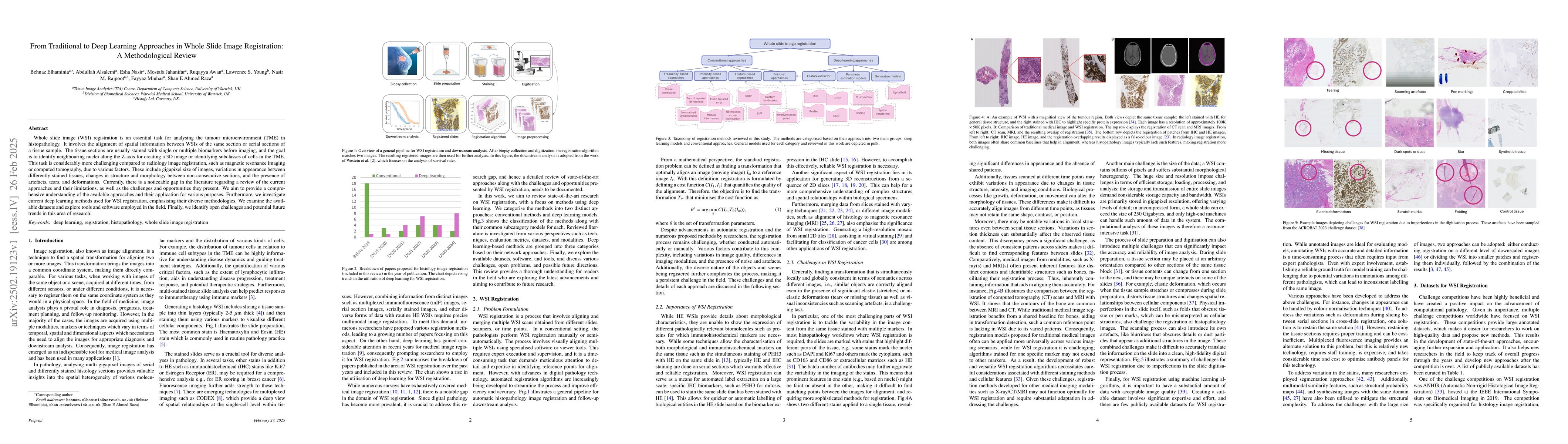
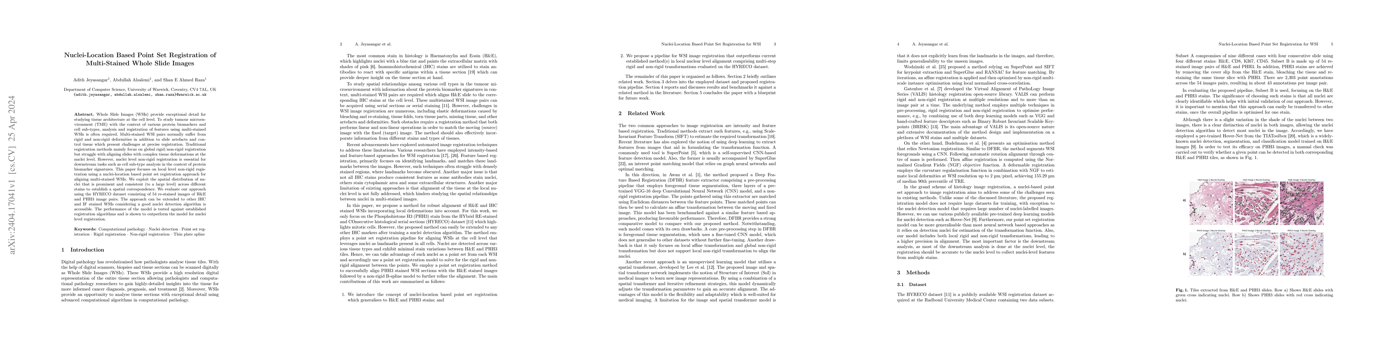
Whole slide image (WSI) registration is an essential task for analysing the tumour microenvironment (TME) in histopathology. It involves the alignment of spatial information between WSIs of the same s...
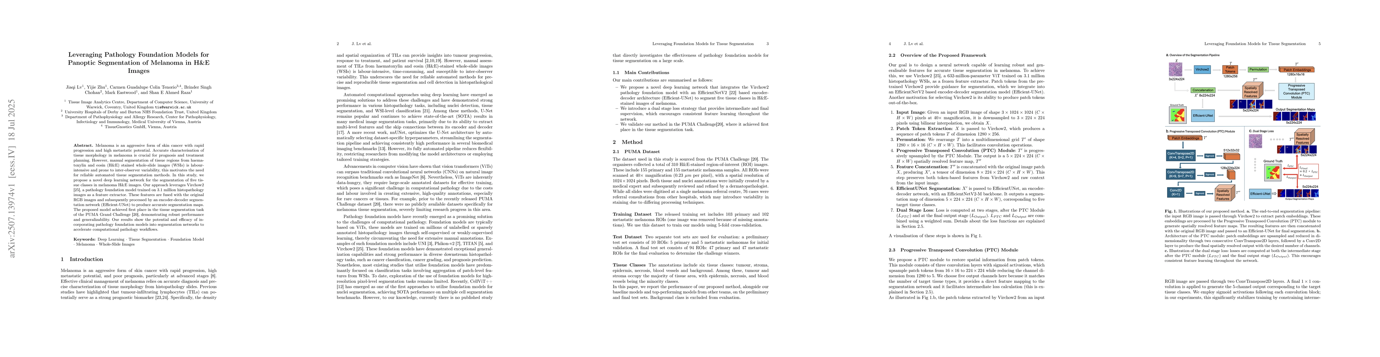
Melanoma is an aggressive form of skin cancer with rapid progression and high metastatic potential. Accurate characterisation of tissue morphology in melanoma is crucial for prognosis and treatment pl...
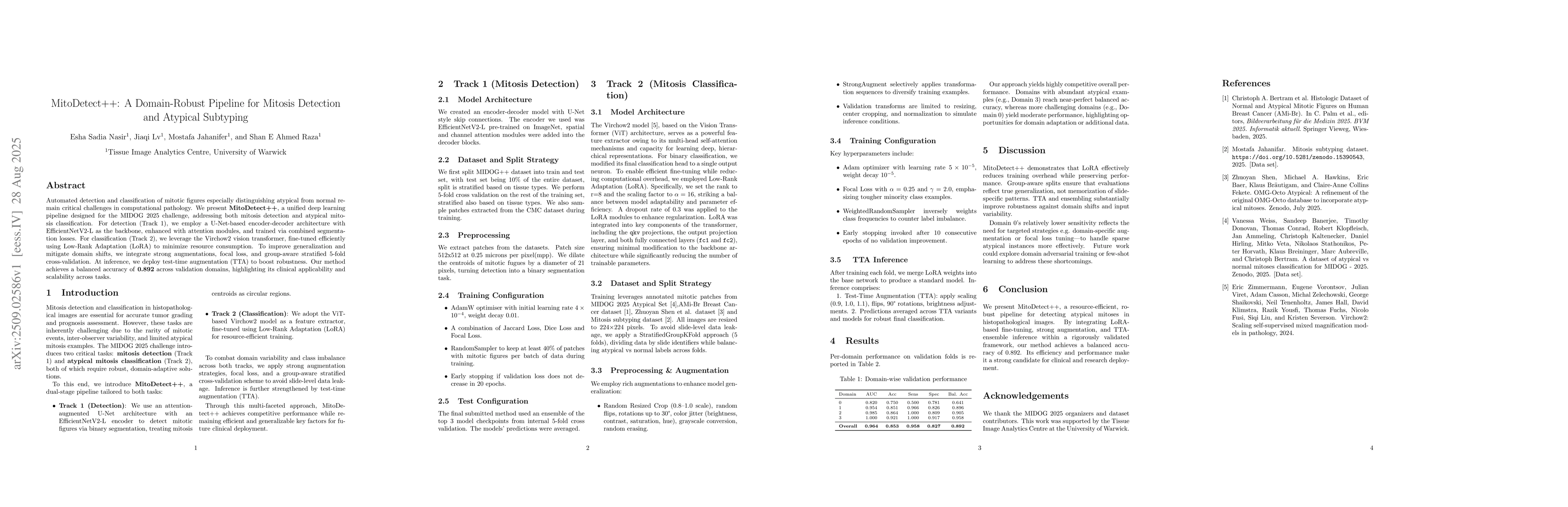
Automated detection and classification of mitotic figures especially distinguishing atypical from normal remain critical challenges in computational pathology. We present MitoDetect++, a unified deep ...
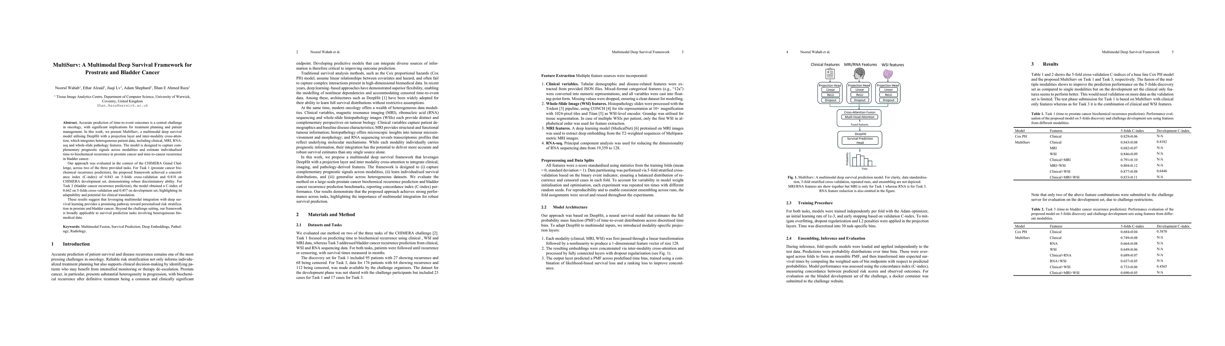
Accurate prediction of time-to-event outcomes is a central challenge in oncology, with significant implications for treatment planning and patient management. In this work, we present MultiSurv, a mul...
Accurate detection and classification of nuclei in histopathology images are critical for diagnostic and research applications. We present KongNet, a multi-headed deep learning architecture featuring ...
Accurate and efficient registration of whole slide images (WSIs) is essential for high-resolution, nuclei-level analysis in multi-stained tissue slides. We propose a novel coarse-to-fine framework COR...
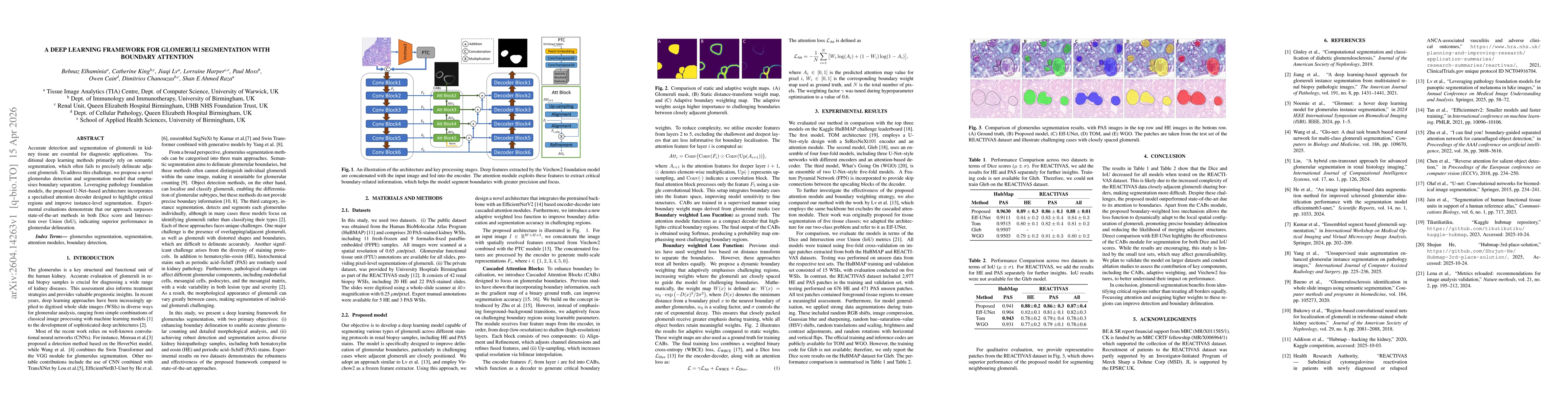
Accurate detection and segmentation of glomeruli in kidney tissue are essential for diagnostic applications. Traditional deep learning methods primarily rely on semantic segmentation, which often fail...
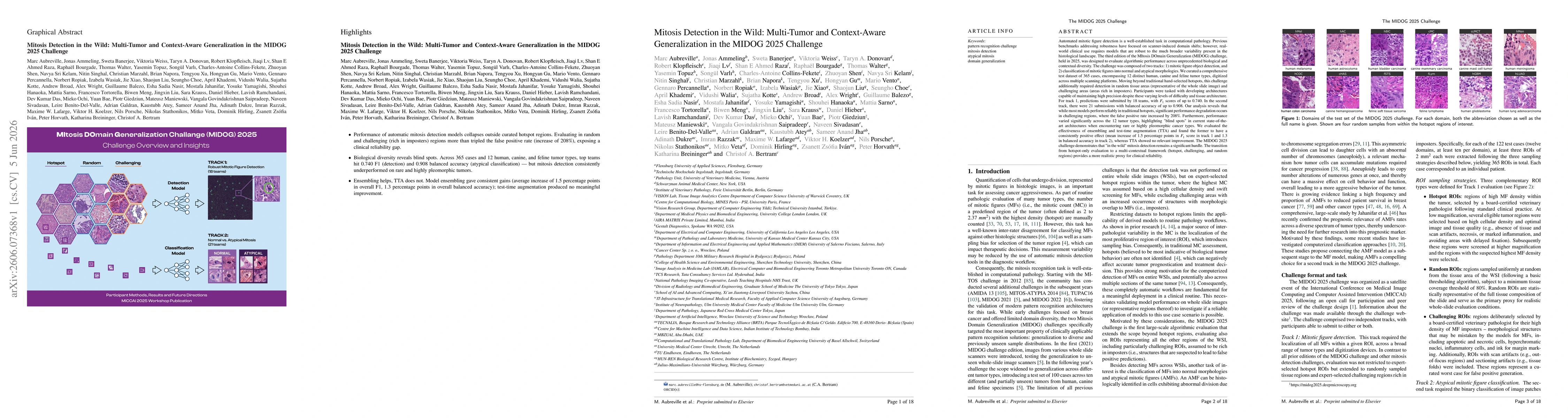
Automated mitosis detection is a well-established task in computational pathology. While previous benchmarks focused on scanner-induced domain shift, clinical "real-world" application requires models ...