Summary

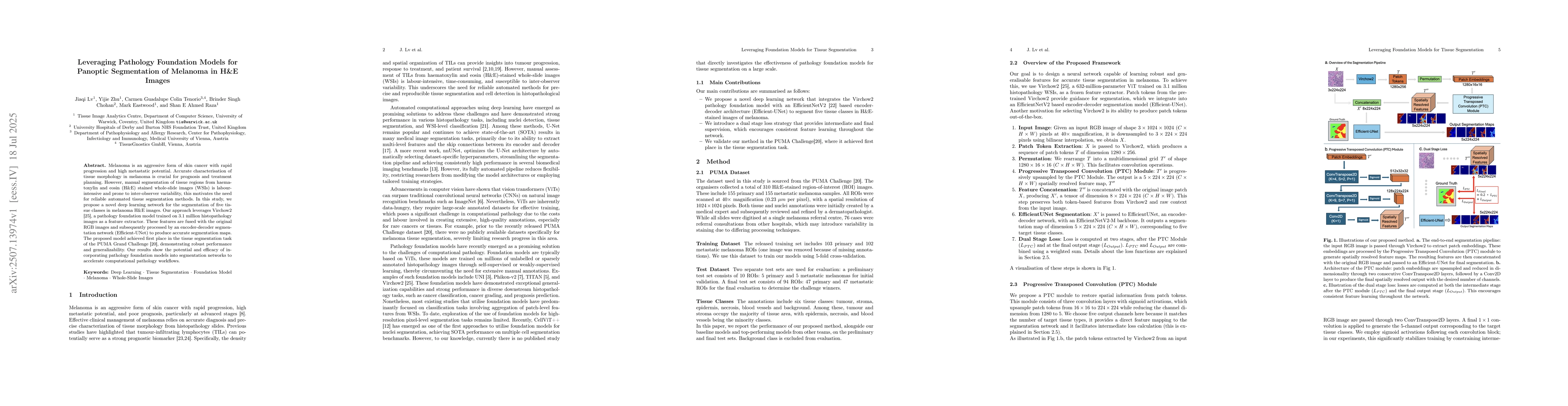
Melanoma is an aggressive form of skin cancer with rapid progression and high metastatic potential. Accurate characterisation of tissue morphology in melanoma is crucial for prognosis and treatment planning. However, manual segmentation of tissue regions from haematoxylin and eosin (H&E) stained whole-slide images (WSIs) is labour-intensive and prone to inter-observer variability, this motivates the need for reliable automated tissue segmentation methods. In this study, we propose a novel deep learning network for the segmentation of five tissue classes in melanoma H&E images. Our approach leverages Virchow2, a pathology foundation model trained on 3.1 million histopathology images as a feature extractor. These features are fused with the original RGB images and subsequently processed by an encoder-decoder segmentation network (Efficient-UNet) to produce accurate segmentation maps. The proposed model achieved first place in the tissue segmentation task of the PUMA Grand Challenge, demonstrating robust performance and generalizability. Our results show the potential and efficacy of incorporating pathology foundation models into segmentation networks to accelerate computational pathology workflows.

AI Key Findings

Generated Sep 03, 2025

Methodology

The research proposes a novel deep learning network for segmenting five tissue classes in melanoma H&E images, leveraging Virchow2, a pathology foundation model, as a feature extractor. The features are fused with RGB images and processed by an Efficient-UNet for accurate segmentation maps.

Key Results

  • The proposed model achieved first place in the tissue segmentation task of the PUMA Grand Challenge.
  • Incorporating pathology foundation models into segmentation networks accelerates computational pathology workflows.
  • The method outperformed baseline models in tissue segmentation, particularly in tumor, stroma, and necrosis regions.

Significance

This research is important for automating tissue segmentation in melanoma H&E images, reducing manual labor and inter-observer variability, which is crucial for prognosis and treatment planning.

Technical Contribution

The introduction of a Progressive Transposed Convolution (PTC) module to transform Virchow2 patch tokens into spatially resolved features, and a dual-stage loss strategy for supervision at intermediate and final stages of the network.

Novelty

The work combines a pathology foundation model with an encoder-decoder segmentation network, employing a dual-stage loss strategy to enhance feature learning and segmentation accuracy in melanoma H&E images.

Limitations

  • The study did not address the generalizability to other types of cancer or tissue types beyond melanoma.
  • Performance degradation was observed on rare tissue classes, possibly due to class imbalance within the dataset.

Future Work

  • Validation on larger datasets such as BCSS and detailed quantitative evaluation using only spatially resolved features.
  • Ablation studies to assess the impact of different weighting configurations in the dual-stage loss strategy.
  • Exploration of alternative pathology foundation models like UNI, Phikon-v2, and TITAN.

Paper Details

PDF Preview

Download PDF

Citation Network

Current paper (gray), citations (green), references (blue)

Display is limited for performance on very large graphs.

Current Paper
Citations
References
Click to view

No citations found for this paper.

Comments (0)

This website uses cookies to ensure you get the best experience. Learn more