Academic Profile
Statistics
Similar Authors
Papers on arXiv
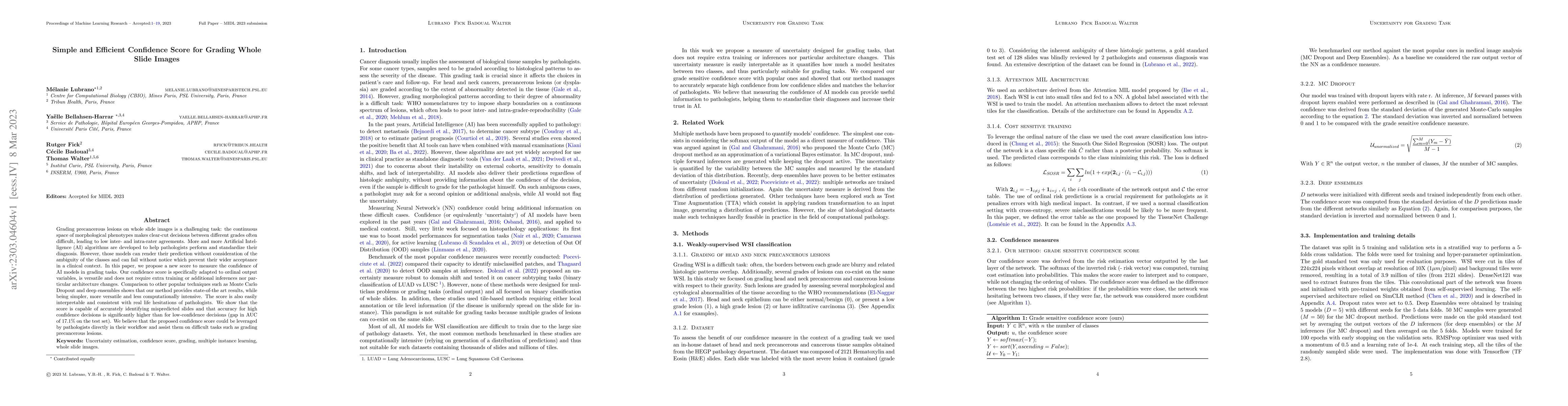
Grading precancerous lesions on whole slide images is a challenging task: the continuous space of morphological phenotypes makes clear-cut decisions between different grades often difficult, leading...
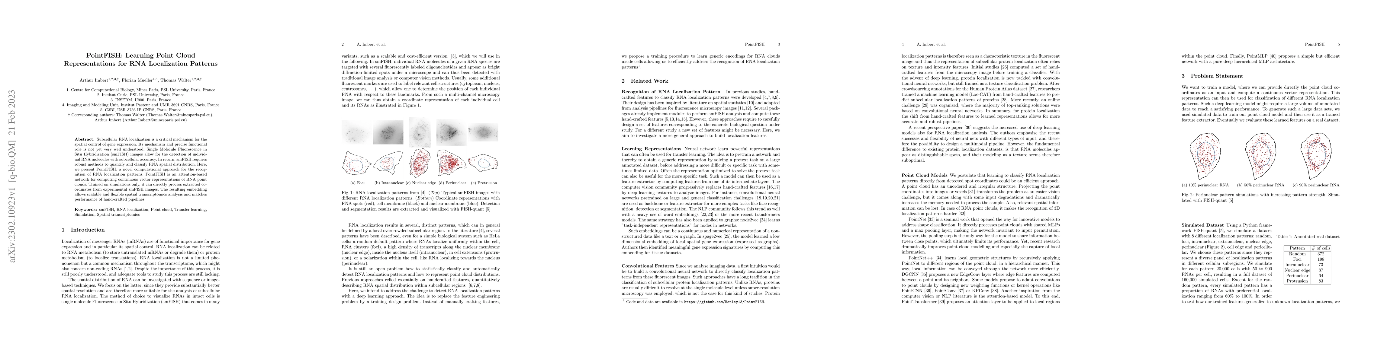
Subcellular RNA localization is a critical mechanism for the spatial control of gene expression. Its mechanism and precise functional role is not yet very well understood. Single Molecule Fluorescen...
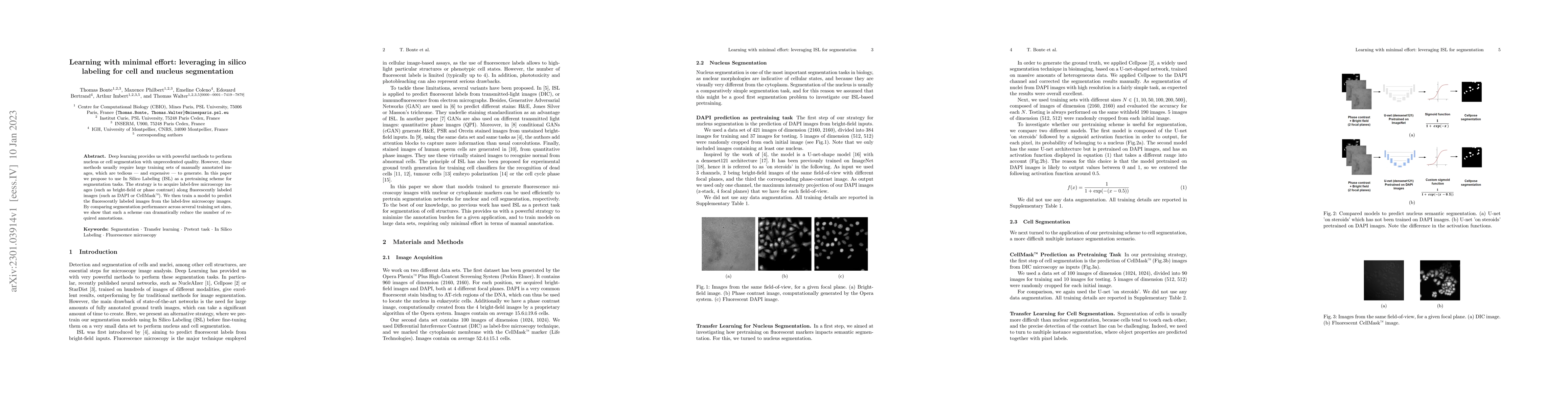
Deep learning provides us with powerful methods to perform nucleus or cell segmentation with unprecedented quality. However, these methods usually require large training sets of manually annotated i...
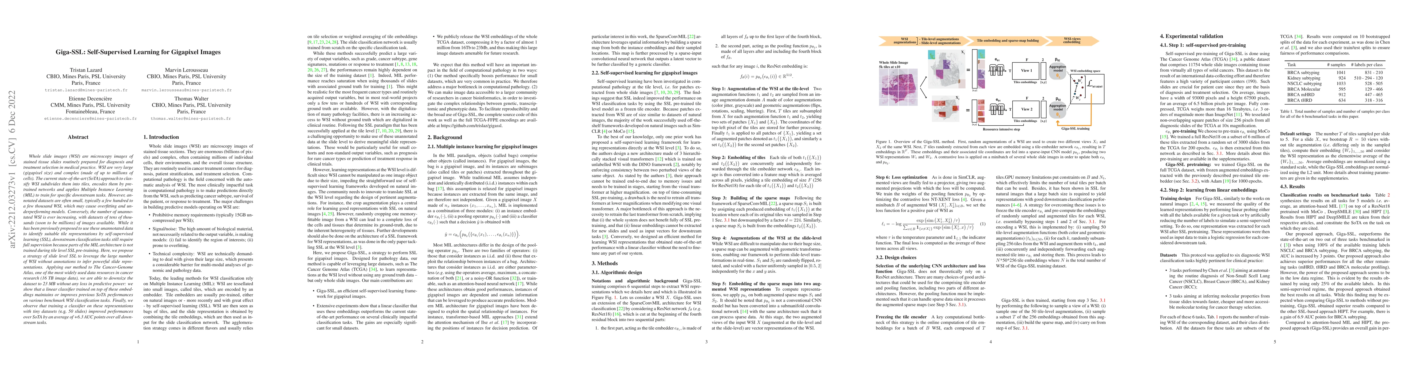
Whole slide images (WSI) are microscopy images of stained tissue slides routinely prepared for diagnosis and treatment selection in medical practice. WSI are very large (gigapixel size) and complex ...
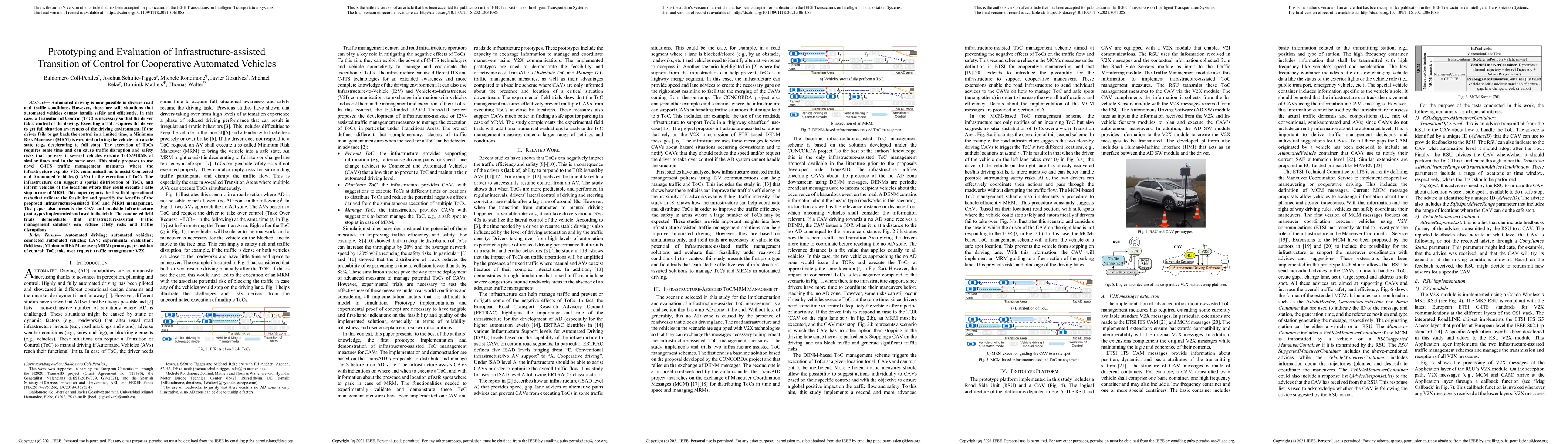
Automated driving is now possible in diverse road and traffic conditions. However, there are still situations that automated vehicles cannot handle safely and efficiently. In this case, a Transition...
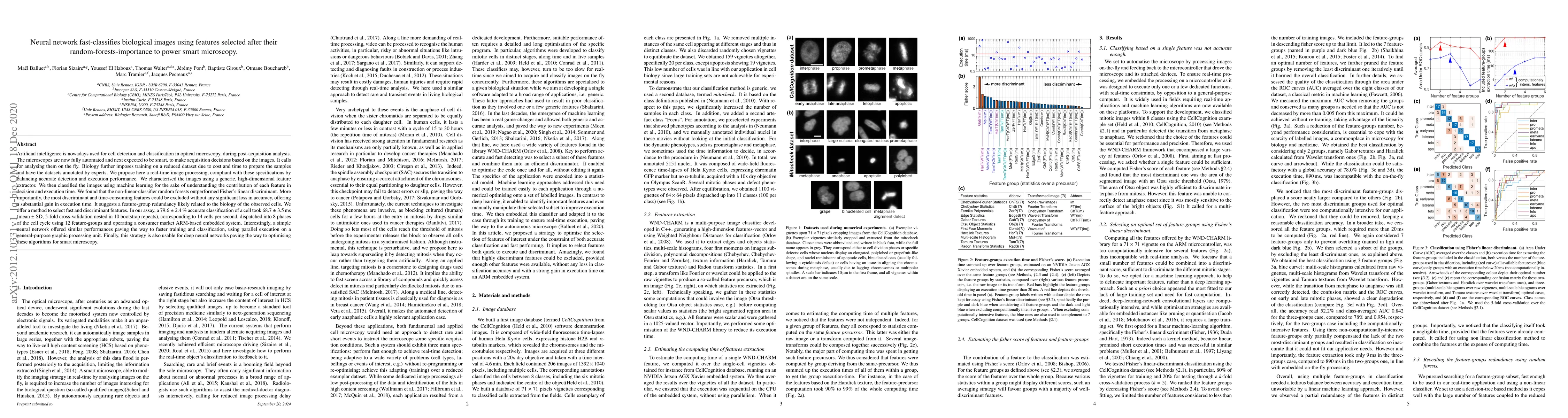
Artificial intelligence is nowadays used for cell detection and classification in optical microscopy, during post-acquisition analysis. The microscopes are now fully automated and next expected to b...
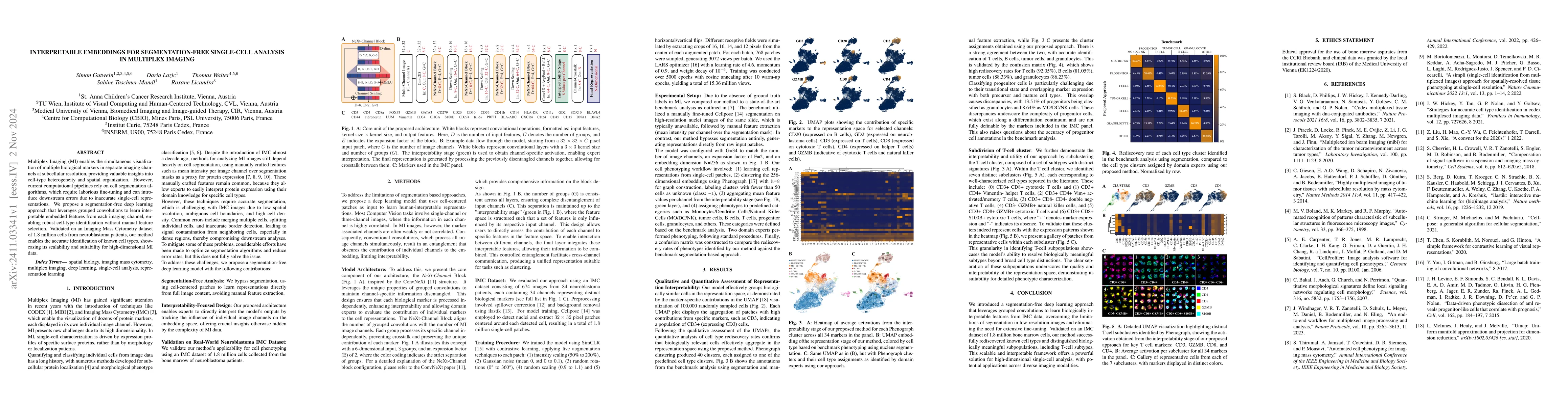
Multiplex Imaging (MI) enables the simultaneous visualization of multiple biological markers in separate imaging channels at subcellular resolution, providing valuable insights into cell-type heteroge...
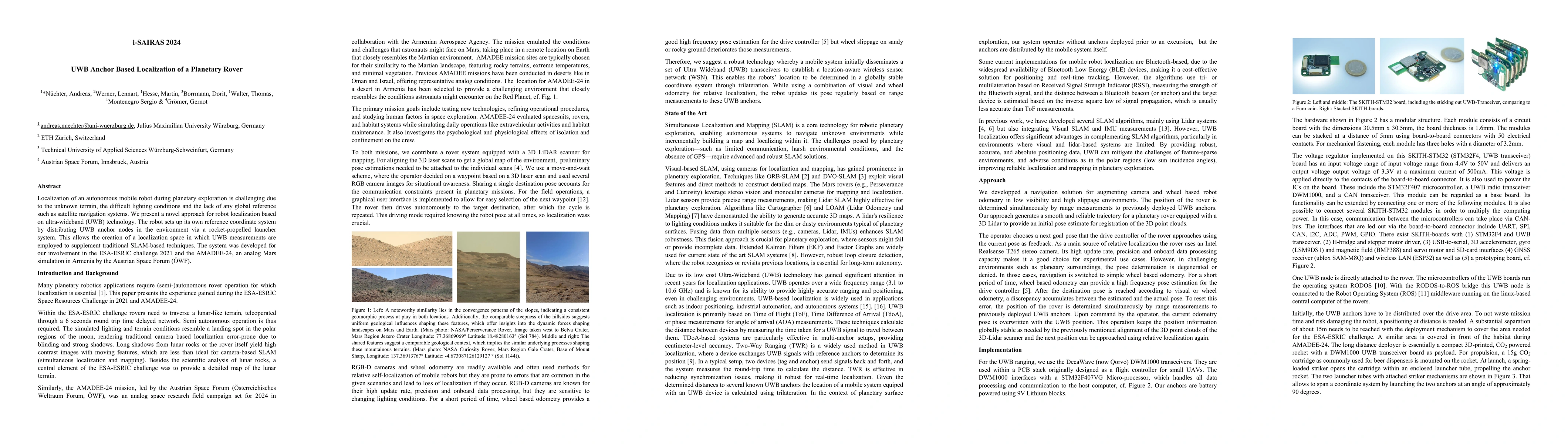
Localization of an autonomous mobile robot during planetary exploration is challenging due to the unknown terrain, the difficult lighting conditions and the lack of any global reference such as satell...
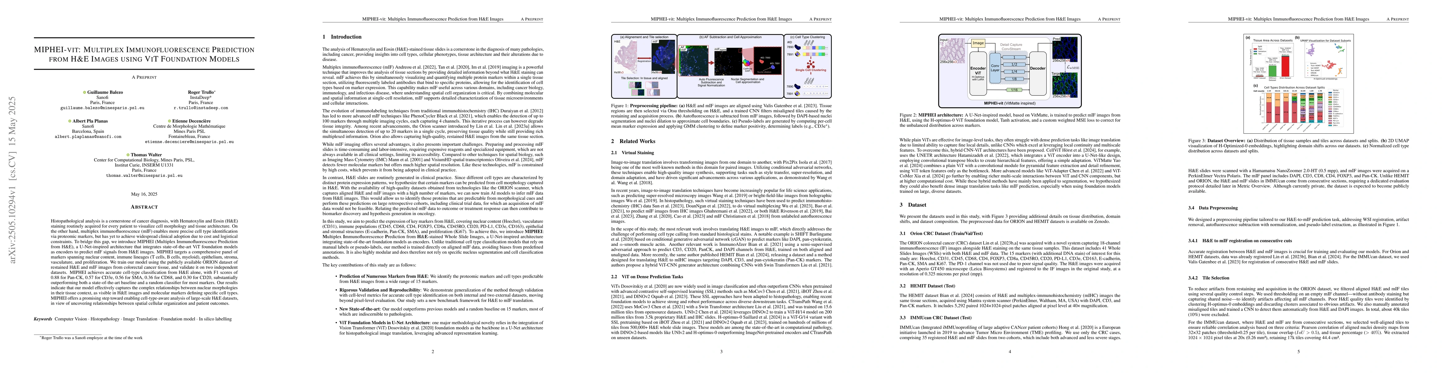
Histopathological analysis is a cornerstone of cancer diagnosis, with Hematoxylin and Eosin (H&E) staining routinely acquired for every patient to visualize cell morphology and tissue architecture. On...
Accurate mitotic figure classification is crucial in computational pathology, as mitotic activity informs cancer grading and patient prognosis. Distinguishing atypical mitotic figures (AMFs), which in...
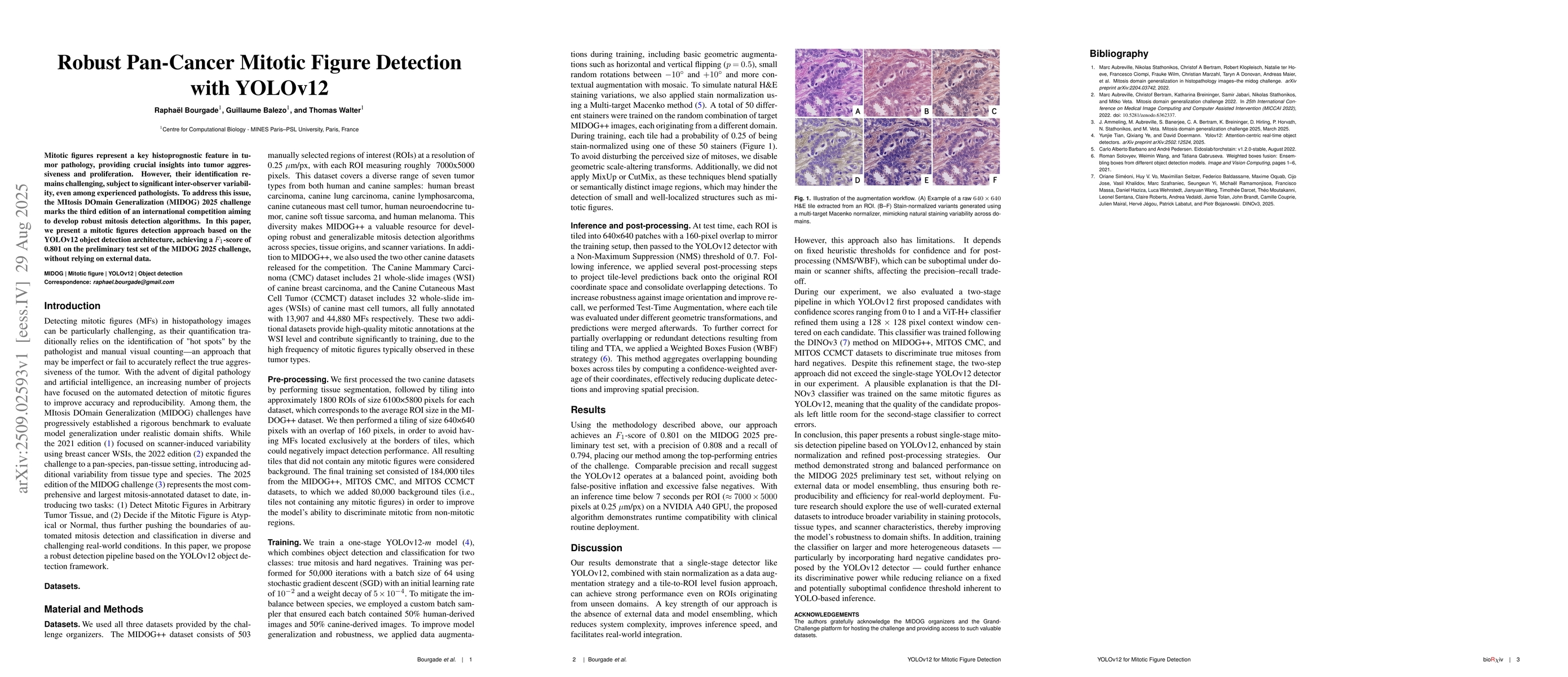
Mitotic figures represent a key histoprognostic feature in tumor pathology, providing crucial insights into tumor aggressiveness and proliferation. However, their identification remains challenging, s...
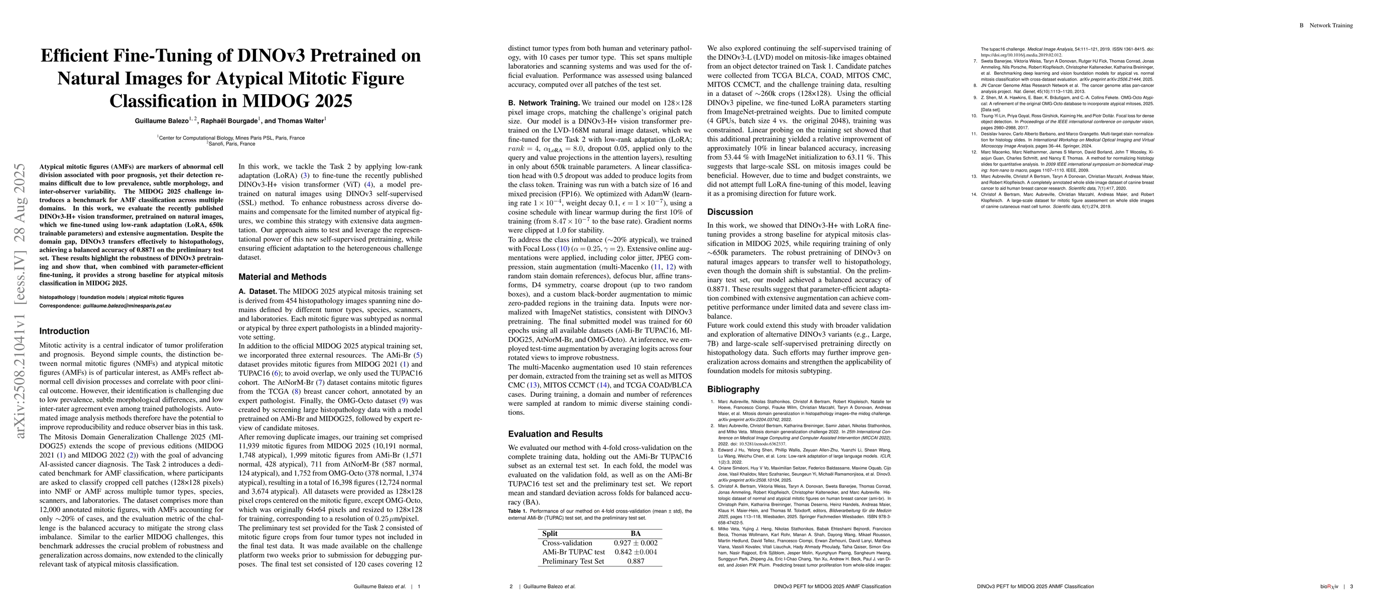
Atypical mitotic figures (AMFs) are markers of abnormal cell division associated with poor prognosis, yet their detection remains difficult due to low prevalence, subtle morphology, and inter-observer...
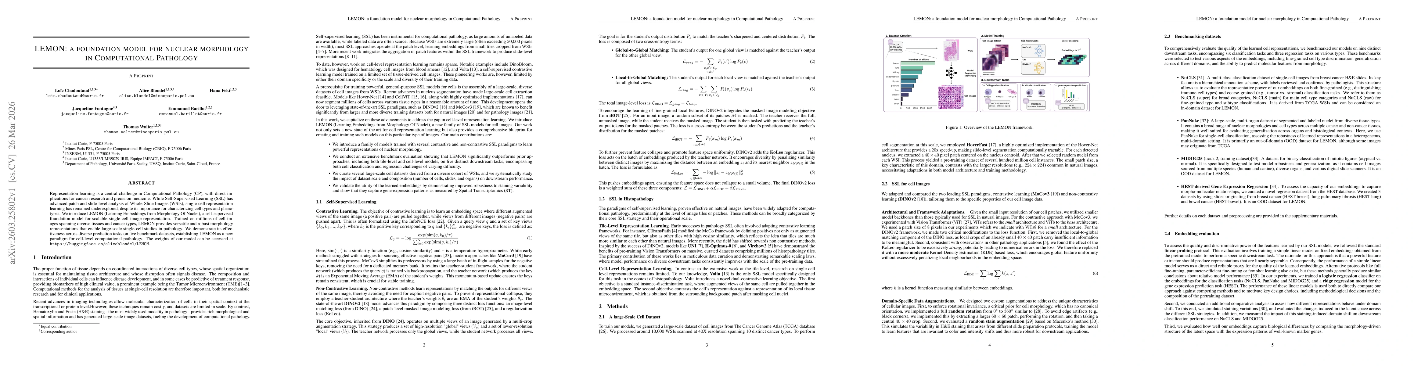
Computational pathology relies on effective representation learning to support cancer research and precision medicine. Although self-supervised learning has driven major progress at the patch and whol...
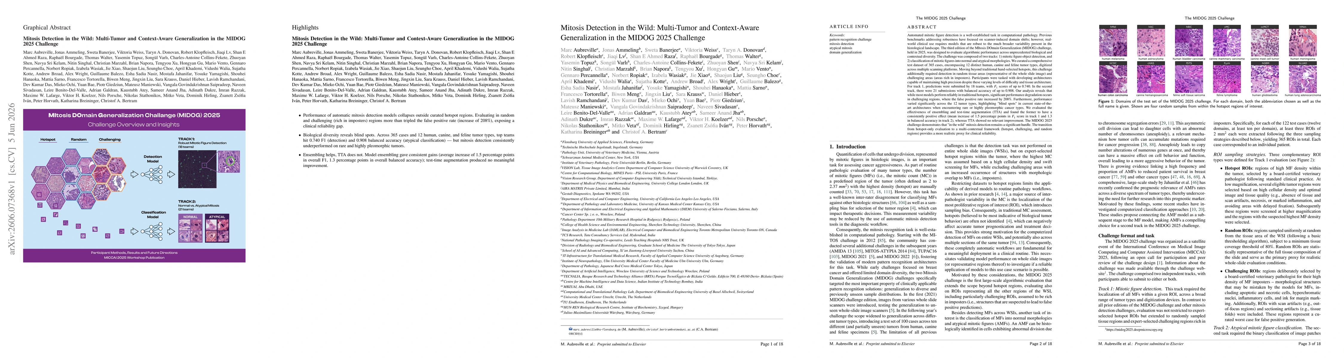
Automated mitosis detection is a well-established task in computational pathology. While previous benchmarks focused on scanner-induced domain shift, clinical "real-world" application requires models ...