Academic Profile
Statistics
Similar Authors
Papers on arXiv
Automated segmentation tools often encounter accuracy and adaptability issues when applied to images of different pathology. The purpose of this study is to explore the feasibility of building a wor...
Uterine leiomyosarcoma (LMS) is a rare but aggressive malignancy. On imaging, it is difficult to differentiate LMS from, for example, degenerated leiomyoma (LM), a prevalent but benign condition. We...
Purpose: This study evaluated the out-of-domain performance and generalization capabilities of automated medical image segmentation models, with a particular focus on adaptation to new image acquisi...
Methods for automatically flag poor performing-predictions are essential for safely implementing machine learning workflows into clinical practice and for identifying difficult cases during model tr...
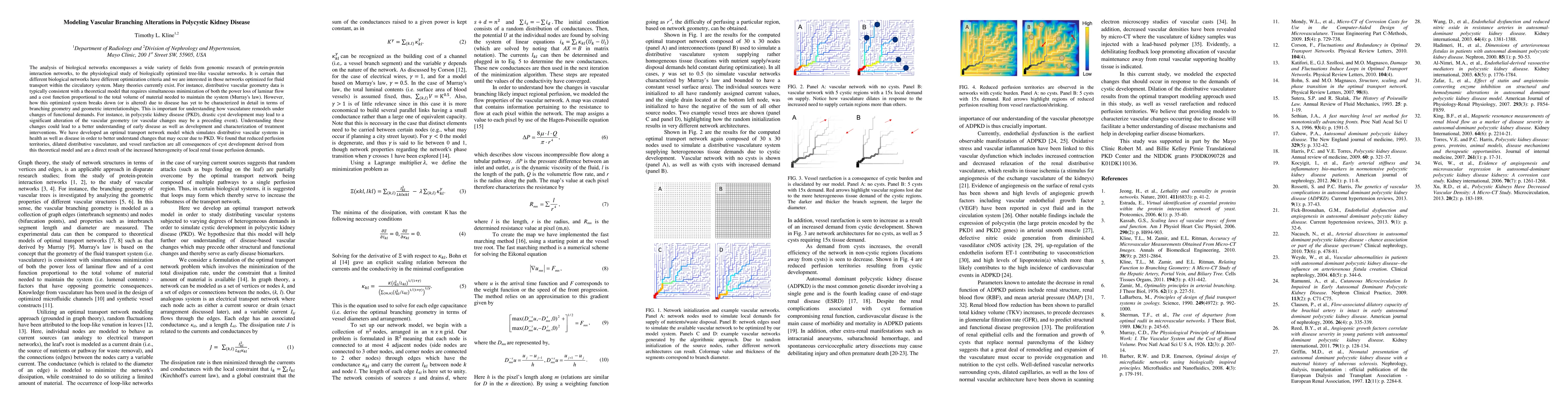
The analysis of biological networks encompasses a wide variety of fields from genomic research of protein-protein interaction networks, to the physiological study of biologically optimized tree-like...
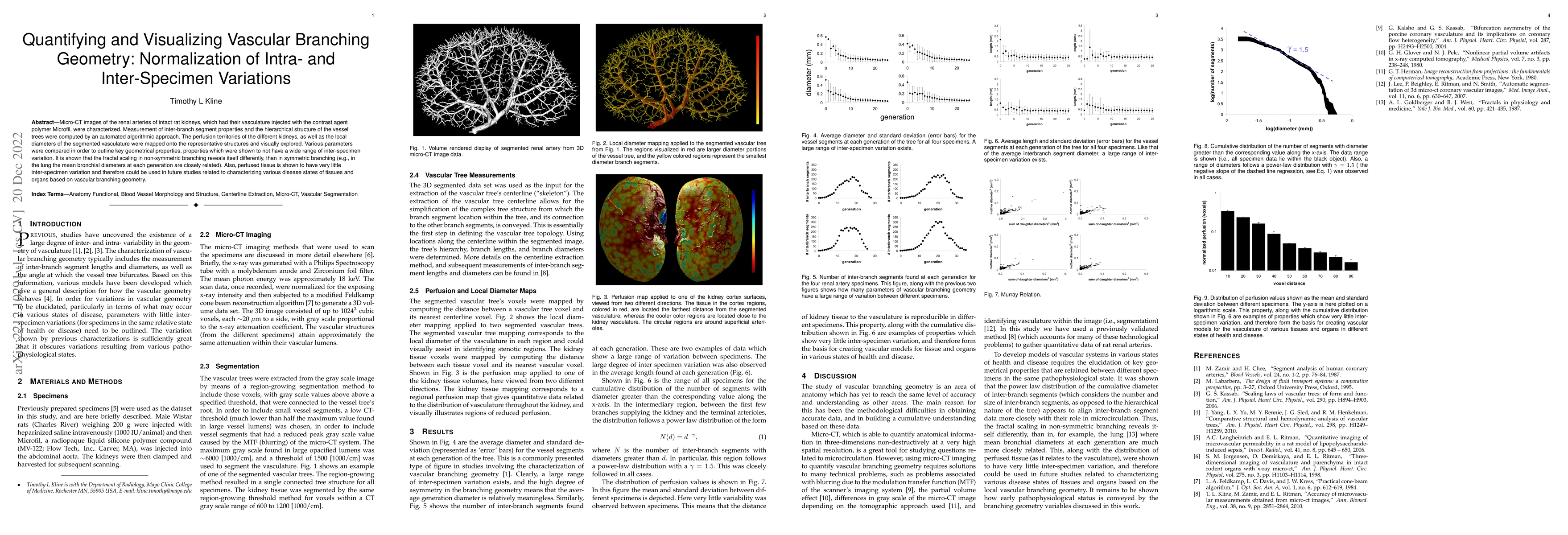
Micro-CT images of the renal arteries of intact rat kidneys, which had their vasculature injected with the contrast agent polymer Microfil, were characterized. Measurement of inter-branch segment pr...
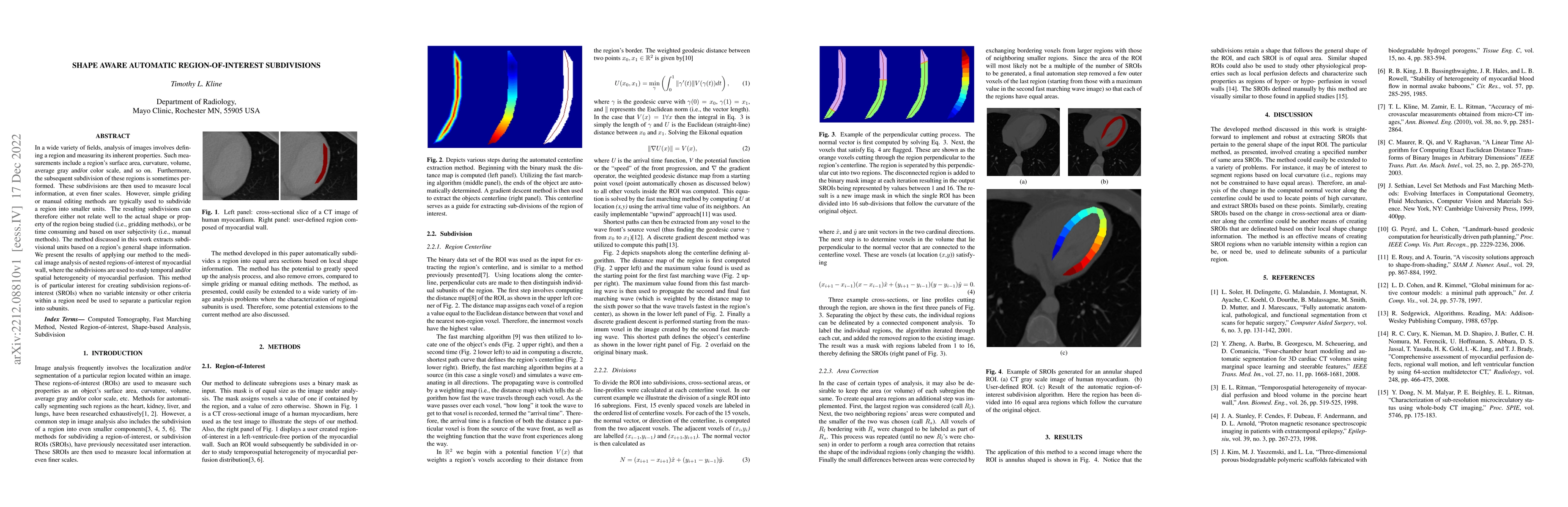
In a wide variety of fields, analysis of images involves defining a region and measuring its inherent properties. Such measurements include a region's surface area, curvature, volume, average gray a...
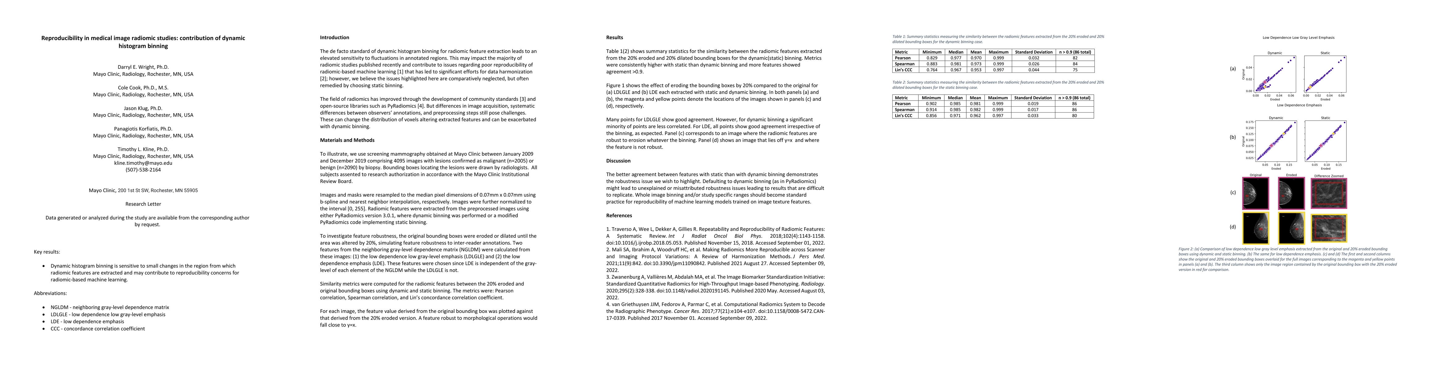
The de facto standard of dynamic histogram binning for radiomic feature extraction leads to an elevated sensitivity to fluctuations in annotated regions. This may impact the majority of radiomic stu...
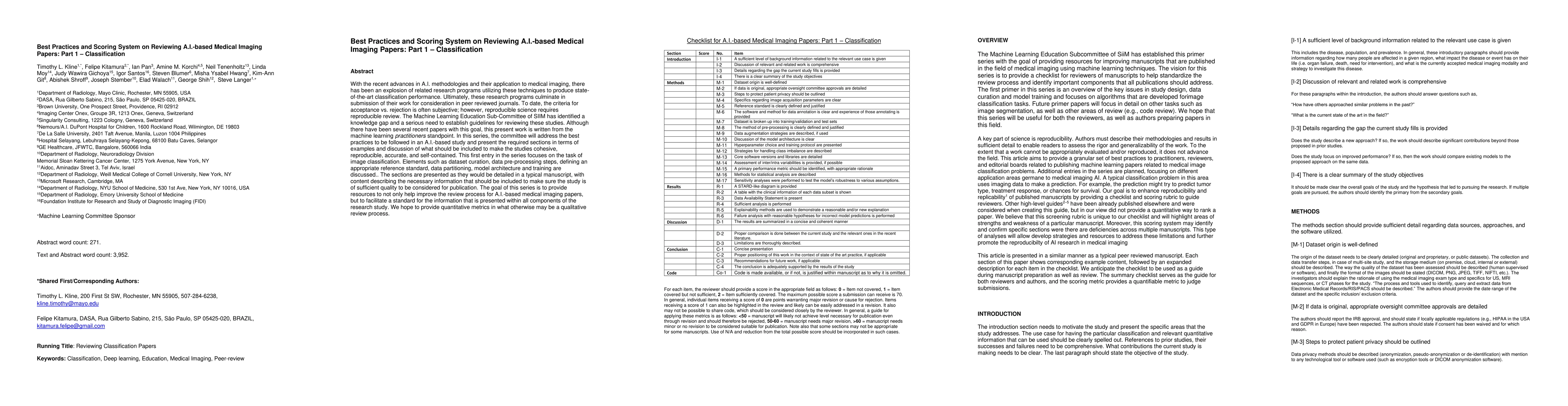
With the recent advances in A.I. methodologies and their application to medical imaging, there has been an explosion of related research programs utilizing these techniques to produce state-of-the-a...
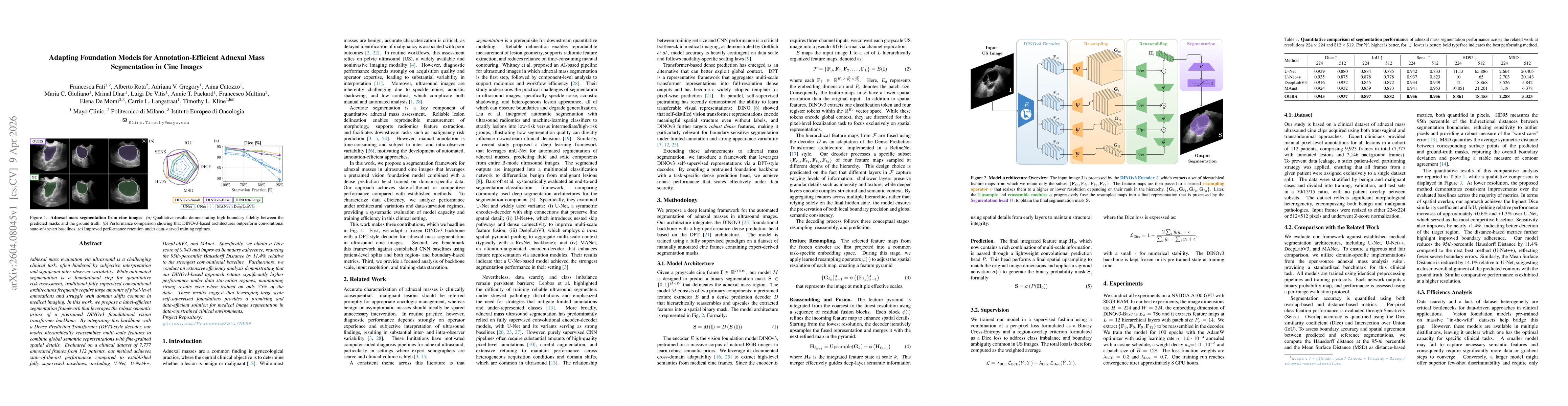
Adnexal mass evaluation via ultrasound is a challenging clinical task, often hindered by subjective interpretation and significant inter-observer variability. While automated segmentation is a foundat...
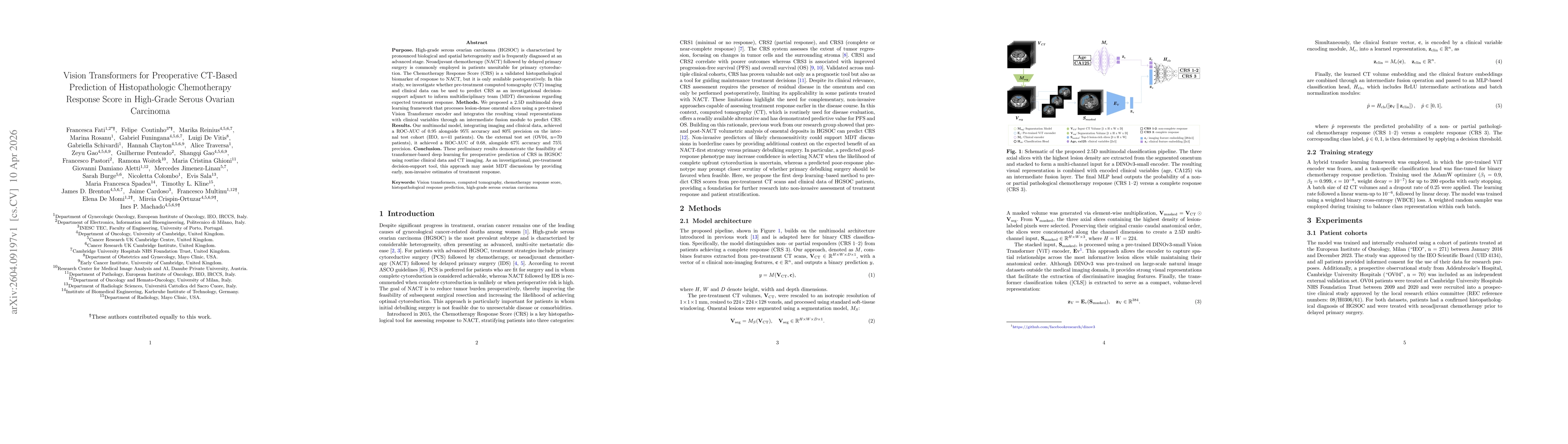
Purpose. High-grade serous ovarian carcinoma (HGSOC) is characterized by pronounced biological and spatial heterogeneity and is frequently diagnosed at an advanced stage. Neoadjuvant chemotherapy (NAC...