Publication
Metrics
AI Quick Summary
This paper proposes a method for automatically subdividing regions of interest in images based on the shape information of the region, aiming to improve the accuracy and efficiency of subsequent measurements. The method is demonstrated in medical image analysis for studying myocardial perfusion, showing its potential in creating precise subdivisions without relying on variable intensity criteria.
Paper Preview
Abstract
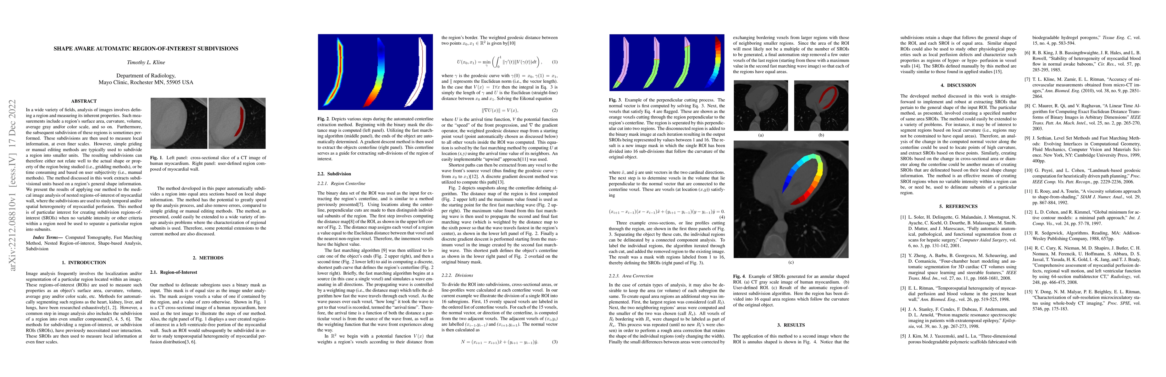
In a wide variety of fields, analysis of images involves defining a region and measuring its inherent properties. Such measurements include a region's surface area, curvature, volume, average gray and/or color scale, and so on. Furthermore, the subsequent subdivision of these regions is sometimes performed. These subdivisions are then used to measure local information, at even finer scales. However, simple griding or manual editing methods are typically used to subdivide a region into smaller units. The resulting subdivisions can therefore either not relate well to the actual shape or property of the region being studied (i.e., gridding methods), or be time consuming and based on user subjectivity (i.e., manual methods). The method discussed in this work extracts subdivisional units based on a region's general shape information. We present the results of applying our method to the medical image analysis of nested regions-of-interest of myocardial wall, where the subdivisions are used to study temporal and/or spatial heterogeneity of myocardial perfusion. This method is of particular interest for creating subdivision regions-of-interest (SROIs) when no variable intensity or other criteria within a region need be used to separate a particular region into subunits.
AI Key Findings
Get AI-generated insights about this paper's methodology, results, significance, and more — seven facets brought into focus.
Impact
Paper Details
Authors
PDF Preview
Key Terms
Citation Network
Current paper (gray), citations (green), references (blue)
Display is limited for performance on very large graphs.
Discussion 0