Academic Profile
Statistics
Similar Authors
Papers on arXiv
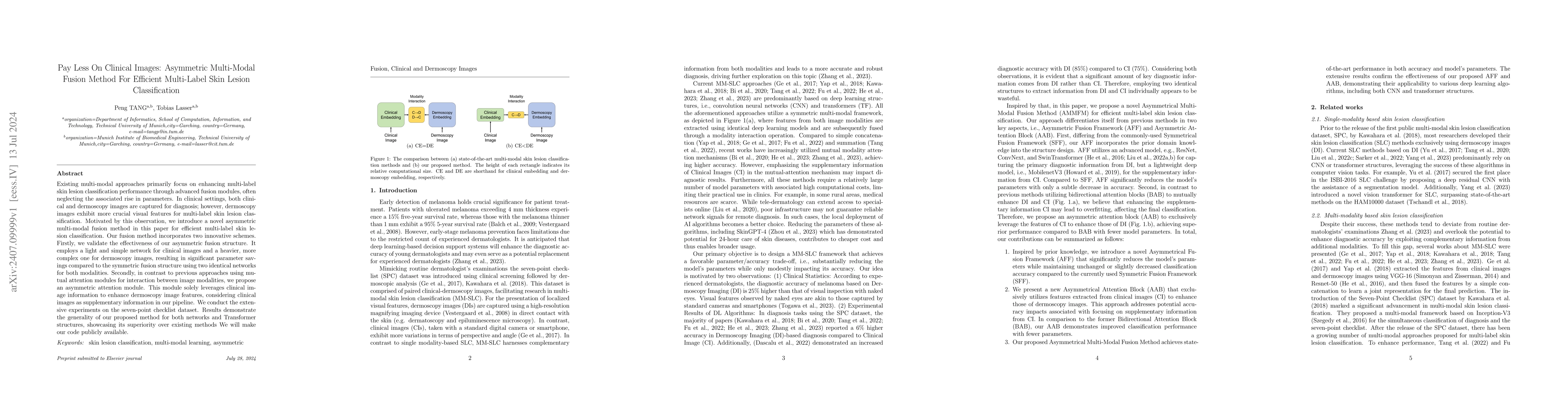
Existing multi-modal approaches primarily focus on enhancing multi-label skin lesion classification performance through advanced fusion modules, often neglecting the associated rise in parameters. In ...
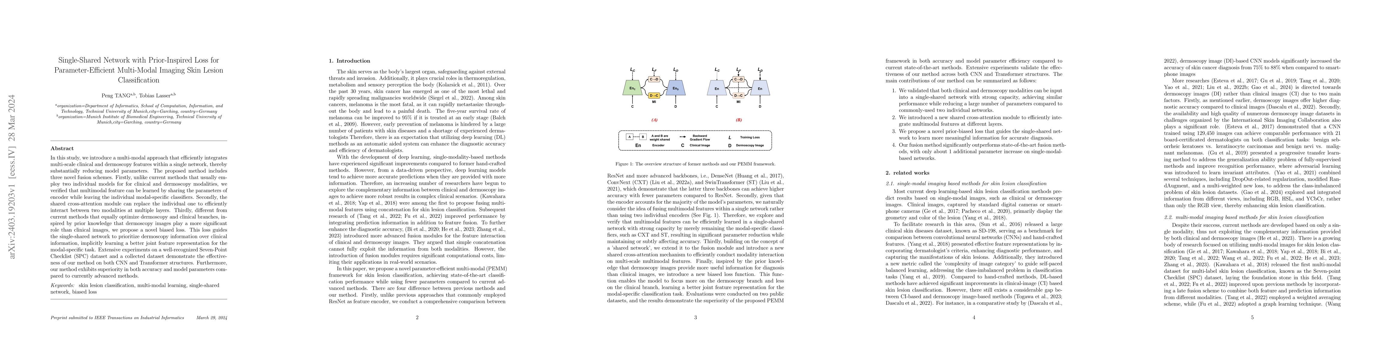
In this study, we introduce a multi-modal approach that efficiently integrates multi-scale clinical and dermoscopy features within a single network, thereby substantially reducing model parameters. ...
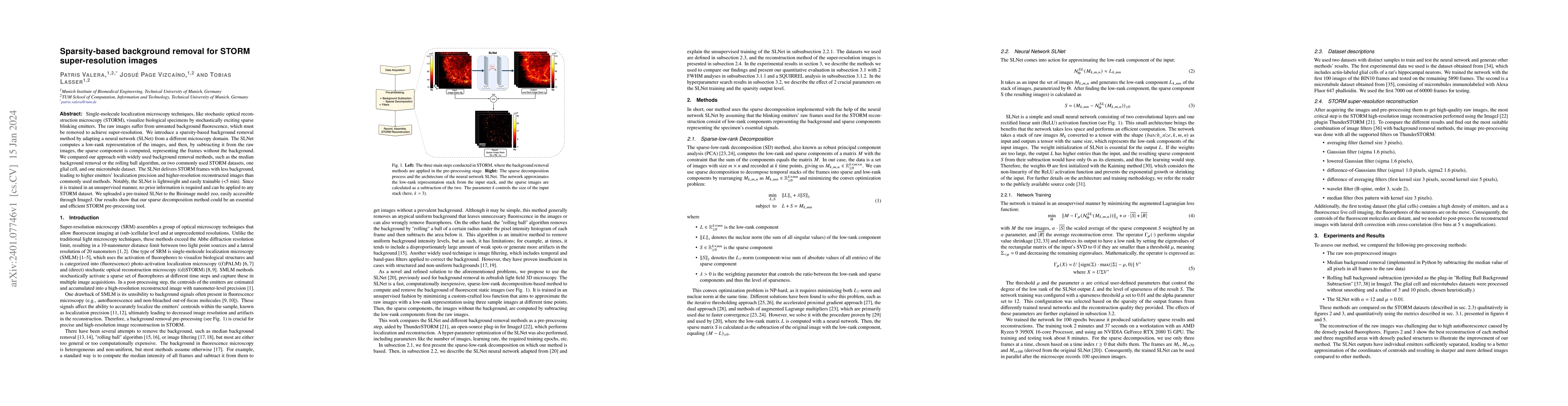
Single-molecule localization microscopy techniques, like stochastic optical reconstruction microscopy (STORM), visualize biological specimens by stochastically exciting sparse blinking emitters. The...
Most convolutional neural network (CNN) based methods for skin cancer classification obtain their results using only dermatological images. Although good classification results have been shown, more...
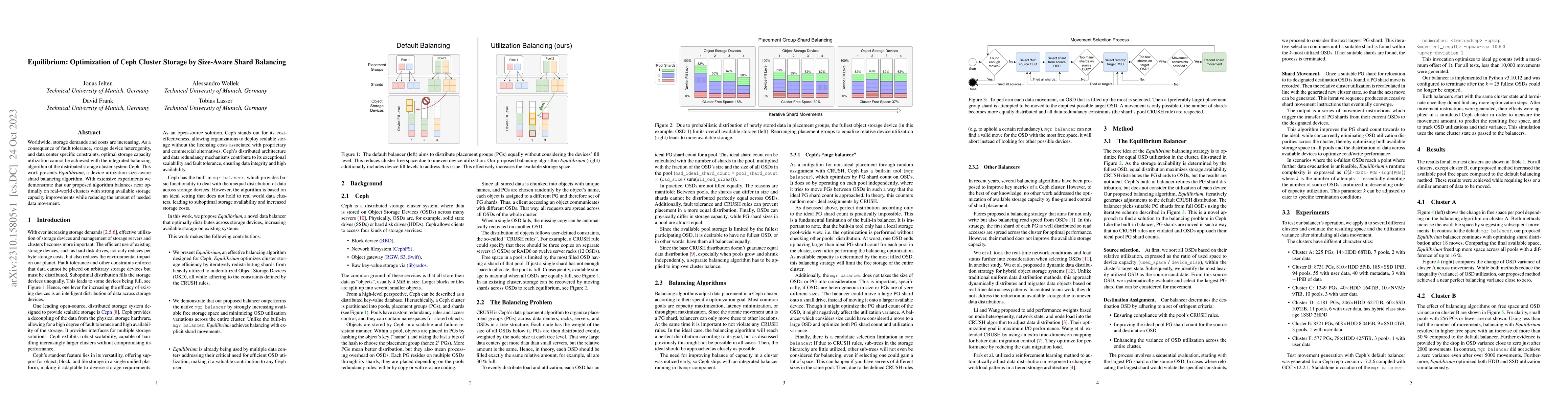
Worldwide, storage demands and costs are increasing. As a consequence of fault tolerance, storage device heterogenity, and data center specific constraints, optimal storage capacity utilization cann...
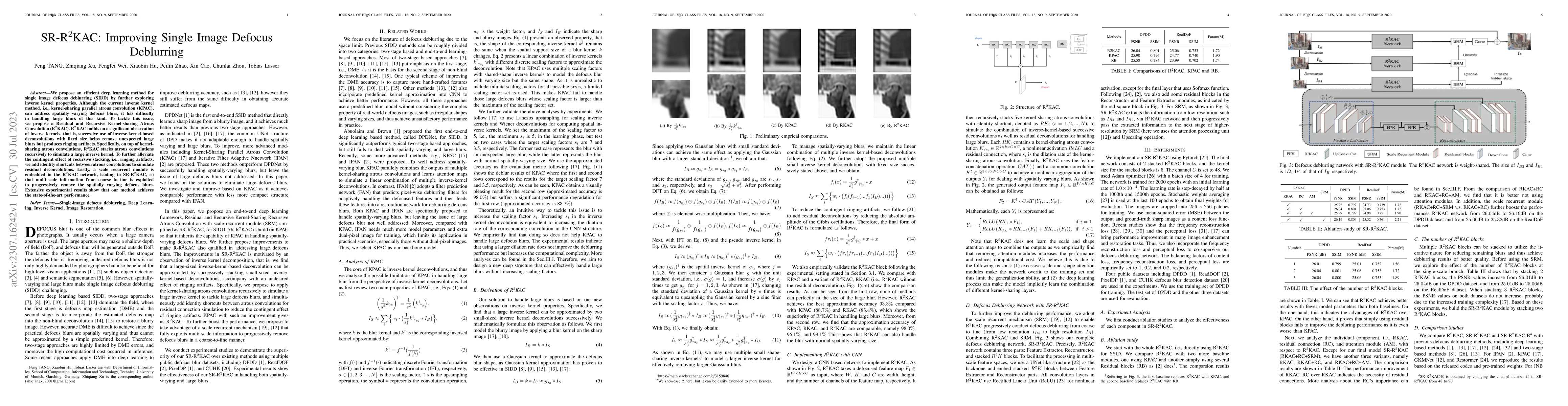
We propose an efficient deep learning method for single image defocus deblurring (SIDD) by further exploring inverse kernel properties. Although the current inverse kernel method, i.e., kernel-shari...
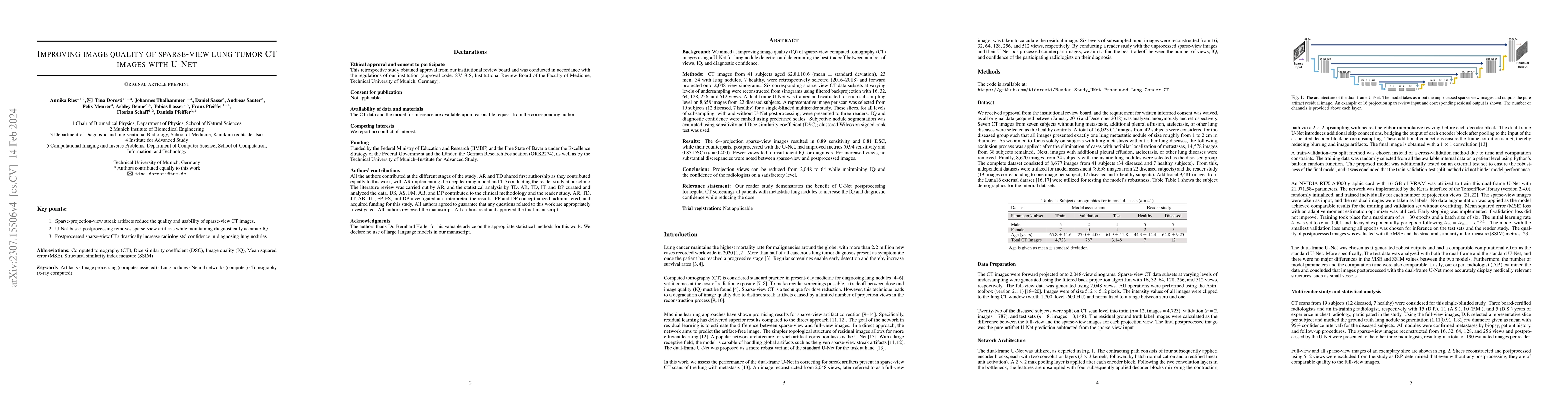
Background: We aimed at improving image quality (IQ) of sparse-view computed tomography (CT) images using a U-Net for lung metastasis detection and determining the best tradeoff between number of vi...
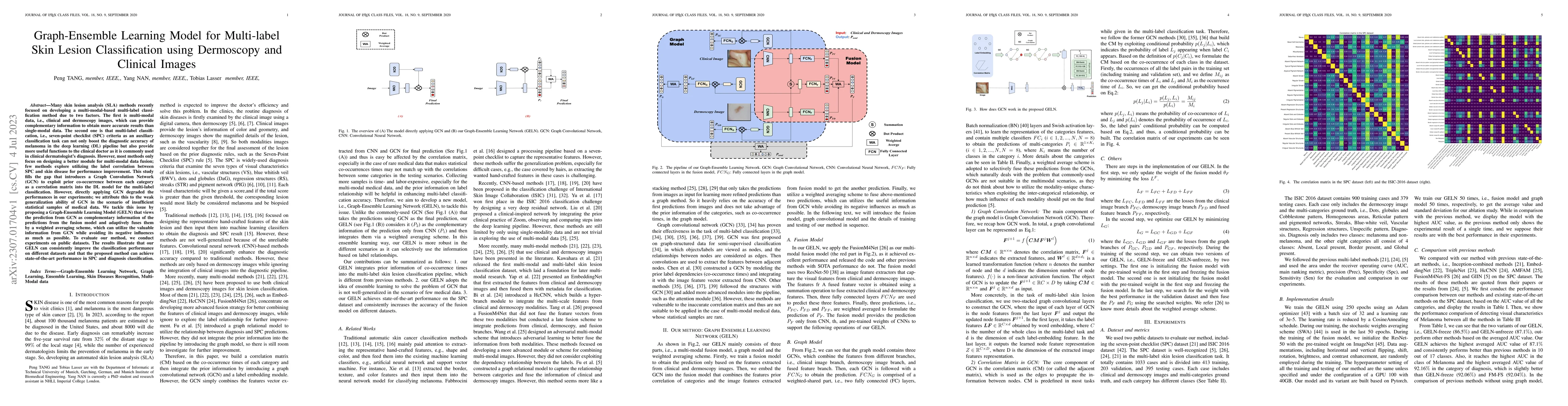
Many skin lesion analysis (SLA) methods recently focused on developing a multi-modal-based multi-label classification method due to two factors. The first is multi-modal data, i.e., clinical and der...
Tomographic imaging systems are expected to work with a wide range of samples that house complex structures and challenging material compositions, which can influence image quality in a bad way. Com...
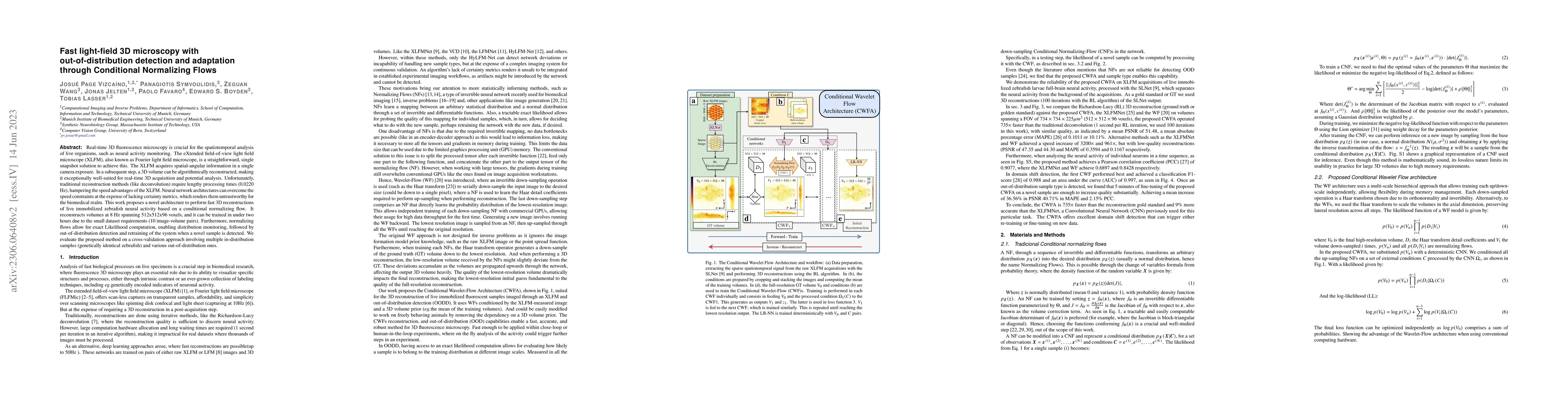
Real-time 3D fluorescence microscopy is crucial for the spatiotemporal analysis of live organisms, such as neural activity monitoring. The eXtended field-of-view light field microscope (XLFM), also ...
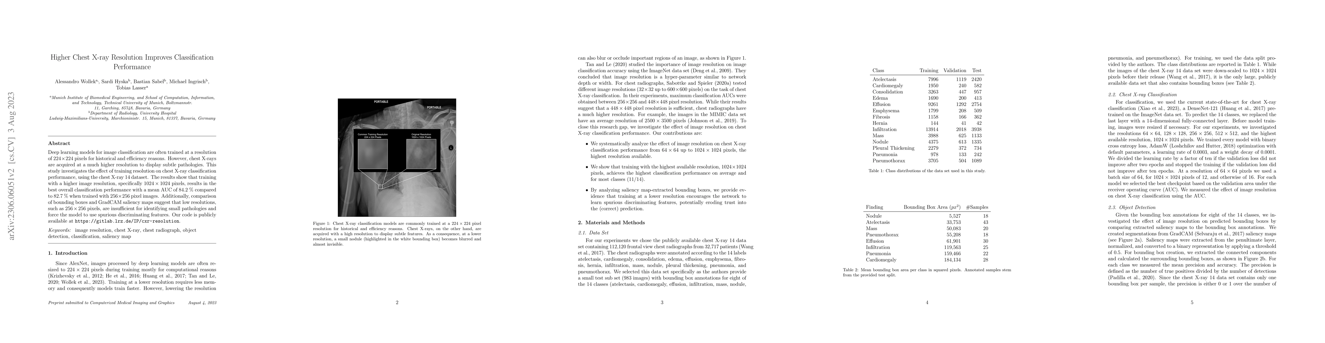
Deep learning models for image classification are often trained at a resolution of 224 x 224 pixels for historical and efficiency reasons. However, chest X-rays are acquired at a much higher resolut...
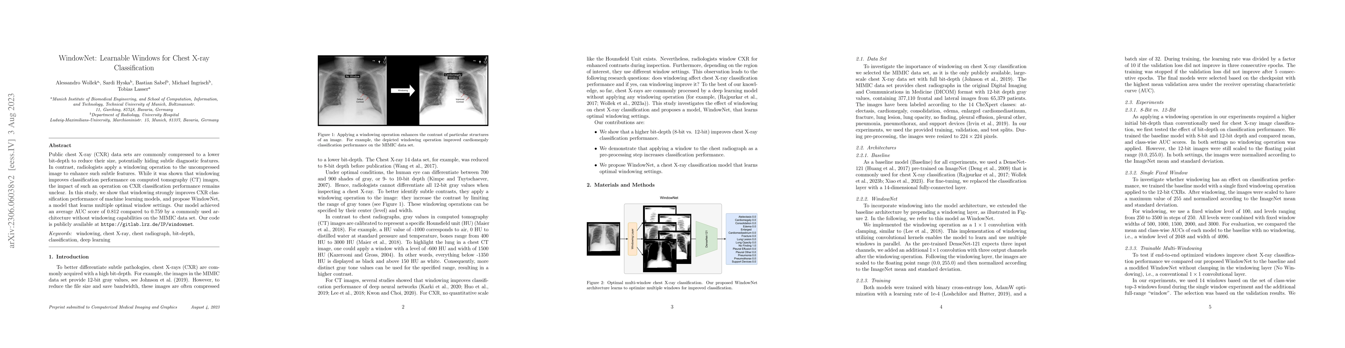
Chest X-ray (CXR) images are commonly compressed to a lower resolution and bit depth to reduce their size, potentially altering subtle diagnostic features. Radiologists use windowing operations to...
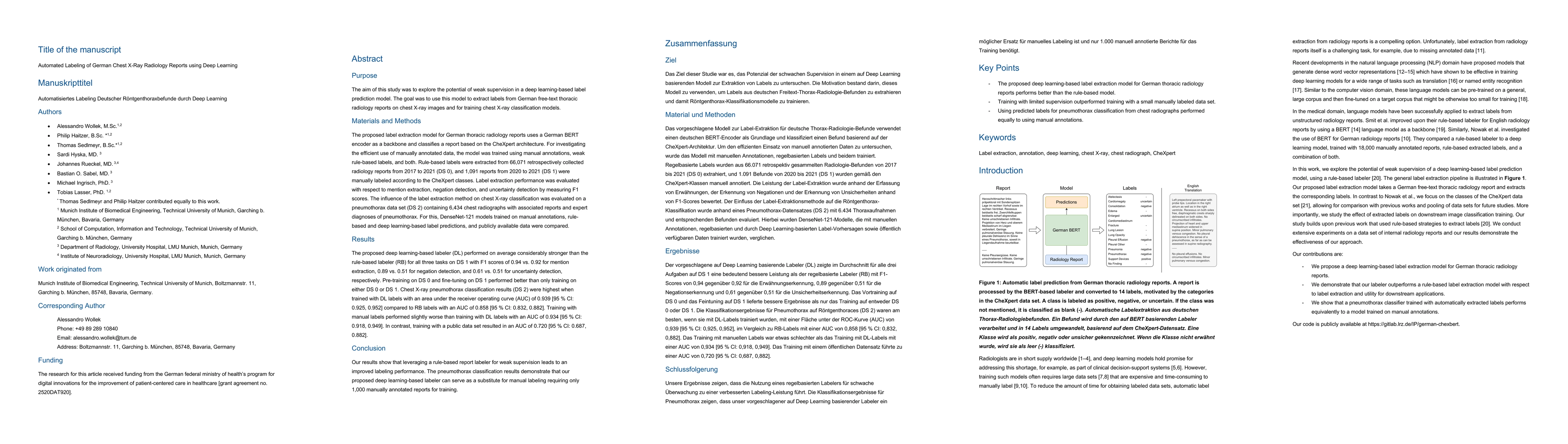
Radiologists are in short supply globally, and deep learning models offer a promising solution to address this shortage as part of clinical decision-support systems. However, training such models of...
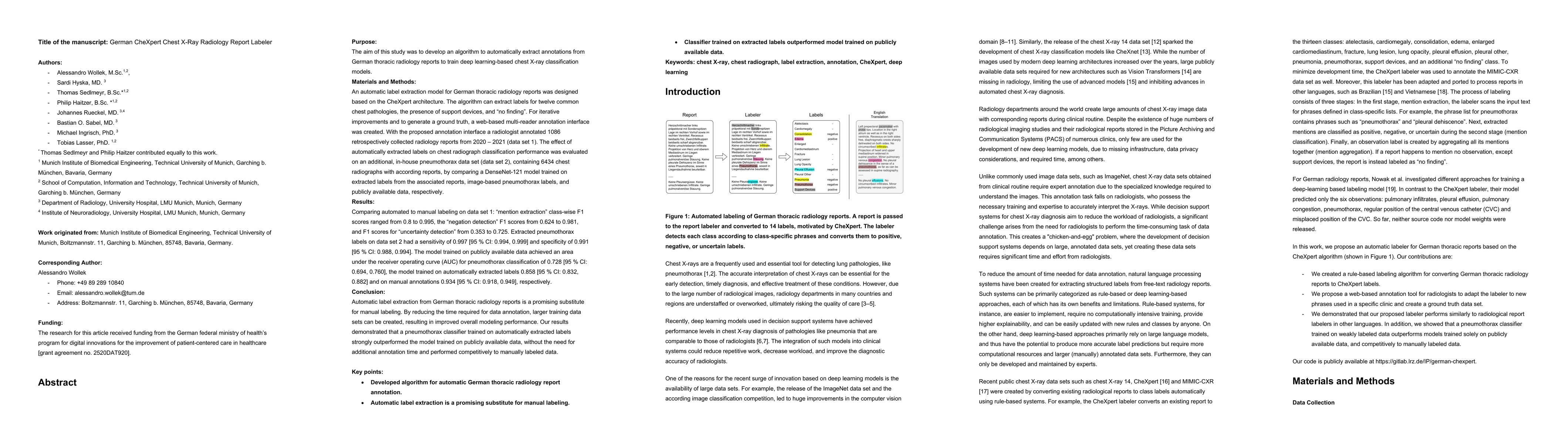
This study aimed to develop an algorithm to automatically extract annotations for chest X-ray classification models from German thoracic radiology reports. An automatic label extraction model was de...
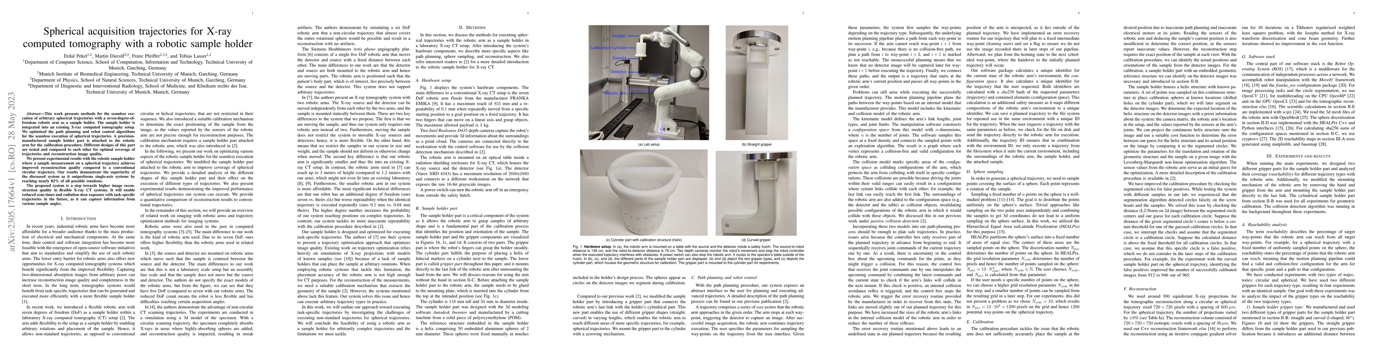
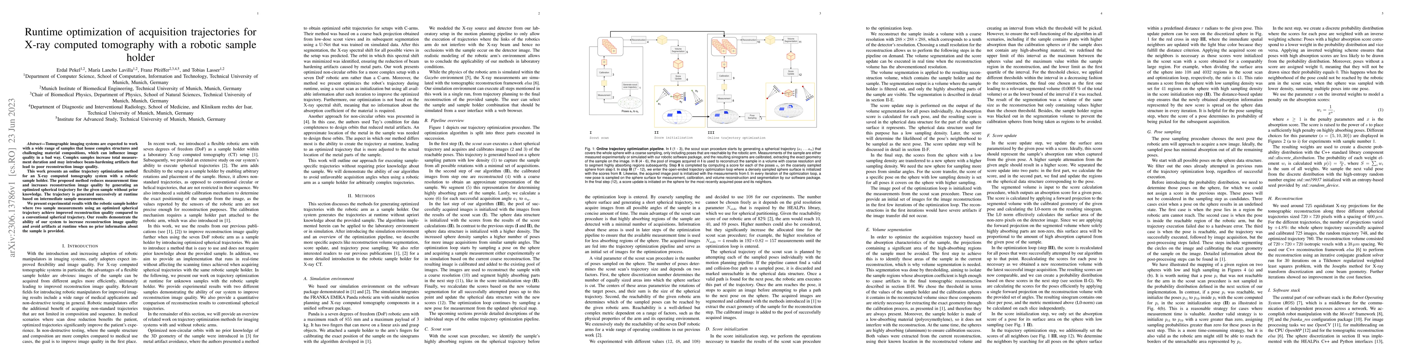
This work presents methods for the seamless execution of arbitrary spherical trajectories with a seven-degree-of-freedom robotic arm as a sample holder. The sample holder is integrated into an exist...
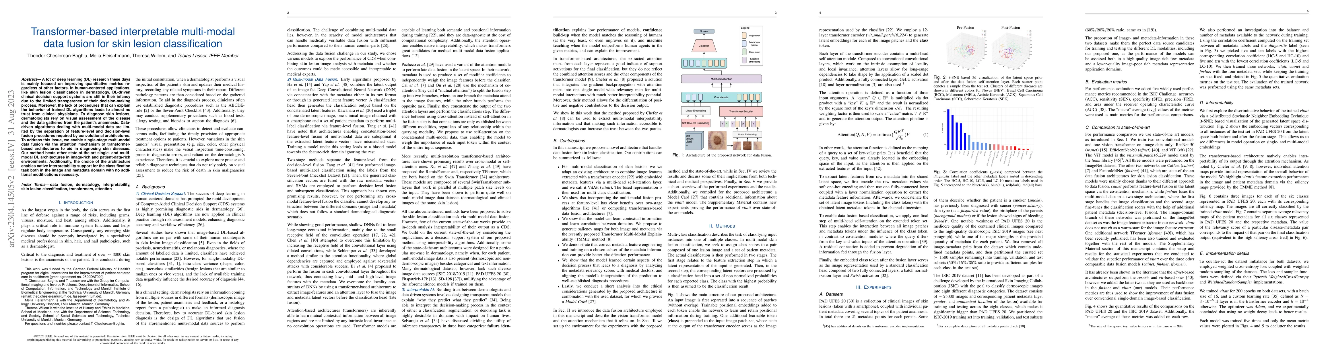
A lot of deep learning (DL) research these days is mainly focused on improving quantitative metrics regardless of other factors. In human-centered applications, like skin lesion classification in de...
Purpose: Sparse-view computed tomography (CT) is an effective way to reduce dose by lowering the total number of views acquired, albeit at the expense of image quality, which, in turn, can impact th...
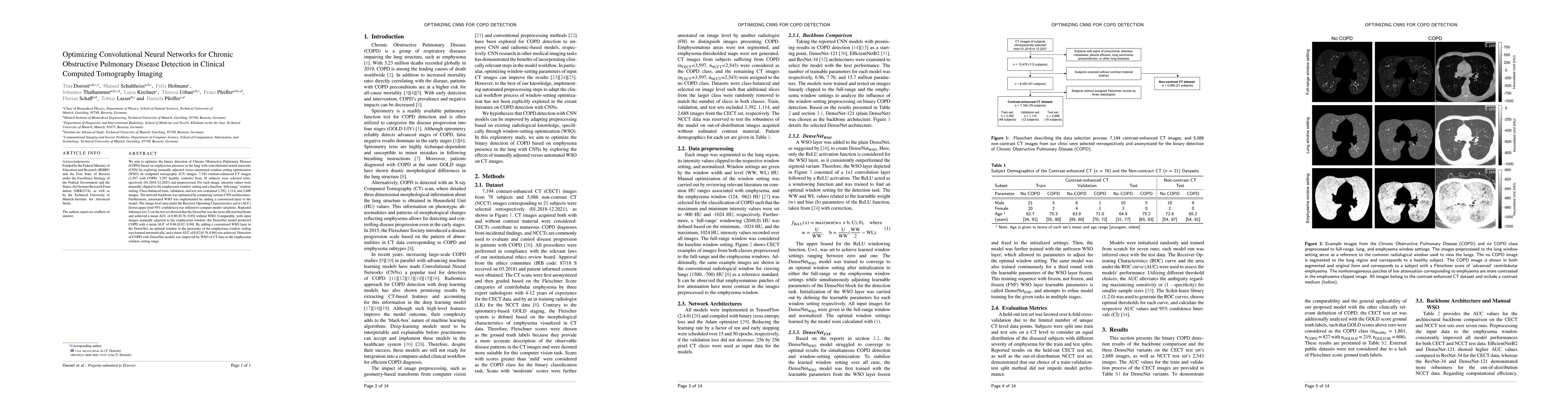
We aim to optimize the binary detection of Chronic Obstructive Pulmonary Disease (COPD) based on emphysema presence in the lung with convolutional neural networks (CNN) by exploring manually adjuste...
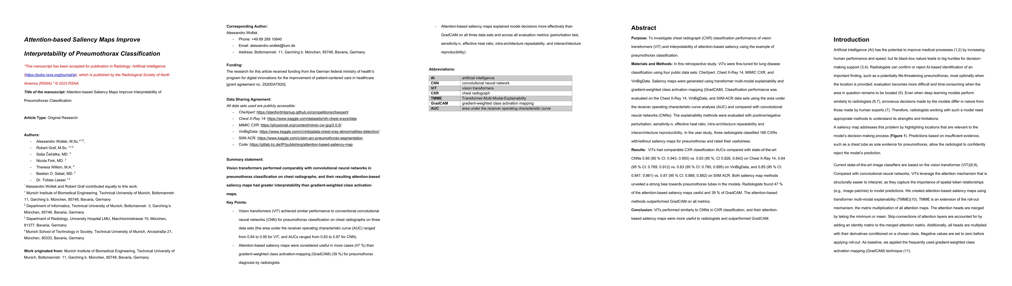
Purpose: To investigate chest radiograph (CXR) classification performance of vision transformers (ViT) and interpretability of attention-based saliency using the example of pneumothorax classificati...
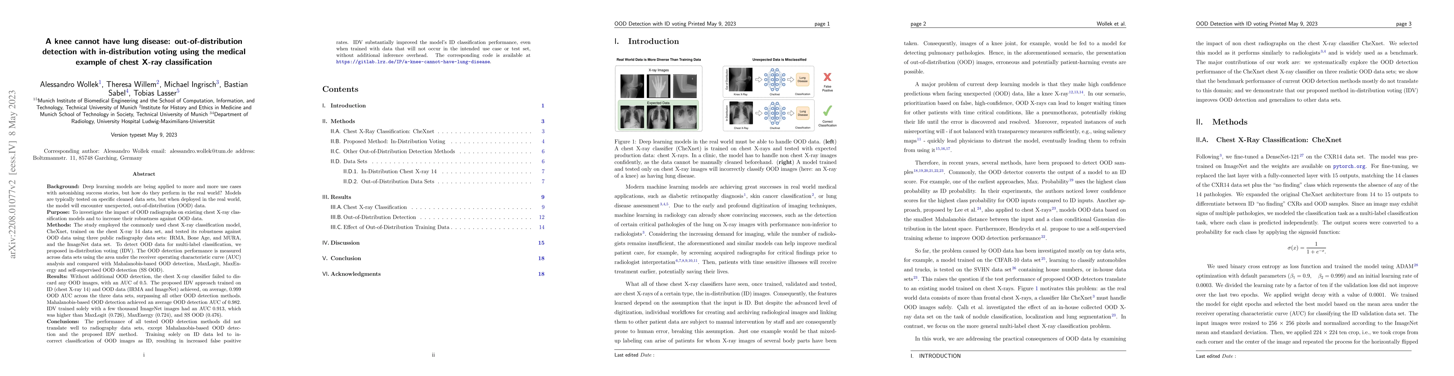
To investigate the impact of OOD radiographs on existing chest X-ray classification models and to increase their robustness against OOD data. The study employed the commonly used chest X-ray classif...
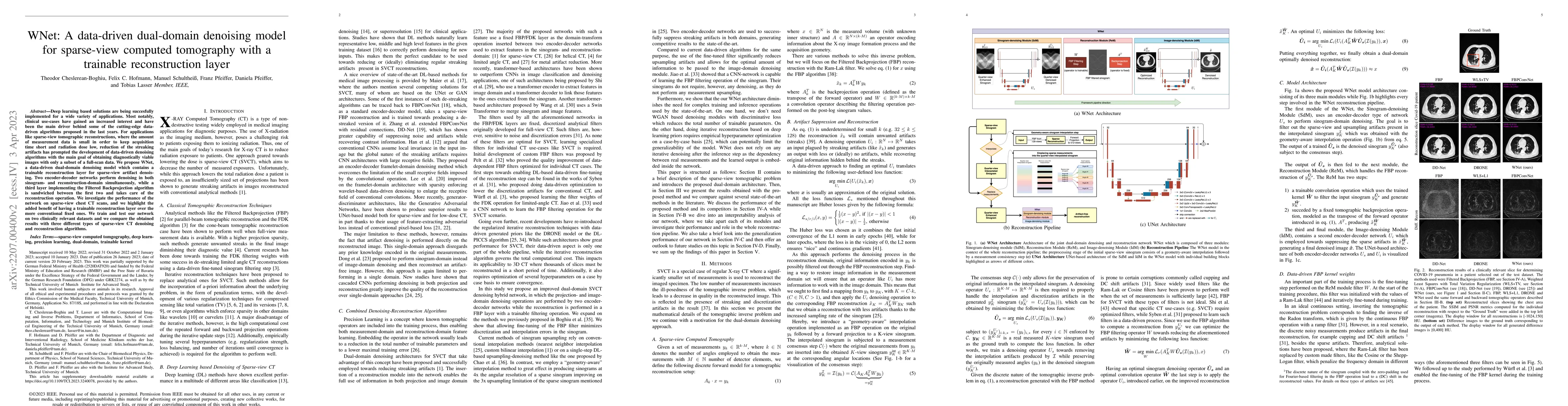
Deep learning based solutions are being succesfully implemented for a wide variety of applications. Most notably, clinical use-cases have gained an increased interest and have been the main driver b...
Anisotropic X-ray Dark-field Tomography (AXDT) is a recently developed imaging modality that enables the visualization of oriented microstructures using lab-based X-ray grating interferometer setups...
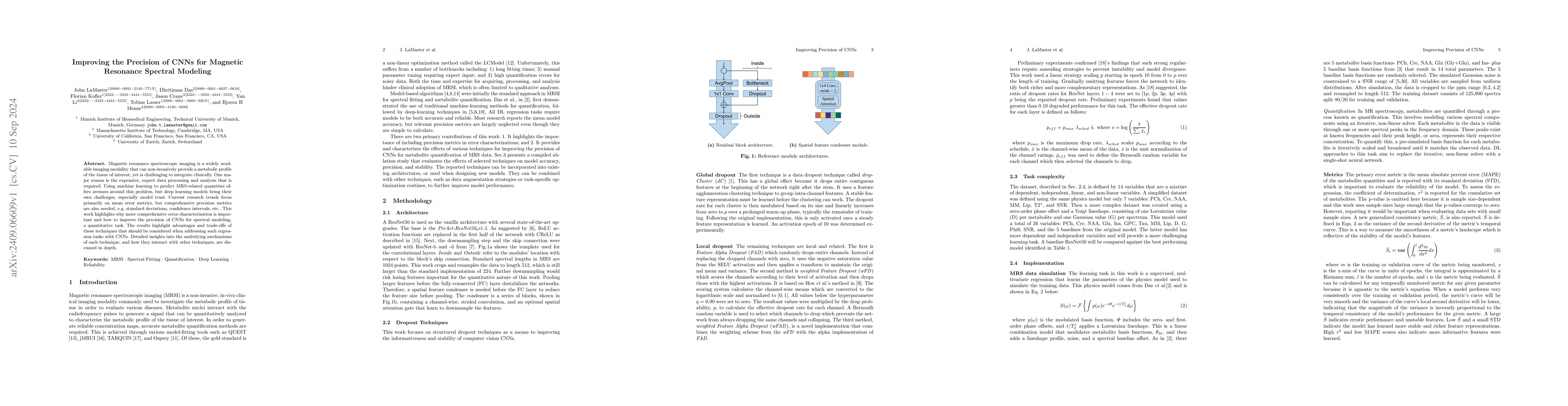
Magnetic resonance spectroscopic imaging is a widely available imaging modality that can non-invasively provide a metabolic profile of the tissue of interest, yet is challenging to integrate clinicall...
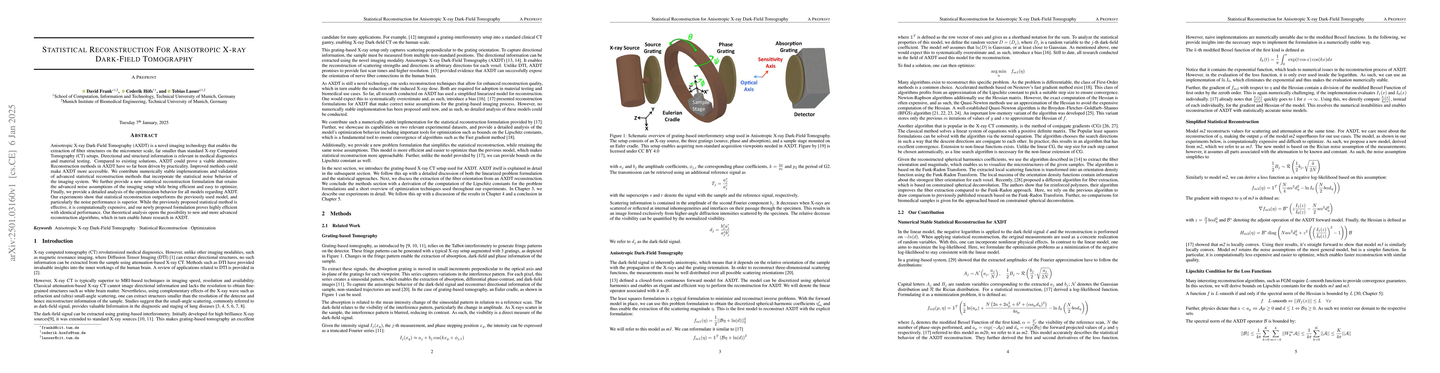
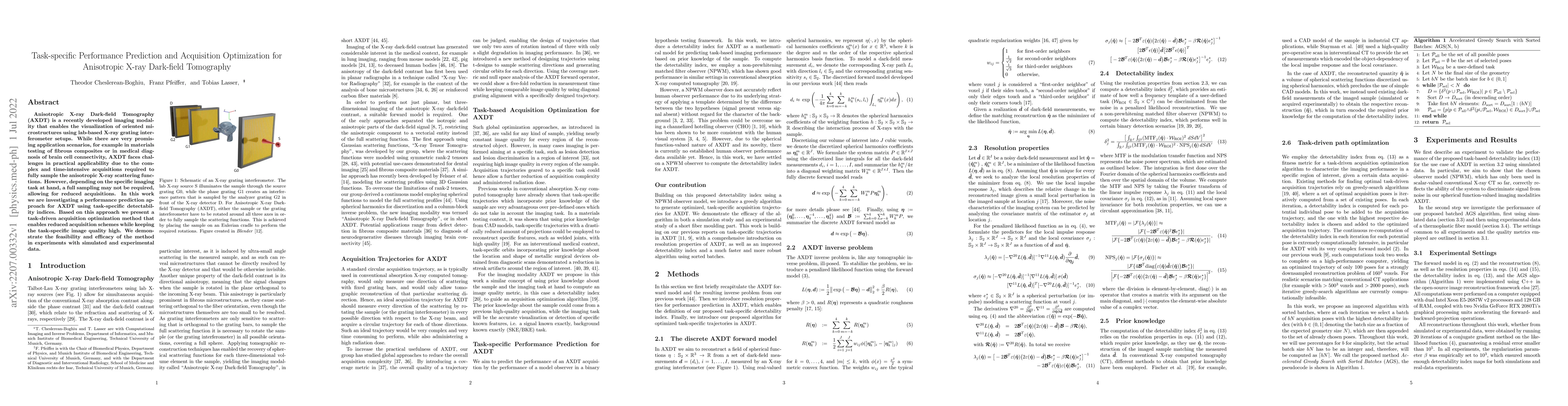
Anisotropic X-ray Dark-Field Tomography (AXDT) is a novel imaging technology that enables the extraction of fiber structures on the micrometer scale, far smaller than standard X-ray Computed Tomograph...
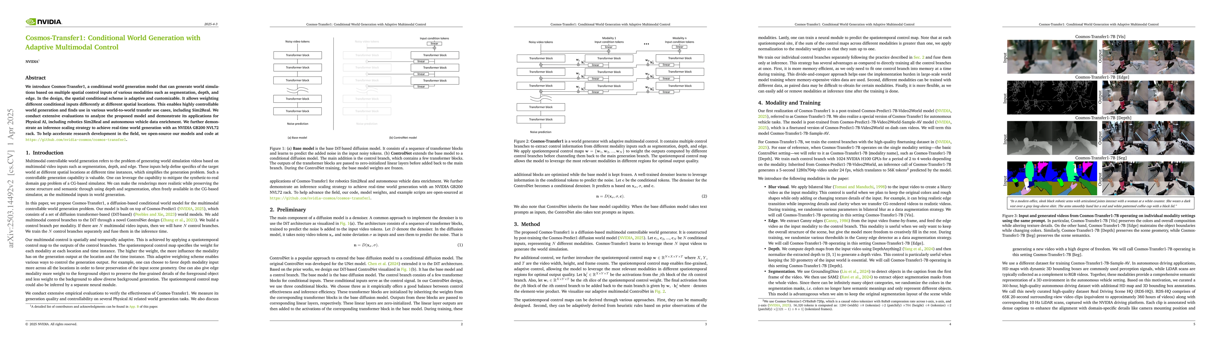
We introduce Cosmos-Transfer, a conditional world generation model that can generate world simulations based on multiple spatial control inputs of various modalities such as segmentation, depth, and e...
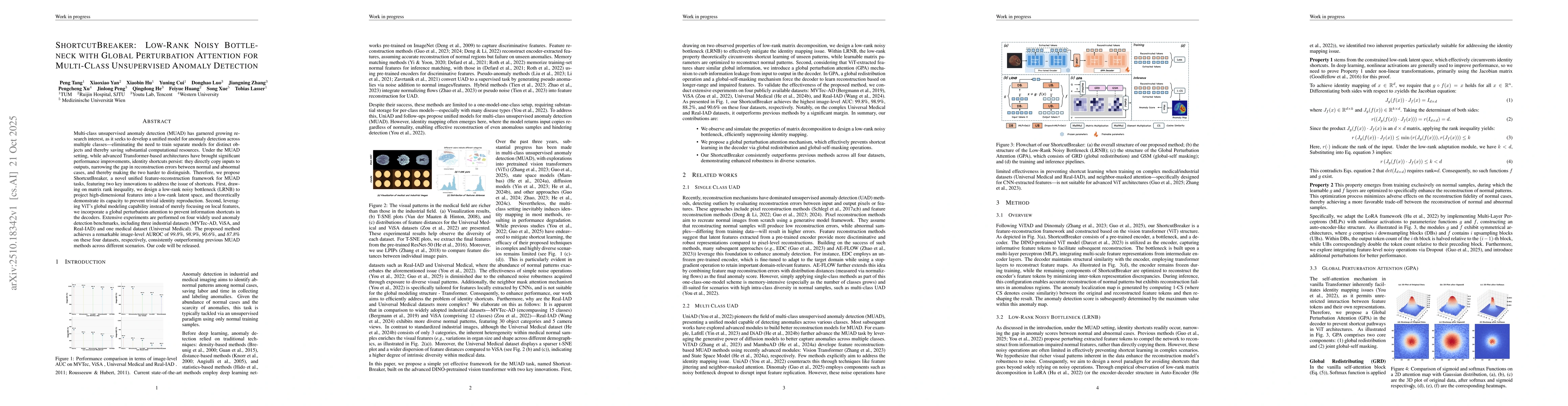
Multi-class unsupervised anomaly detection (MUAD) has garnered growing research interest, as it seeks to develop a unified model for anomaly detection across multiple classes, i.e., eliminating the ne...

Robust and accurate odometry estimation is essential in modern robotics. In environments characterized by highly dynamic motion and sensor noise, odometry estimation becomes increasingly challenging. ...