Publication
Metrics
AI Quick Summary
This study compares vision transformers (ViTs) to convolutional neural networks (CNNs) for chest radiograph classification, finding ViTs perform similarly with higher interpretability through attention-based saliency maps, which were rated more useful by radiologists compared to GradCAM.
Paper Preview
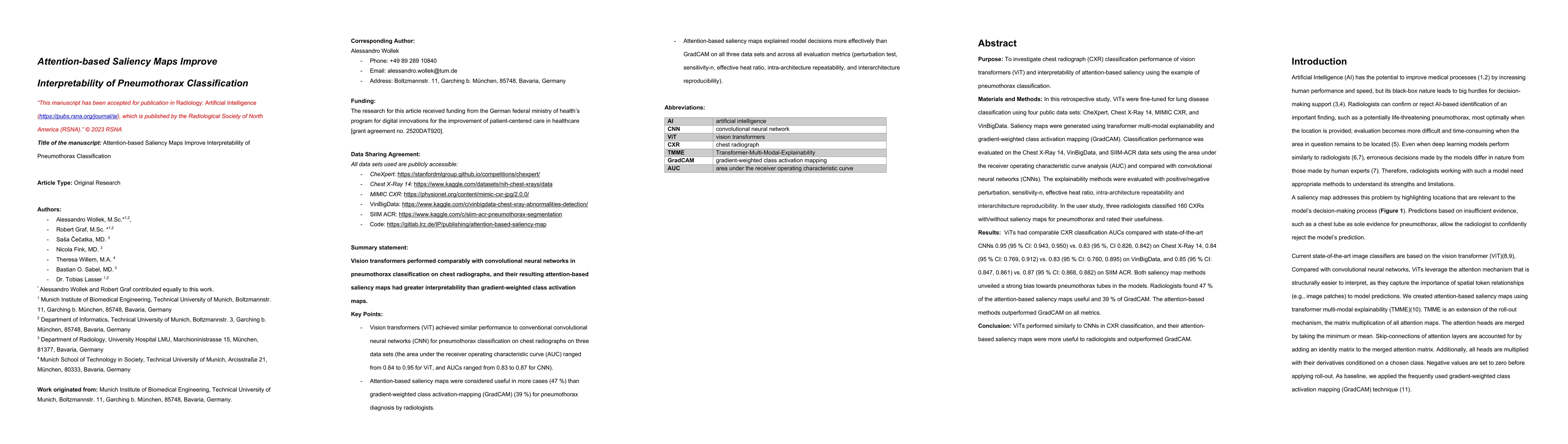
Abstract
Purpose: To investigate chest radiograph (CXR) classification performance of vision transformers (ViT) and interpretability of attention-based saliency using the example of pneumothorax classification. Materials and Methods: In this retrospective study, ViTs were fine-tuned for lung disease classification using four public data sets: CheXpert, Chest X-Ray 14, MIMIC CXR, and VinBigData. Saliency maps were generated using transformer multimodal explainability and gradient-weighted class activation mapping (GradCAM). Classification performance was evaluated on the Chest X-Ray 14, VinBigData, and SIIM-ACR data sets using the area under the receiver operating characteristic curve analysis (AUC) and compared with convolutional neural networks (CNNs). The explainability methods were evaluated with positive/negative perturbation, sensitivity-n, effective heat ratio, intra-architecture repeatability and interarchitecture reproducibility. In the user study, three radiologists classified 160 CXRs with/without saliency maps for pneumothorax and rated their usefulness. Results: ViTs had comparable CXR classification AUCs compared with state-of-the-art CNNs 0.95 (95% CI: 0.943, 0.950) versus 0.83 (95%, CI 0.826, 0.842) on Chest X-Ray 14, 0.84 (95% CI: 0.769, 0.912) versus 0.83 (95% CI: 0.760, 0.895) on VinBigData, and 0.85 (95% CI: 0.847, 0.861) versus 0.87 (95% CI: 0.868, 0.882) on SIIM ACR. Both saliency map methods unveiled a strong bias toward pneumothorax tubes in the models. Radiologists found 47% of the attention-based saliency maps useful and 39% of GradCAM. The attention-based methods outperformed GradCAM on all metrics. Conclusion: ViTs performed similarly to CNNs in CXR classification, and their attention-based saliency maps were more useful to radiologists and outperformed GradCAM.
AI Key Findings
Get AI-generated insights about this paper's methodology, results, significance, and more — seven facets brought into focus.
Impact
Paper Details
Authors
PDF Preview
Key Terms
Citation Network
Current paper (gray), citations (green), references (blue)
Display is limited for performance on very large graphs.
Discussion 0