Academic Profile
Statistics
Similar Authors
Papers on arXiv
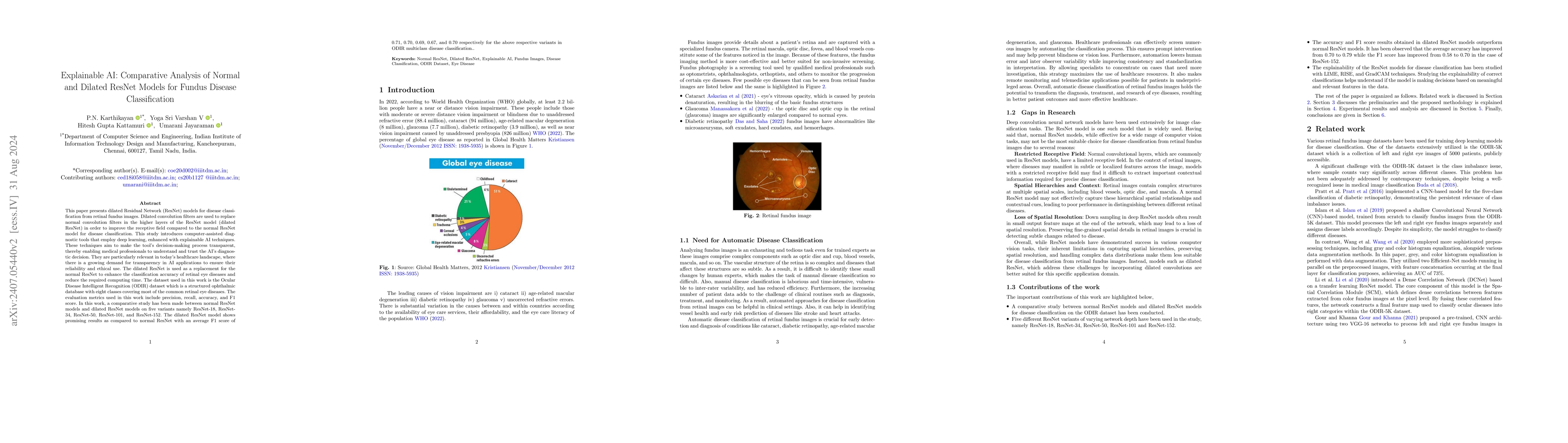
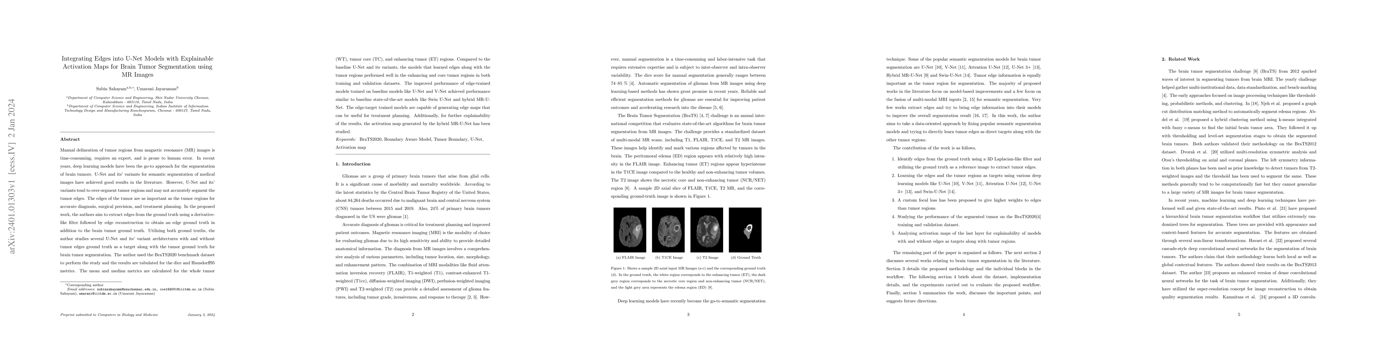
This paper presents dilated Residual Network (ResNet) models for disease classification from retinal fundus images. Dilated convolution filters are used to replace normal convolution filters in the hi...
Manual delineation of tumor regions from magnetic resonance (MR) images is time-consuming, requires an expert, and is prone to human error. In recent years, deep learning models have been the go-to ...
In deep learning, mini-batch training is commonly used to optimize network parameters. However, the traditional mini-batch method may not learn the under-represented samples and complex patterns in ...
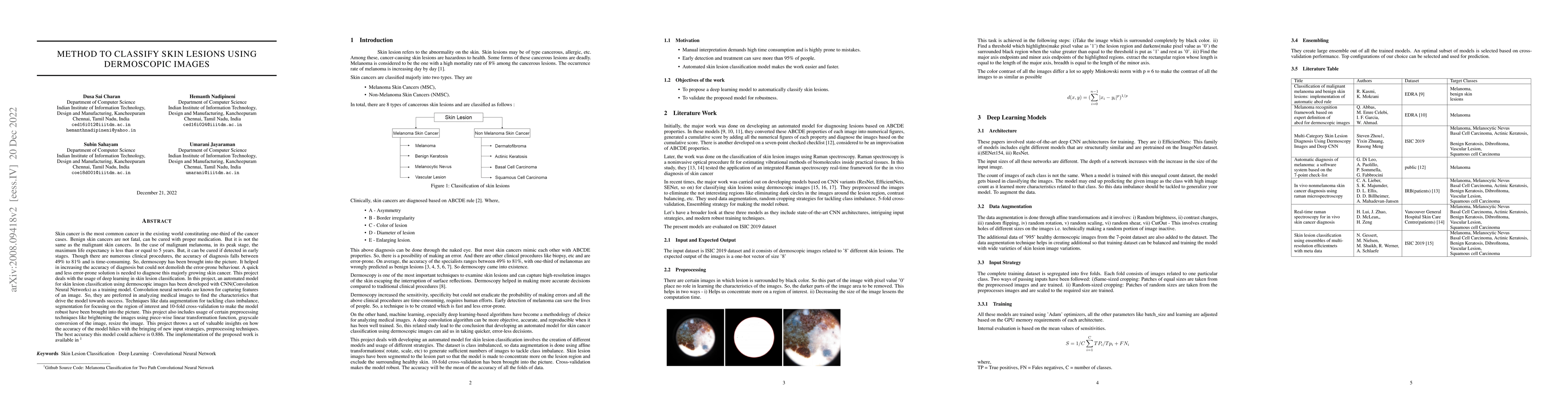
Skin cancer is the most common cancer in the existing world constituting one-third of the cancer cases. Benign skin cancers are not fatal, can be cured with proper medication. But it is not the same...
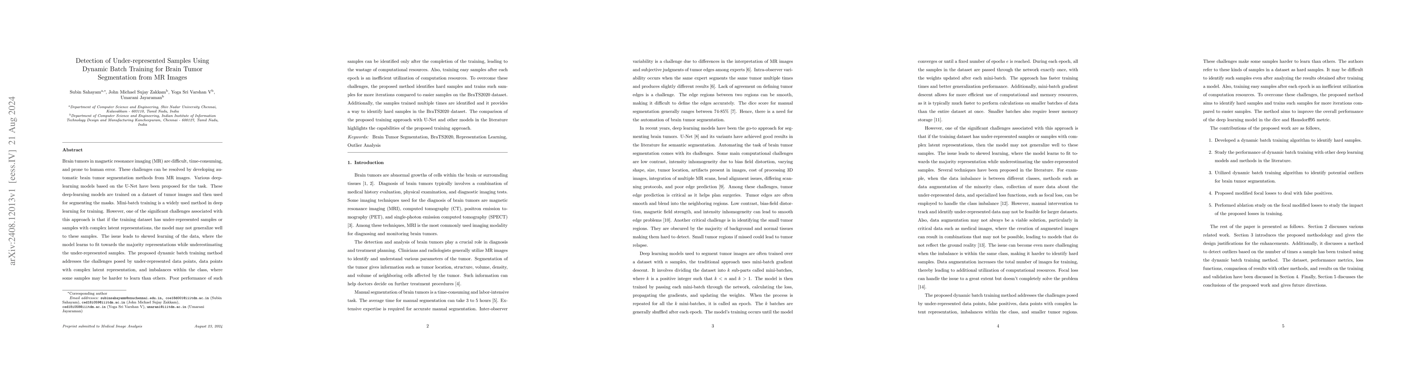
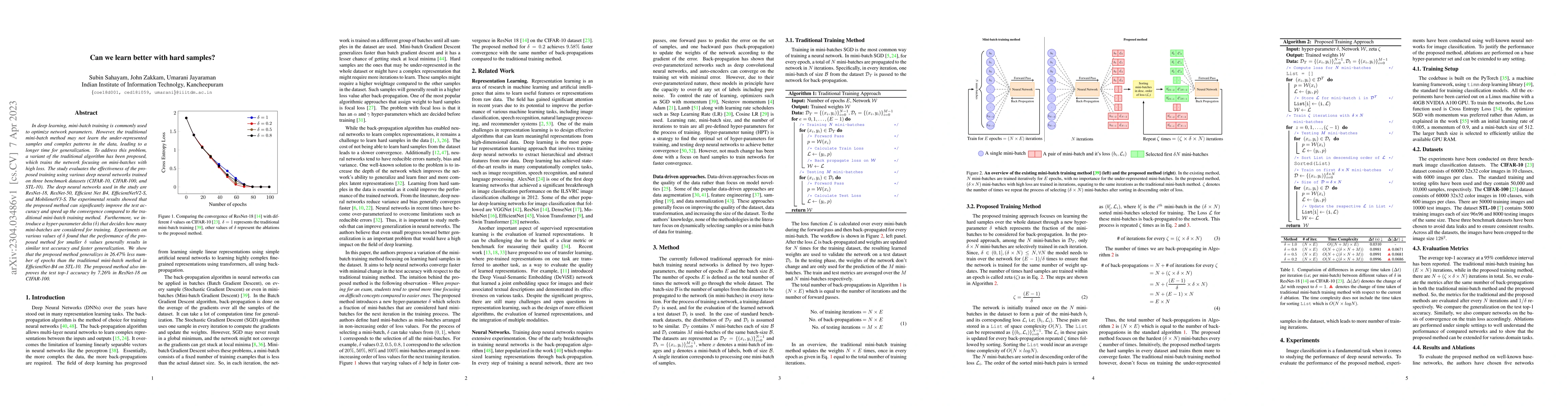
Brain tumors in magnetic resonance imaging (MR) are difficult, time-consuming, and prone to human error. These challenges can be resolved by developing automatic brain tumor segmentation methods from ...
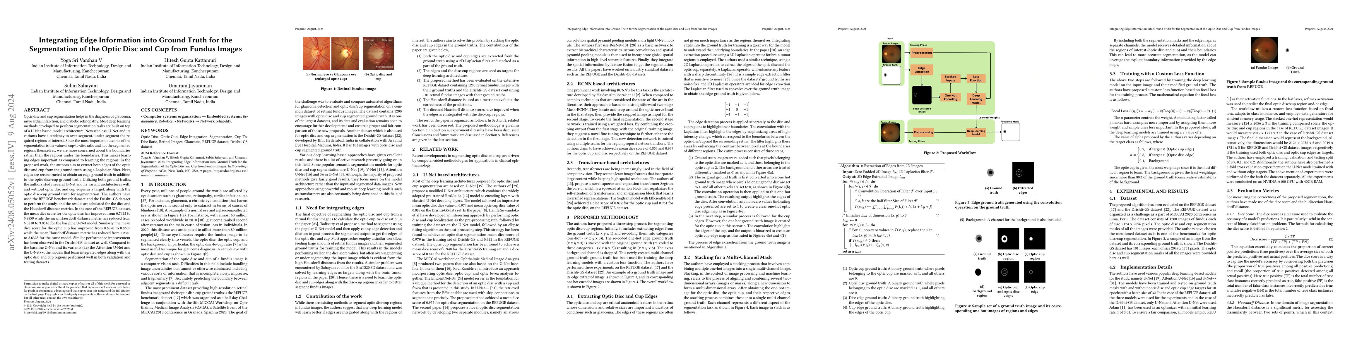
Optic disc and cup segmentation helps in the diagnosis of glaucoma, myocardial infarction, and diabetic retinopathy. Most deep learning methods developed to perform segmentation tasks are built on top...
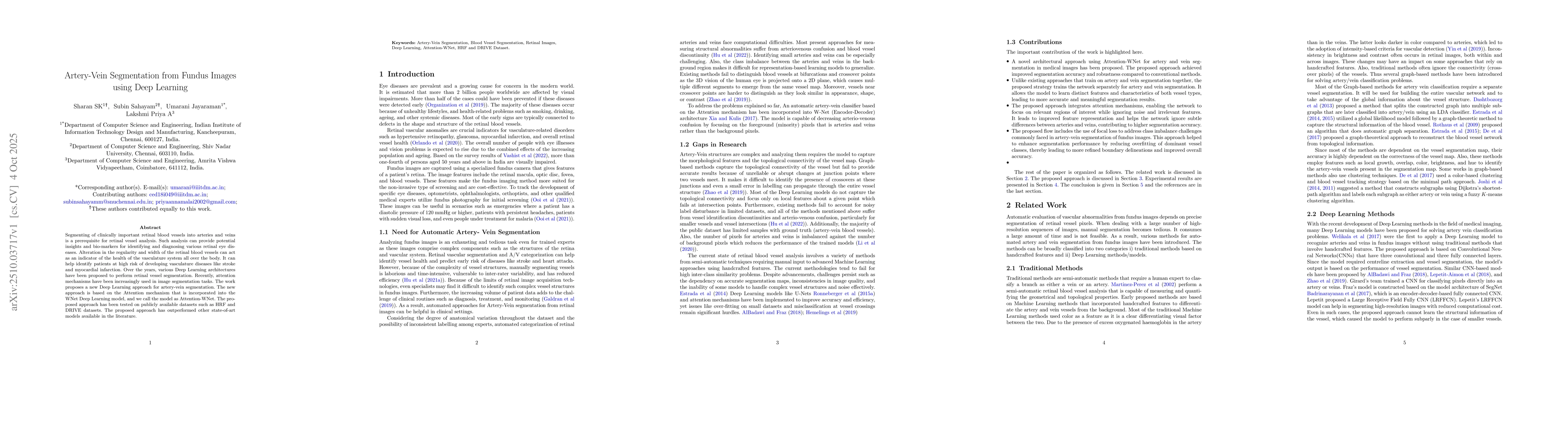
Segmenting of clinically important retinal blood vessels into arteries and veins is a prerequisite for retinal vessel analysis. Such analysis can provide potential insights and bio-markers for identif...