Integrating Edges into U-Net Models with Explainable Activation Maps for Brain Tumor Segmentation using MR Images
Publication
Metrics
AI Quick Summary
This paper proposes integrating tumor edges into U-Net models to improve brain tumor segmentation accuracy using MR images. The study shows that models trained with edge ground truth outperform baseline U-Net variants, achieving results comparable to state-of-the-art models, and generate useful edge maps for treatment planning.
Paper Preview
Abstract
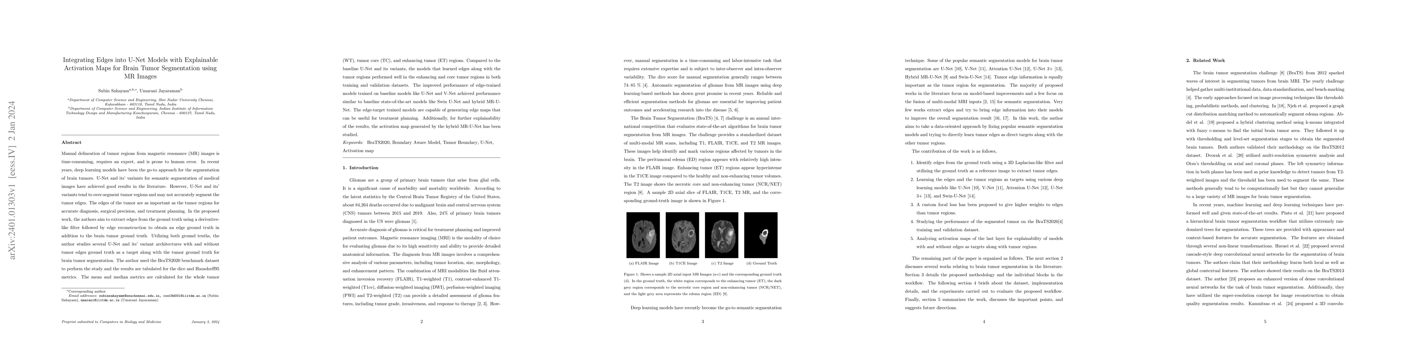
Manual delineation of tumor regions from magnetic resonance (MR) images is time-consuming, requires an expert, and is prone to human error. In recent years, deep learning models have been the go-to approach for the segmentation of brain tumors. U-Net and its' variants for semantic segmentation of medical images have achieved good results in the literature. However, U-Net and its' variants tend to over-segment tumor regions and may not accurately segment the tumor edges. The edges of the tumor are as important as the tumor regions for accurate diagnosis, surgical precision, and treatment planning. In the proposed work, the authors aim to extract edges from the ground truth using a derivative-like filter followed by edge reconstruction to obtain an edge ground truth in addition to the brain tumor ground truth. Utilizing both ground truths, the author studies several U-Net and its' variant architectures with and without tumor edges ground truth as a target along with the tumor ground truth for brain tumor segmentation. The author used the BraTS2020 benchmark dataset to perform the study and the results are tabulated for the dice and Hausdorff95 metrics. The mean and median metrics are calculated for the whole tumor (WT), tumor core (TC), and enhancing tumor (ET) regions. Compared to the baseline U-Net and its variants, the models that learned edges along with the tumor regions performed well in core tumor regions in both training and validation datasets. The improved performance of edge-trained models trained on baseline models like U-Net and V-Net achieved performance similar to baseline state-of-the-art models like Swin U-Net and hybrid MR-U-Net. The edge-target trained models are capable of generating edge maps that can be useful for treatment planning. Additionally, for further explainability of the results, the activation map generated by the hybrid MR-U-Net has been studied.
AI Key Findings
Get AI-generated insights about this paper's methodology, results, significance, and more — seven facets brought into focus.
Impact
Paper Details
Authors
PDF Preview
Key Terms
Citation Network
Current paper (gray), citations (green), references (blue)
Display is limited for performance on very large graphs.
Discussion 0