Academic Profile
Statistics
Similar Authors
Papers on arXiv
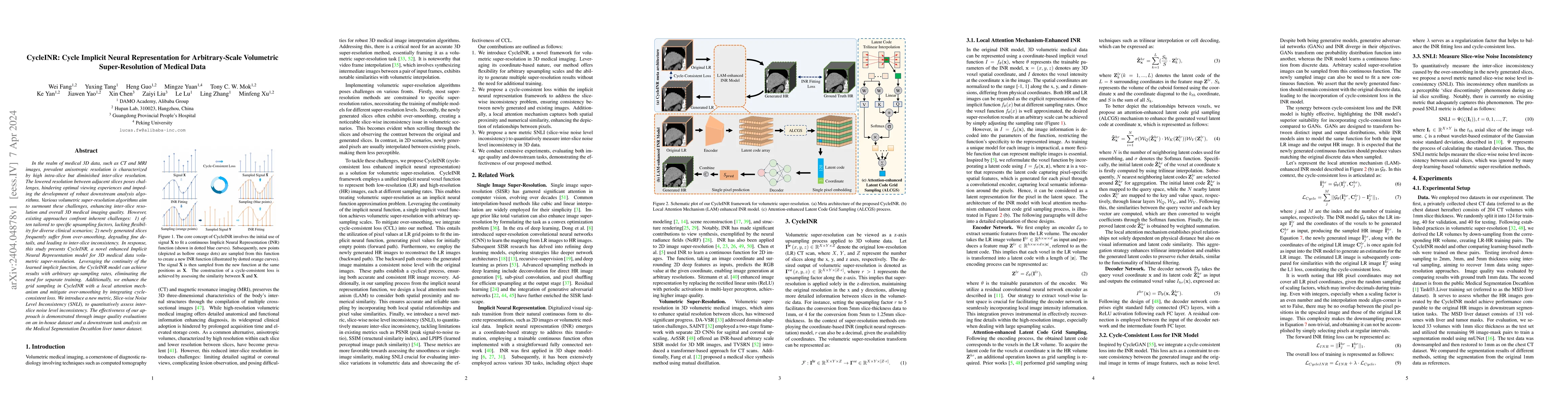
In the realm of medical 3D data, such as CT and MRI images, prevalent anisotropic resolution is characterized by high intra-slice but diminished inter-slice resolution. The lowered resolution betwee...
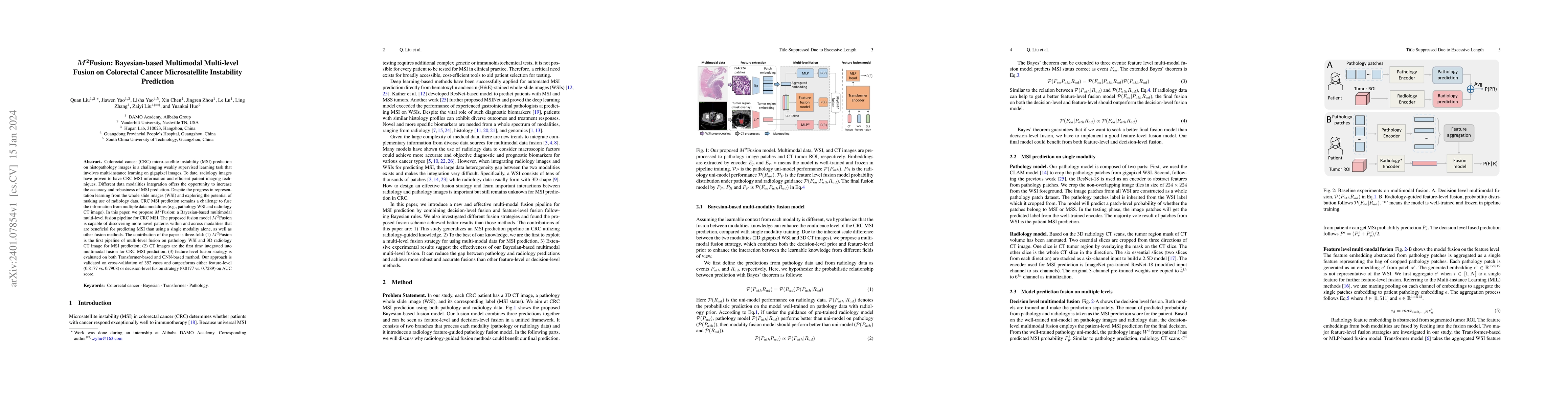
Colorectal cancer (CRC) micro-satellite instability (MSI) prediction on histopathology images is a challenging weakly supervised learning task that involves multi-instance learning on gigapixel imag...
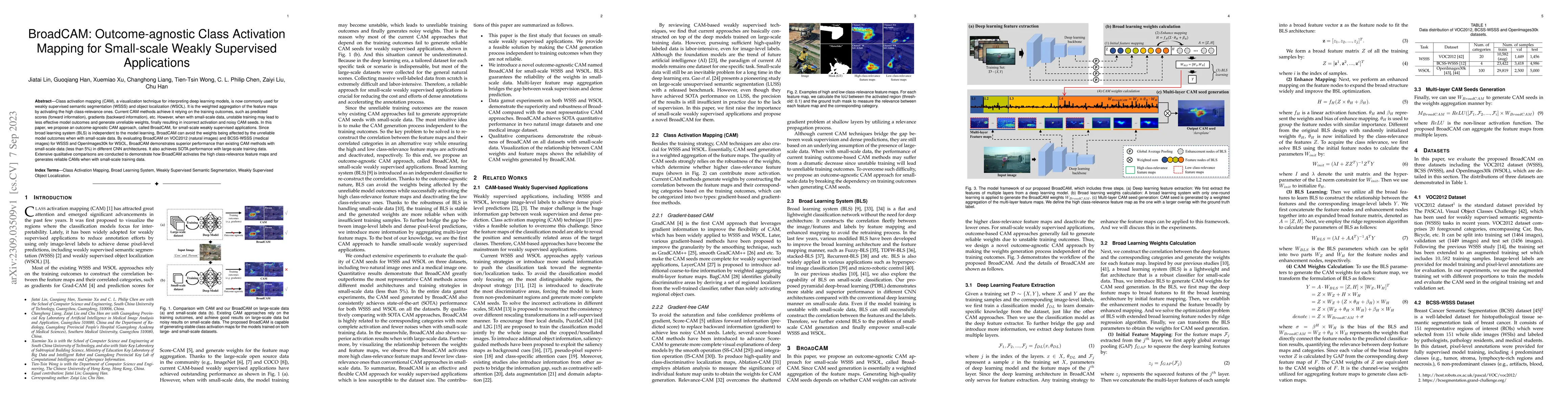
Class activation mapping~(CAM), a visualization technique for interpreting deep learning models, is now commonly used for weakly supervised semantic segmentation~(WSSS) and object localization~(WSOL...
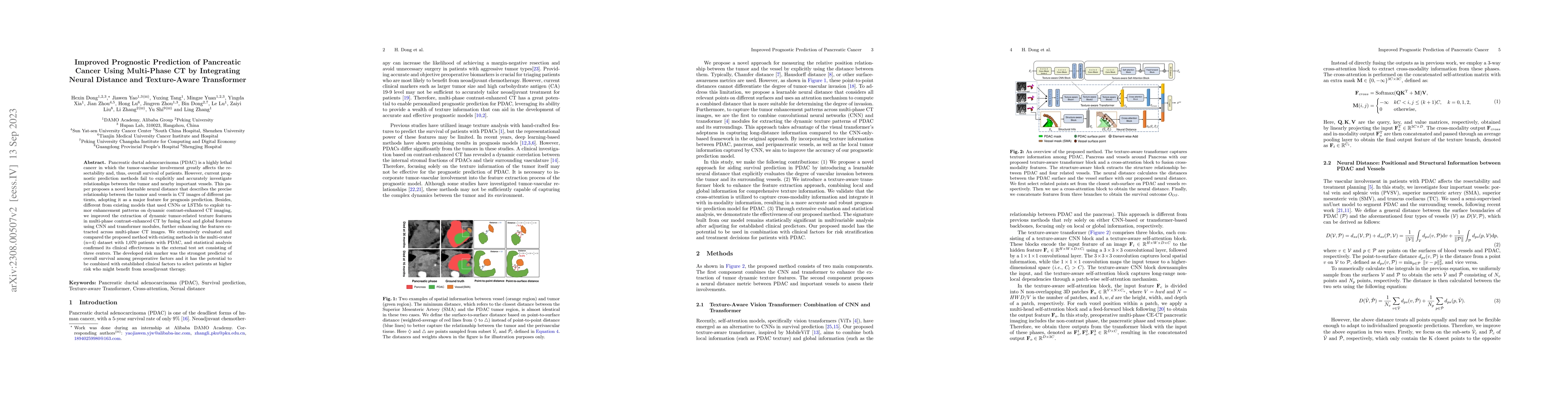
Pancreatic ductal adenocarcinoma (PDAC) is a highly lethal cancer in which the tumor-vascular involvement greatly affects the resectability and, thus, overall survival of patients. However, current ...
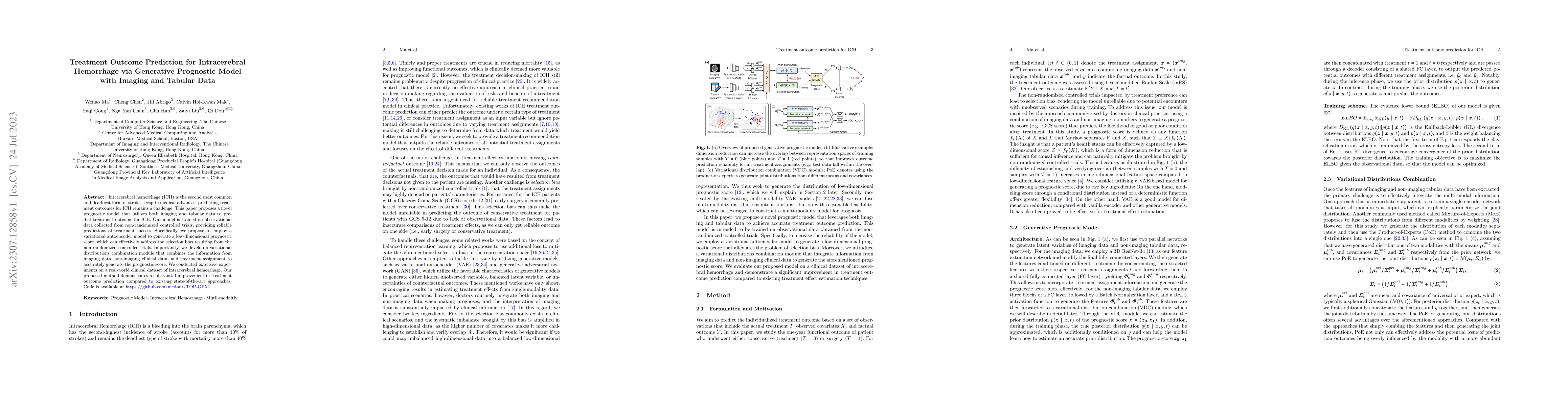
Intracerebral hemorrhage (ICH) is the second most common and deadliest form of stroke. Despite medical advances, predicting treat ment outcomes for ICH remains a challenge. This paper proposes a nov...
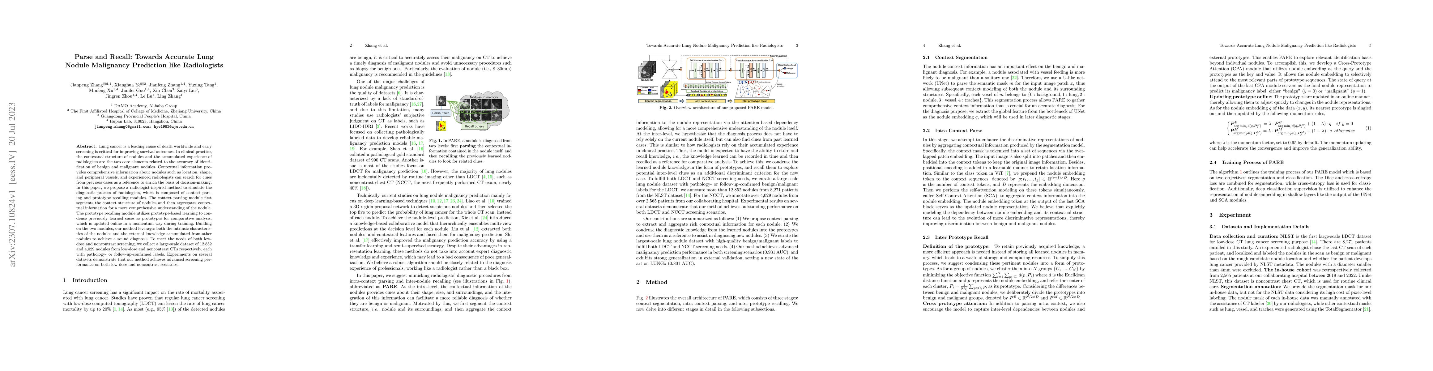
Lung cancer is a leading cause of death worldwide and early screening is critical for improving survival outcomes. In clinical practice, the contextual structure of nodules and the accumulated exper...
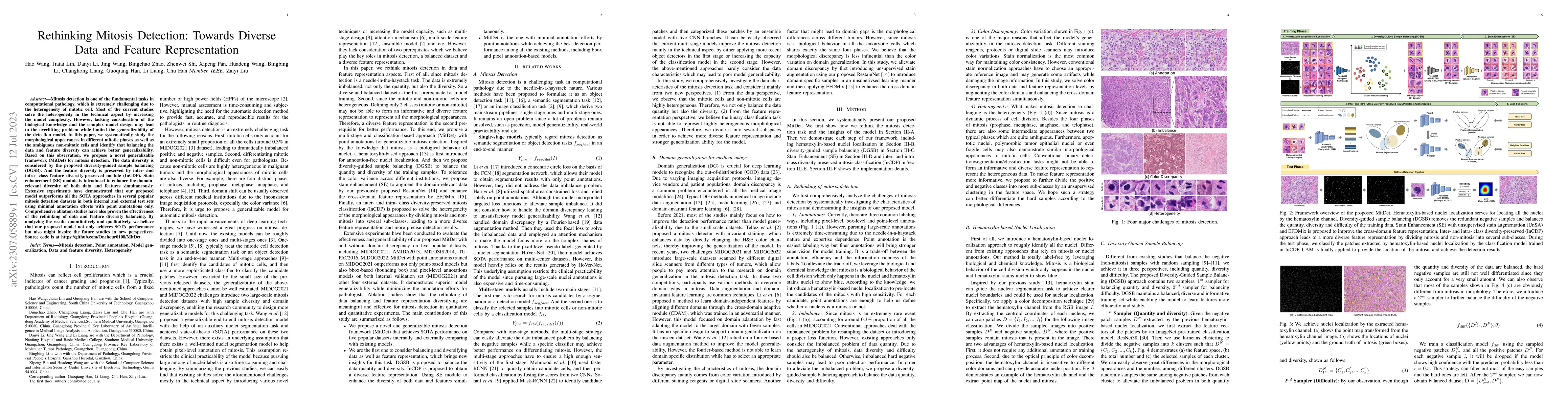
Mitosis detection is one of the fundamental tasks in computational pathology, which is extremely challenging due to the heterogeneity of mitotic cell. Most of the current studies solve the heterogen...
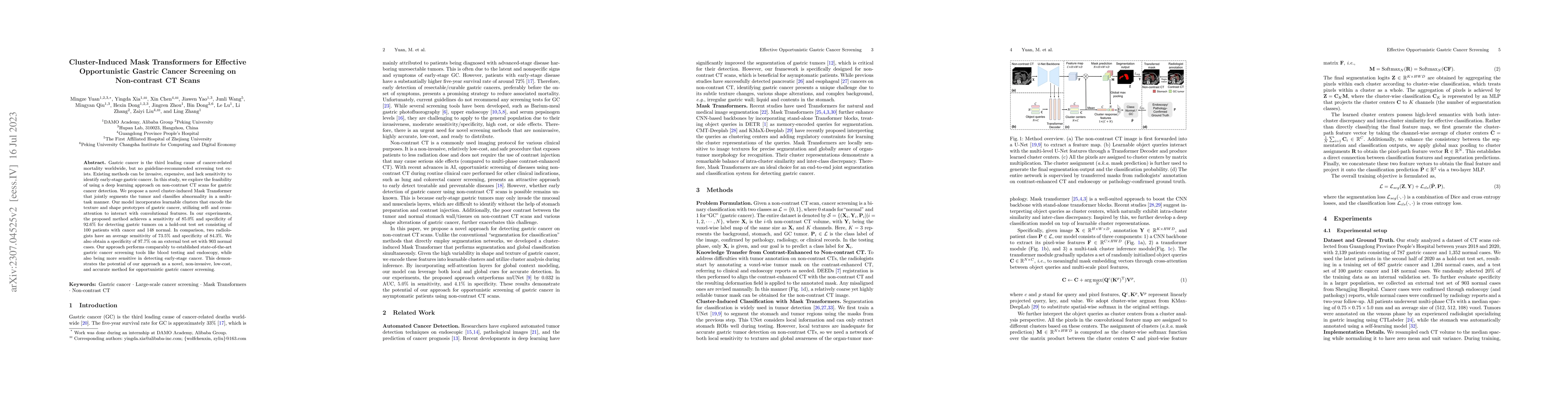
Gastric cancer is the third leading cause of cancer-related mortality worldwide, but no guideline-recommended screening test exists. Existing methods can be invasive, expensive, and lack sensitivity...
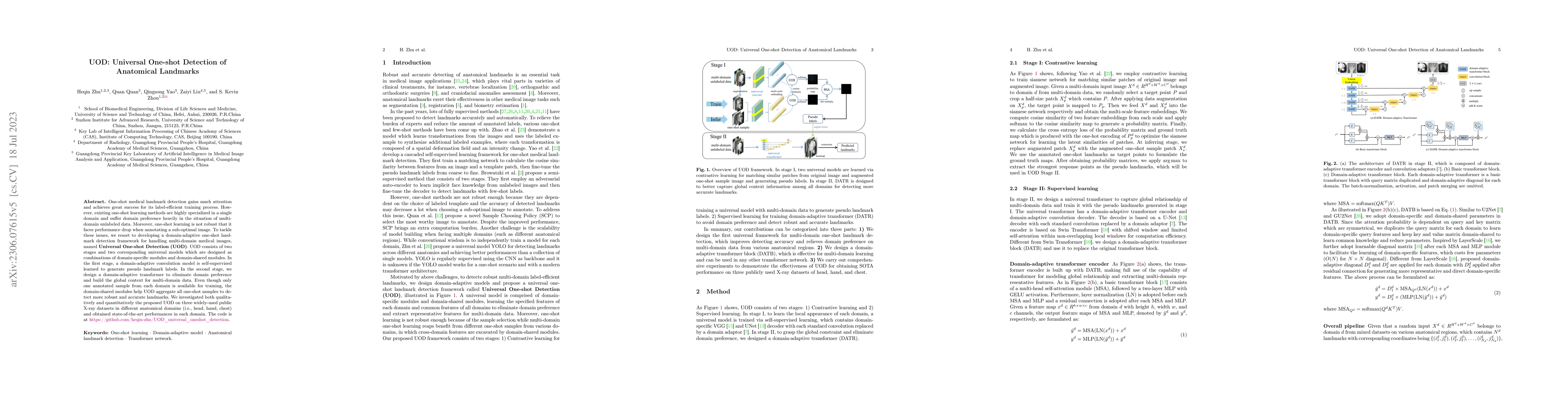
One-shot medical landmark detection gains much attention and achieves great success for its label-efficient training process. However, existing one-shot learning methods are highly specialized in a ...
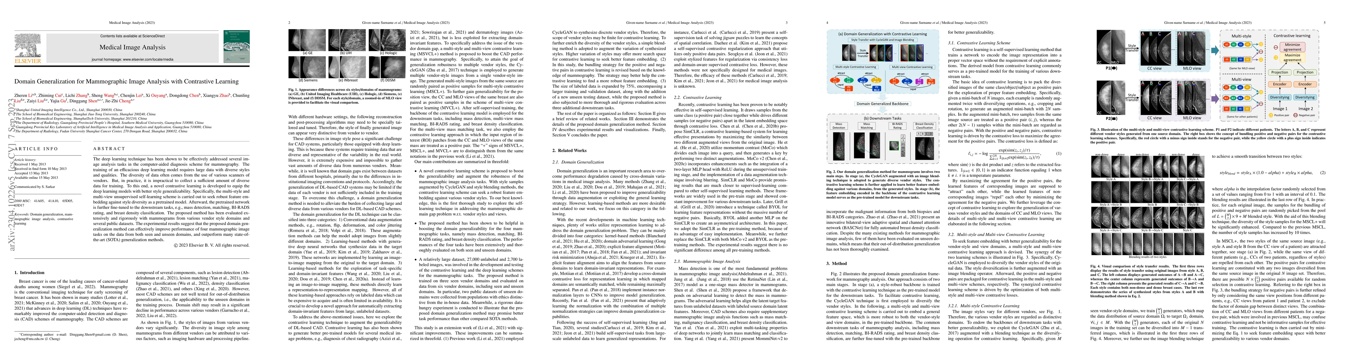
The deep learning technique has been shown to be effectively addressed several image analysis tasks in the computer-aided diagnosis scheme for mammography. The training of an efficacious deep learni...
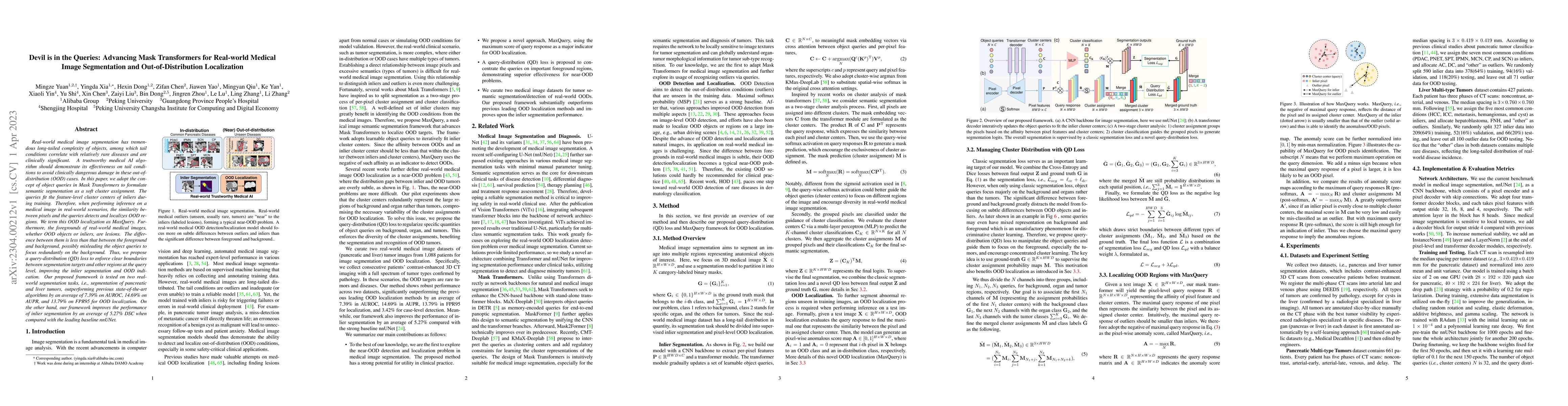
Real-world medical image segmentation has tremendous long-tailed complexity of objects, among which tail conditions correlate with relatively rare diseases and are clinically significant. A trustwor...
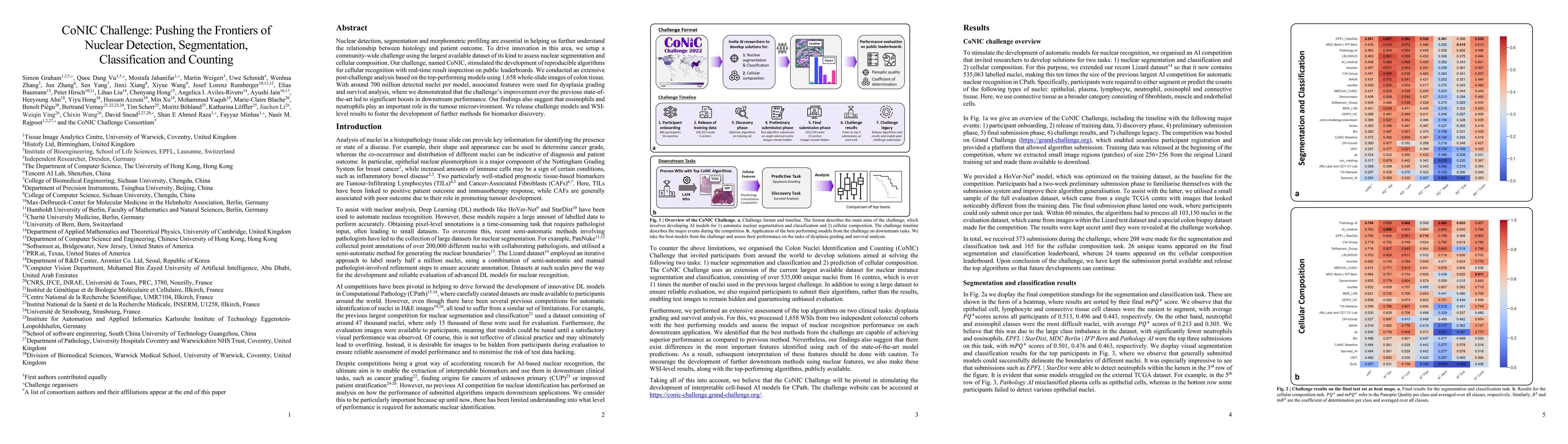
Nuclear detection, segmentation and morphometric profiling are essential in helping us further understand the relationship between histology and patient outcome. To drive innovation in this area, we...
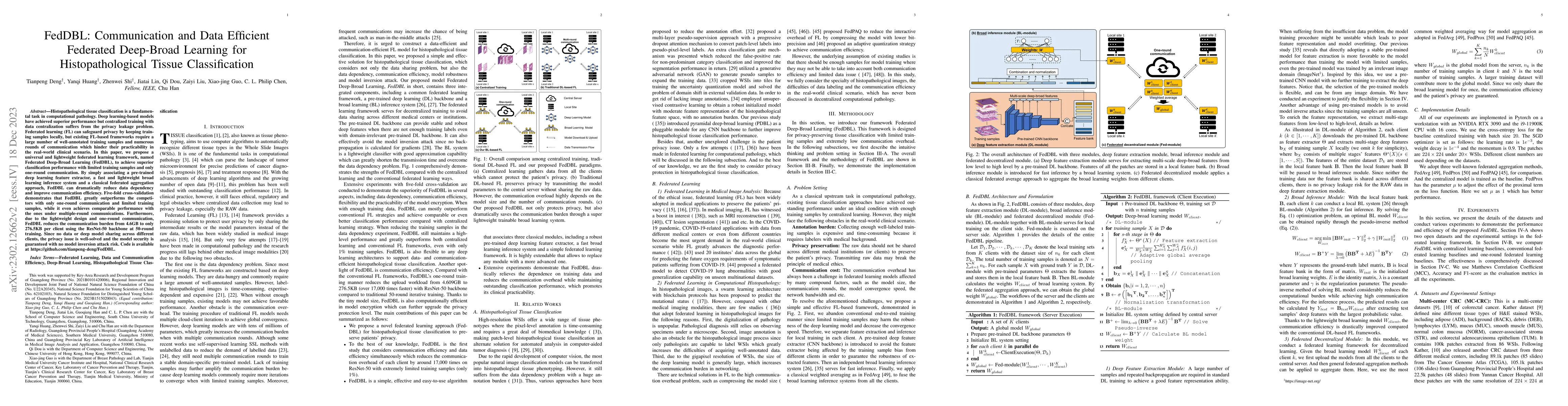
Histopathological tissue classification is a fundamental task in computational pathology. Deep learning-based models have achieved superior performance but centralized training with data centralizat...
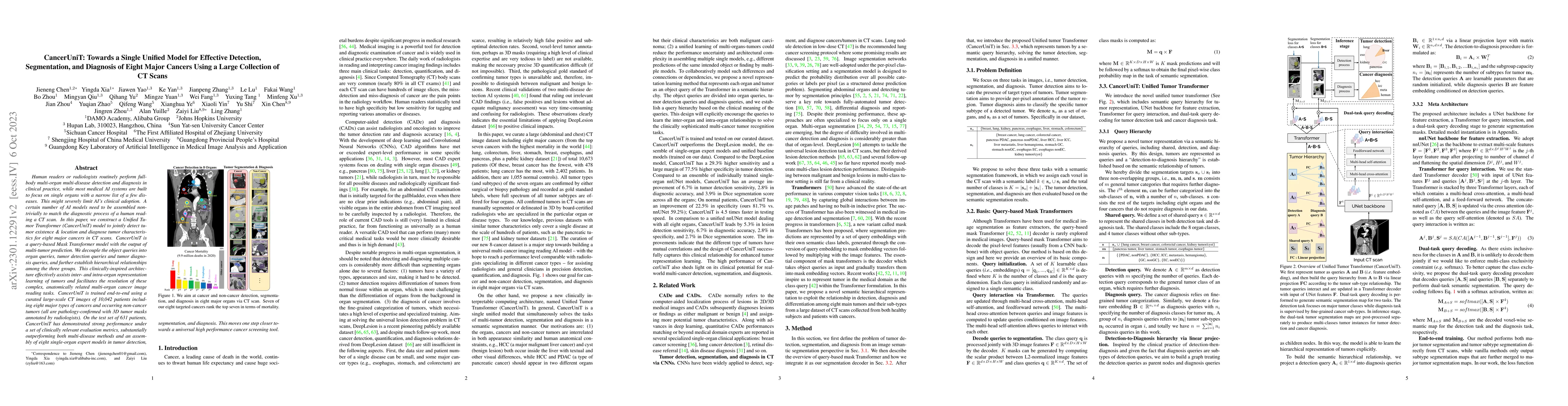
Human readers or radiologists routinely perform full-body multi-organ multi-disease detection and diagnosis in clinical practice, while most medical AI systems are built to focus on single organs wi...
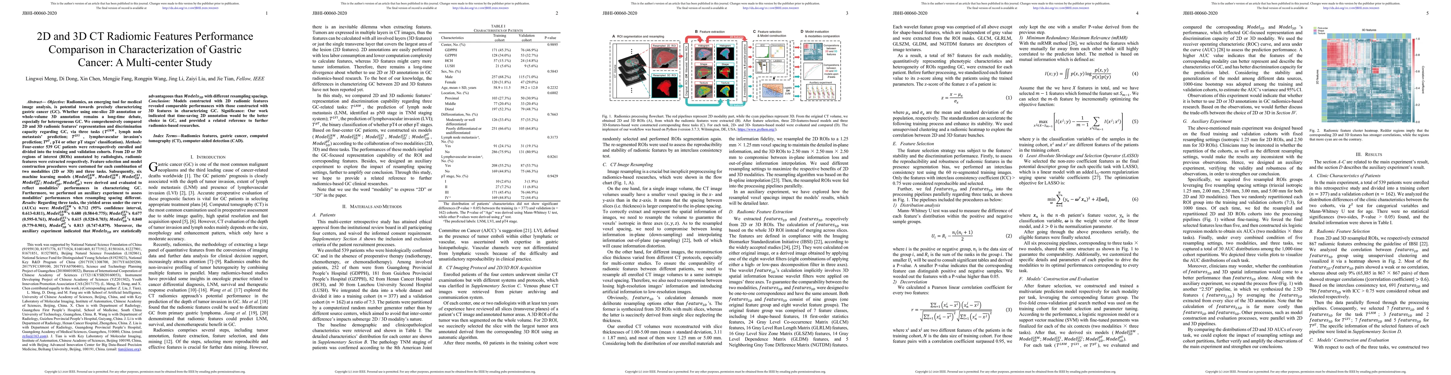
Objective: Radiomics, an emerging tool for medical image analysis, is potential towards precisely characterizing gastric cancer (GC). Whether using one-slice 2D annotation or whole-volume 3D annotat...
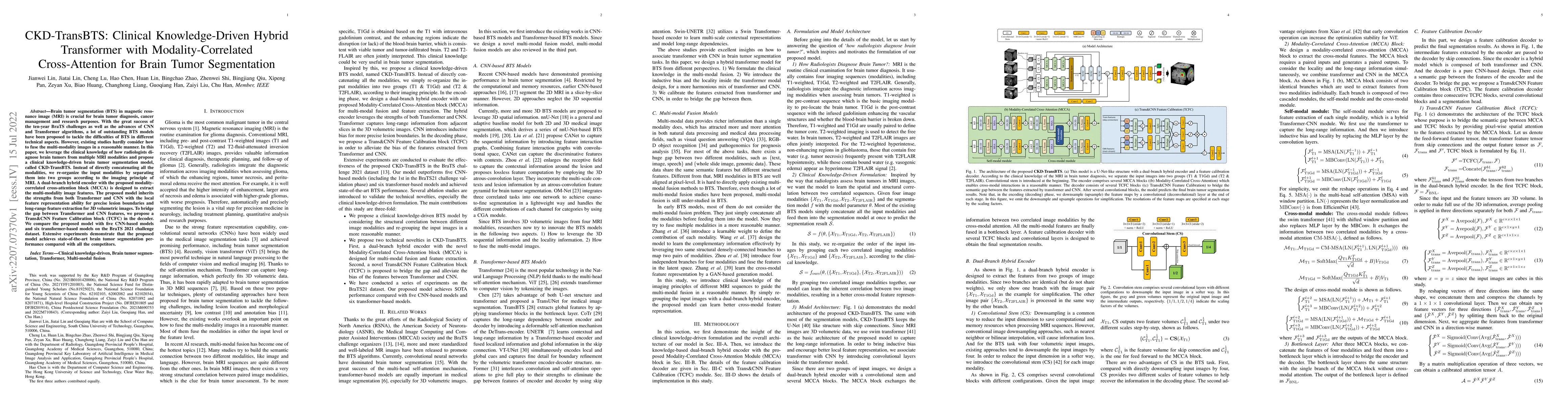
Brain tumor segmentation (BTS) in magnetic resonance image (MRI) is crucial for brain tumor diagnosis, cancer management and research purposes. With the great success of the ten-year BraTS challenge...
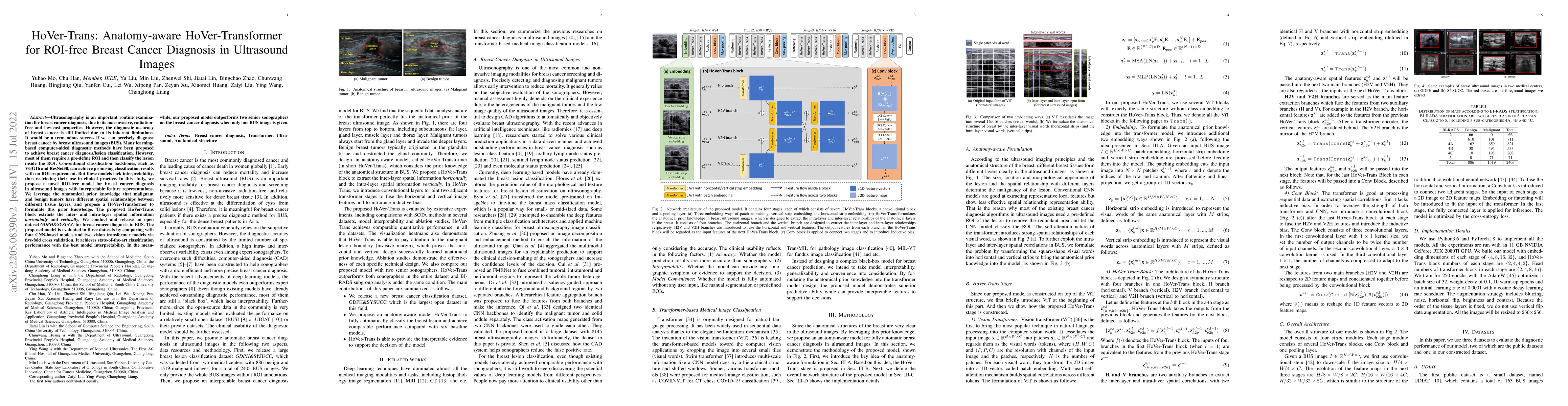
Ultrasonography is an important routine examination for breast cancer diagnosis, due to its non-invasive, radiation-free and low-cost properties. However, the diagnostic accuracy of breast cancer is...
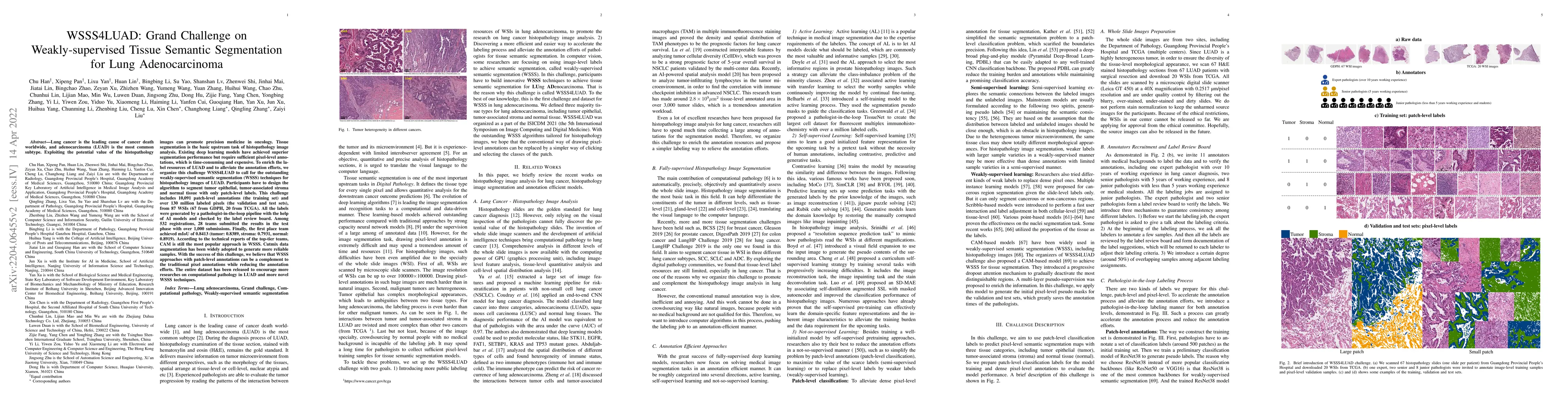
Lung cancer is the leading cause of cancer death worldwide, and adenocarcinoma (LUAD) is the most common subtype. Exploiting the potential value of the histopathology images can promote precision me...
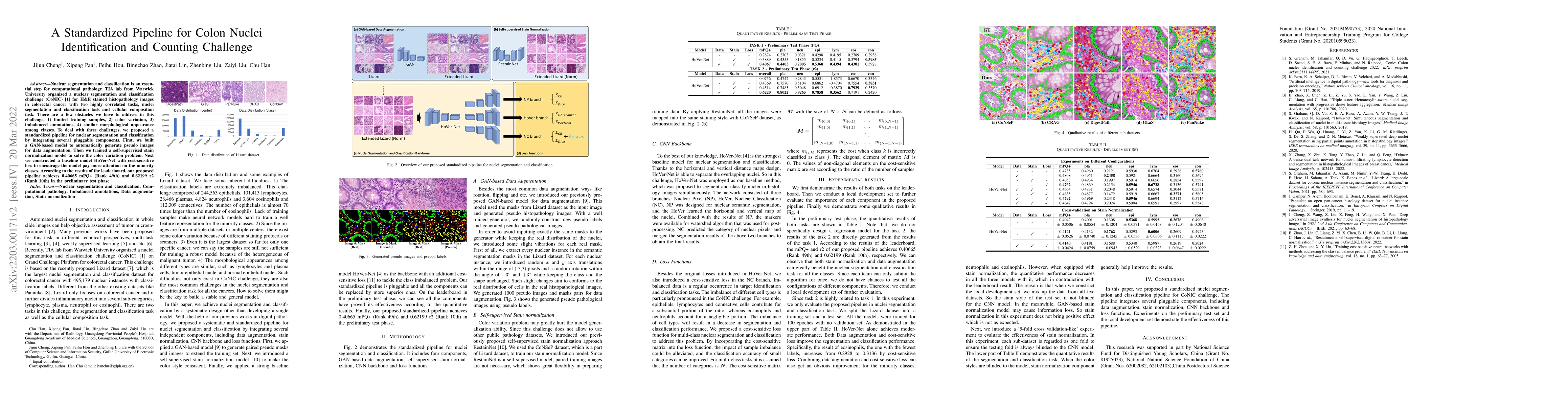
Nuclear segmentation and classification is an essential step for computational pathology. TIA lab from Warwick University organized a nuclear segmentation and classification challenge (CoNIC) for H&...
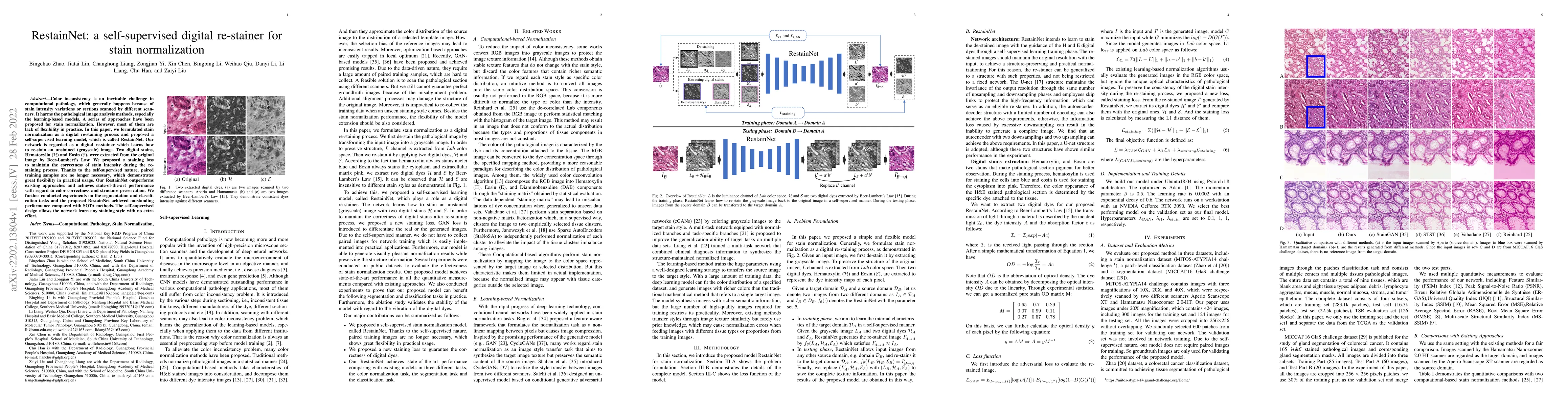
Color inconsistency is an inevitable challenge in computational pathology, which generally happens because of stain intensity variations or sections scanned by different scanners. It harms the patho...
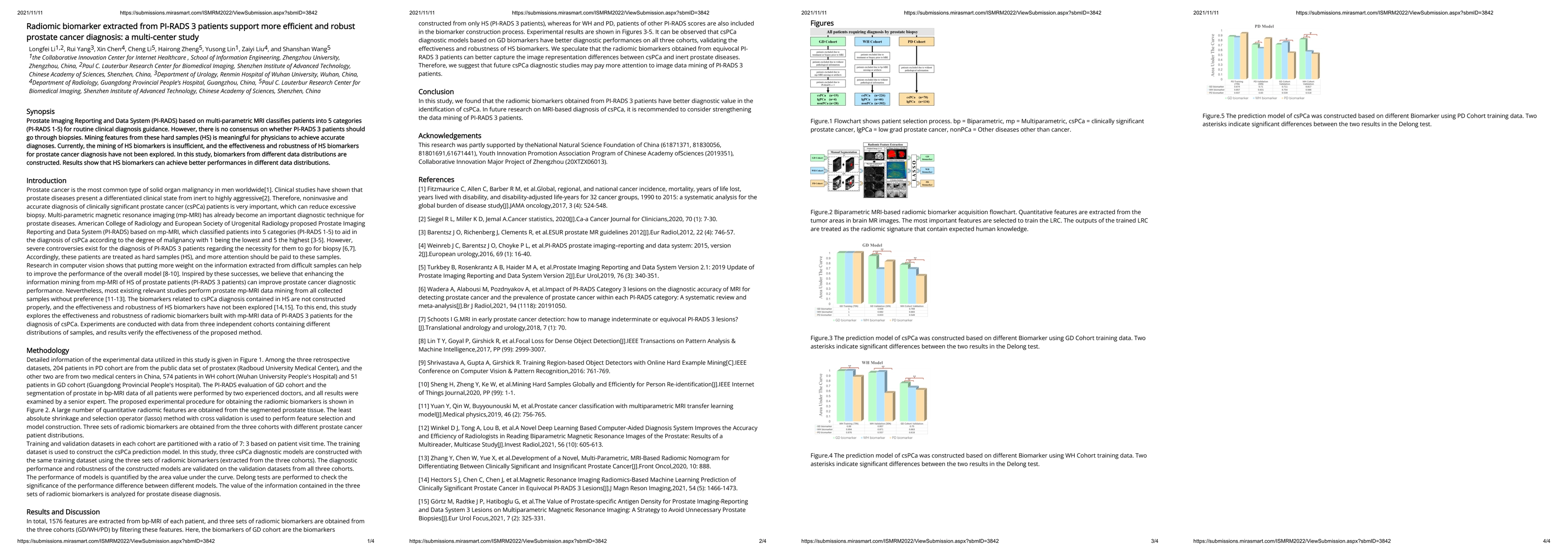
Prostate Imaging Reporting and Data System (PI-RADS) based on multi-parametric MRI classi\^ees patients into 5 categories (PI-RADS 1-5) for routine clinical diagnosis guidance. However, there is no ...
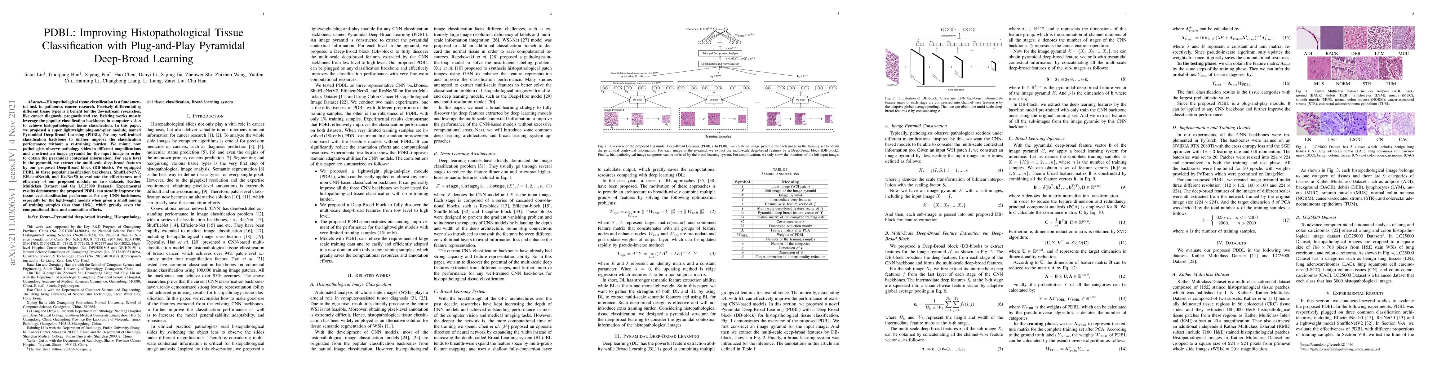
Histopathological tissue classification is a fundamental task in pathomics cancer research. Precisely differentiating different tissue types is a benefit for the downstream researches, like cancer d...
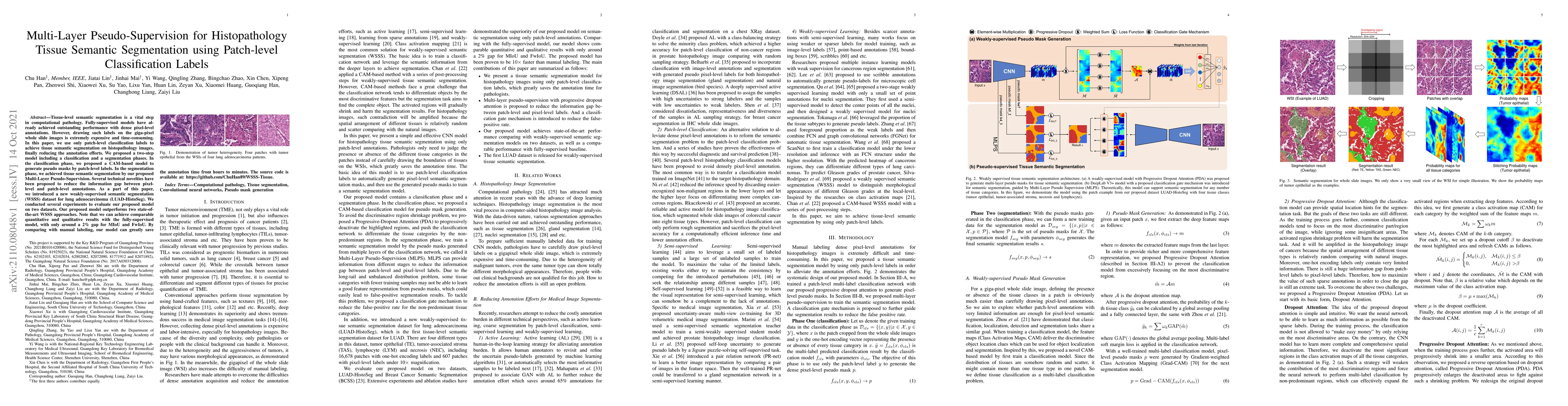
Tissue-level semantic segmentation is a vital step in computational pathology. Fully-supervised models have already achieved outstanding performance with dense pixel-level annotations. However, draw...
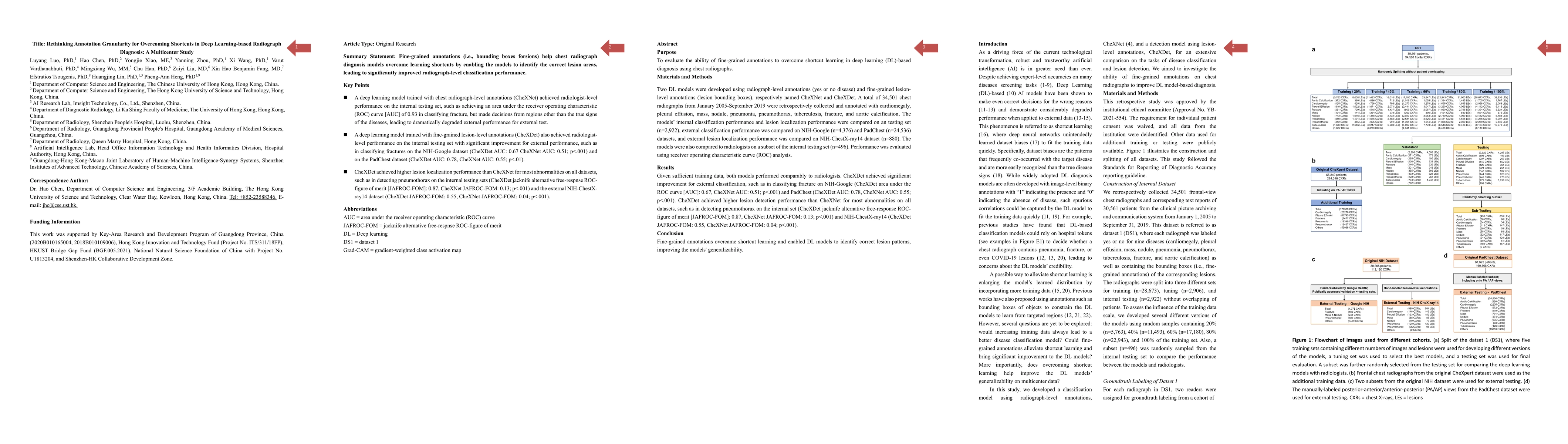
Two DL models were developed using radiograph-level annotations (yes or no disease) and fine-grained lesion-level annotations (lesion bounding boxes), respectively named CheXNet and CheXDet. The mod...
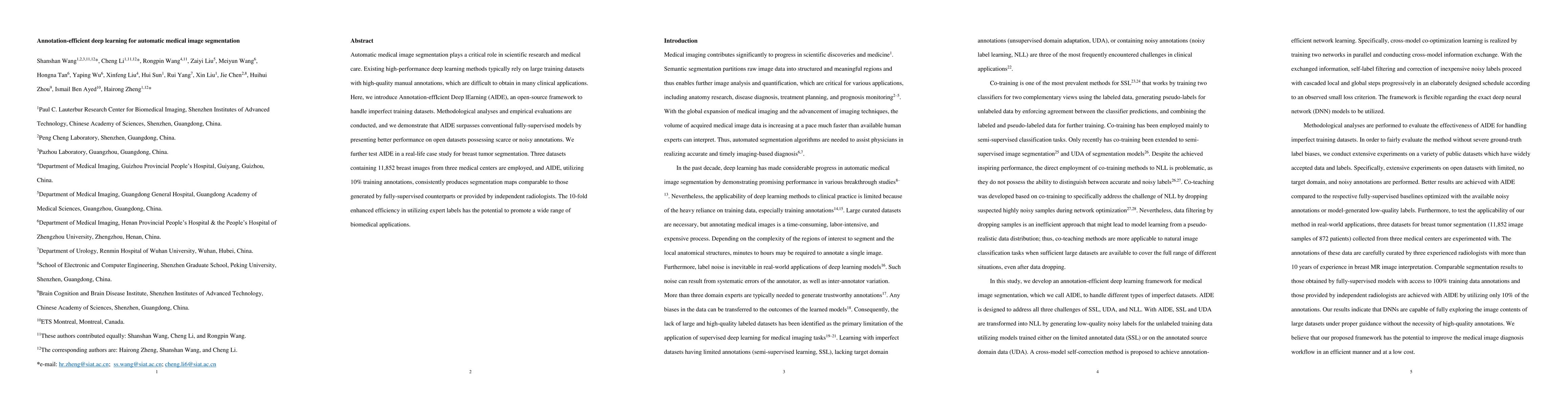
Automatic medical image segmentation plays a critical role in scientific research and medical care. Existing high-performance deep learning methods typically rely on large training datasets with hig...
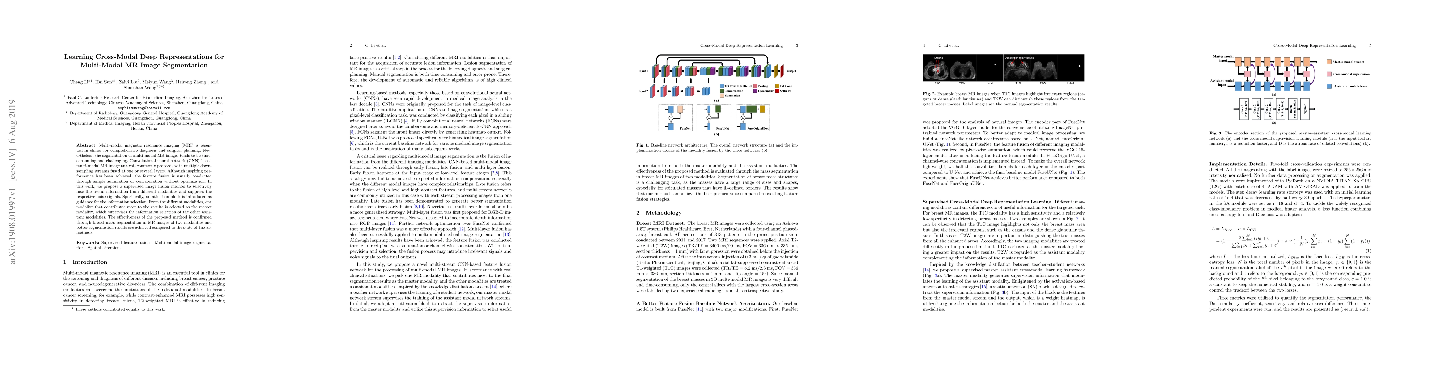
Multi-modal magnetic resonance imaging (MRI) is essential in clinics for comprehensive diagnosis and surgical planning. Nevertheless, the segmentation of multi-modal MR images tends to be time-consu...
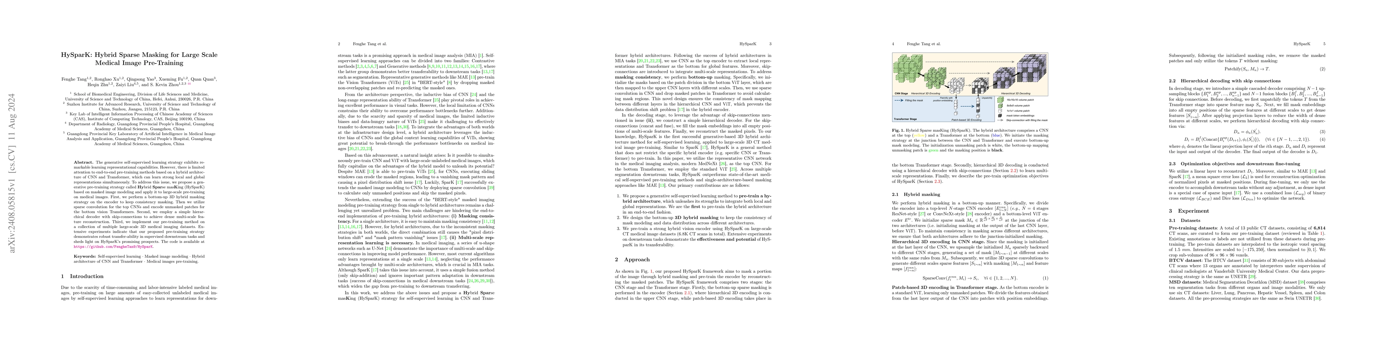
The generative self-supervised learning strategy exhibits remarkable learning representational capabilities. However, there is limited attention to end-to-end pre-training methods based on a hybrid ar...
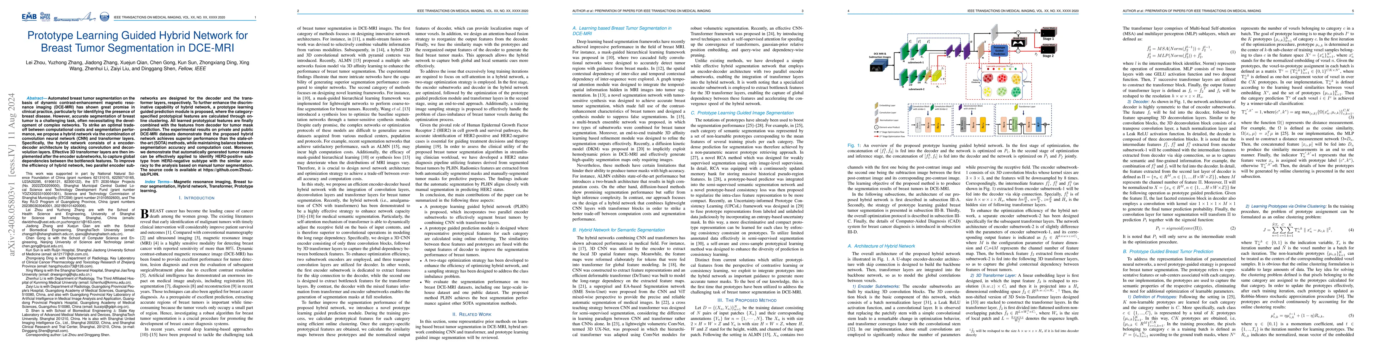
Automated breast tumor segmentation on the basis of dynamic contrast-enhancement magnetic resonance imaging (DCE-MRI) has shown great promise in clinical practice, particularly for identifying the pre...
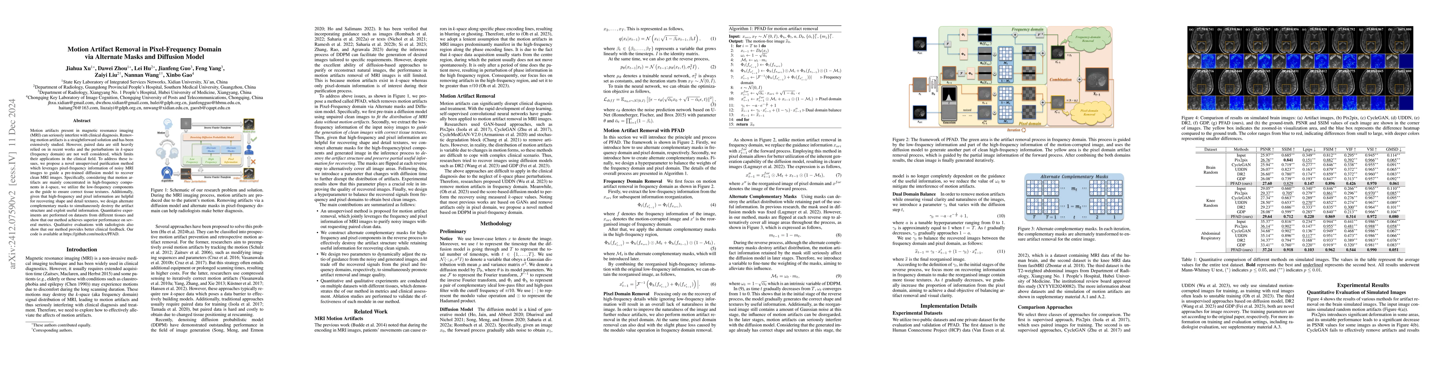
Motion artifacts present in magnetic resonance imaging (MRI) can seriously interfere with clinical diagnosis. Removing motion artifacts is a straightforward solution and has been extensively studied. ...
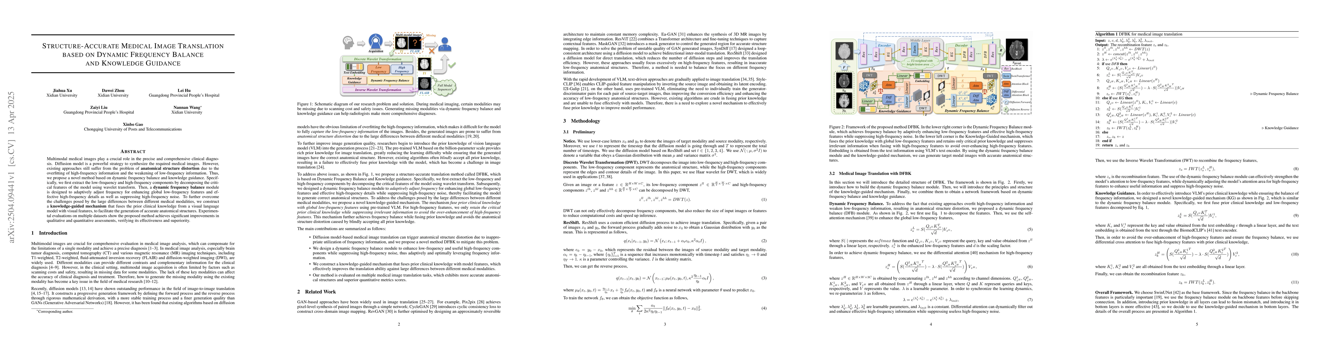
Multimodal medical images play a crucial role in the precise and comprehensive clinical diagnosis. Diffusion model is a powerful strategy to synthesize the required medical images. However, existing a...
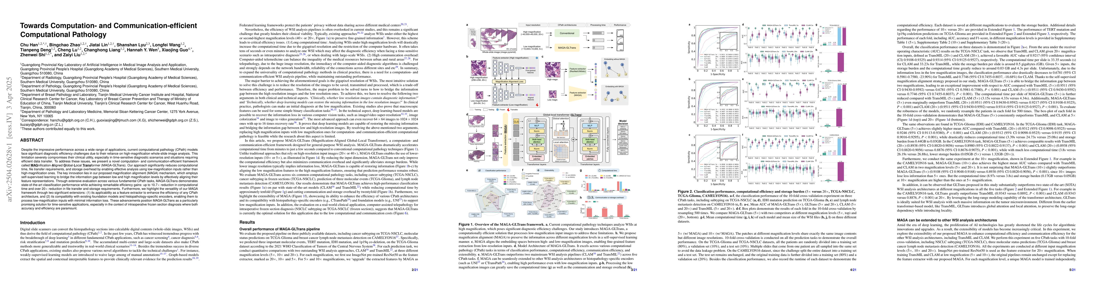
Despite the impressive performance across a wide range of applications, current computational pathology models face significant diagnostic efficiency challenges due to their reliance on high-magnifica...
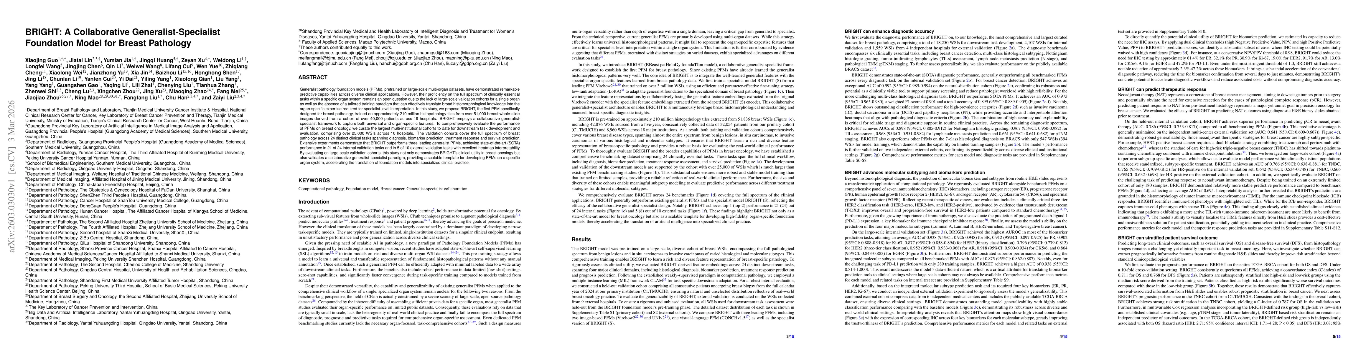
Generalist pathology foundation models (PFMs), pretrained on large-scale multi-organ datasets, have demonstrated remarkable predictive capabilities across diverse clinical applications. However, their...
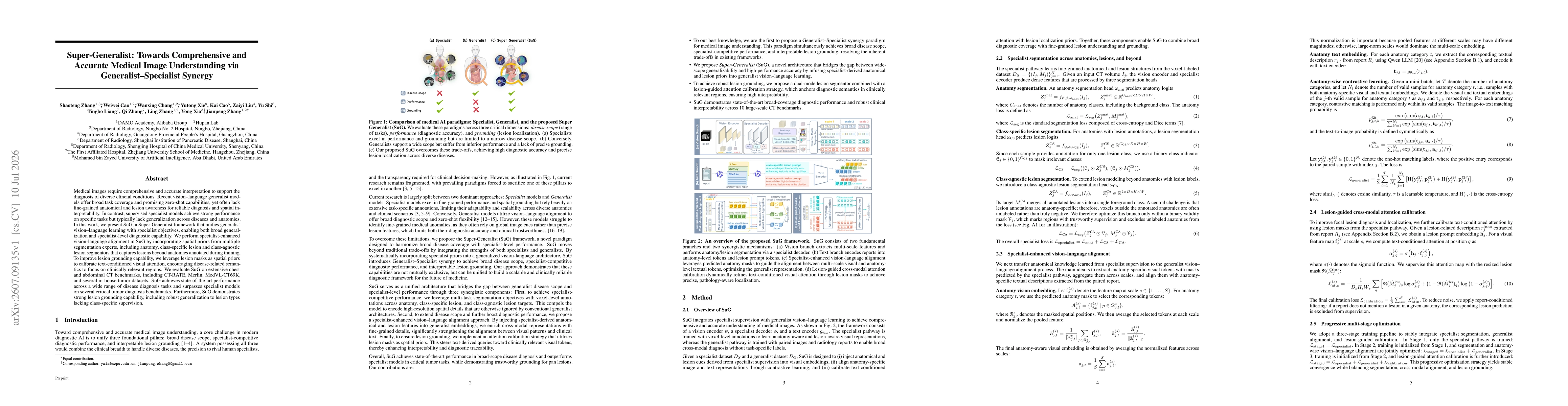
Medical images require comprehensive and accurate interpretation to support the diagnosis of diverse clincial conditions. Recent vision-language generalist models offer broad task coverage and promisi...