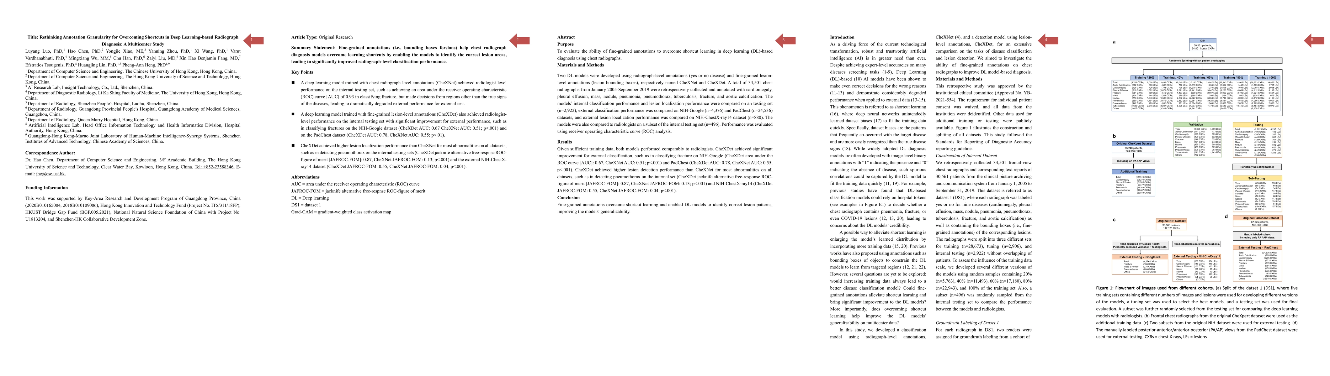
Rethinking Annotation Granularity for Overcoming Shortcuts in Deep Learning-based Radiograph Diagnosis: A Multicenter Study
Publication
Metrics
AI Quick Summary
This study compares two deep learning models trained with different annotation granularities for radiograph diagnosis: CheXNet (radiograph-level) and CheXDet (fine-grained lesion-level). CheXDet showed superior external classification and lesion localization performance, indicating that fine-grained annotations enhance model generalizability and reduce shortcut learning.
Paper Preview
Abstract
Two DL models were developed using radiograph-level annotations (yes or no disease) and fine-grained lesion-level annotations (lesion bounding boxes), respectively named CheXNet and CheXDet. The models' internal classification performance and lesion localization performance were compared on a testing set (n=2,922), external classification performance was compared on NIH-Google (n=4,376) and PadChest (n=24,536) datasets, and external lesion localization performance was compared on NIH-ChestX-ray14 dataset (n=880). The models were also compared to radiologists on a subset of the internal testing set (n=496). Given sufficient training data, both models performed comparably to radiologists. CheXDet achieved significant improvement for external classification, such as in classifying fracture on NIH-Google (CheXDet area under the ROC curve [AUC]: 0.67, CheXNet AUC: 0.51; p<.001) and PadChest (CheXDet AUC: 0.78, CheXNet AUC: 0.55; p<.001). CheXDet achieved higher lesion detection performance than CheXNet for most abnormalities on all datasets, such as in detecting pneumothorax on the internal set (CheXDet jacknife alternative free-response ROC-figure of merit [JAFROC-FOM]: 0.87, CheXNet JAFROC-FOM: 0.13; p<.001) and NIH-ChestX-ray14 (CheXDet JAFROC-FOM: 0.55, CheXNet JAFROC-FOM: 0.04; p<.001). To summarize, fine-grained annotations overcame shortcut learning and enabled DL models to identify correct lesion patterns, improving the models' generalizability.
AI Key Findings
Get AI-generated insights about this paper's methodology, results, significance, and more — seven facets brought into focus.
Impact
Paper Details
Authors
PDF Preview
Key Terms
Citation Network
Current paper (gray), citations (green), references (blue)
Display is limited for performance on very large graphs.
Discussion 0