3D Skin Segmentation Methods in Medical Imaging: A Comparison
Publication
Metrics
Paper Preview
Abstract
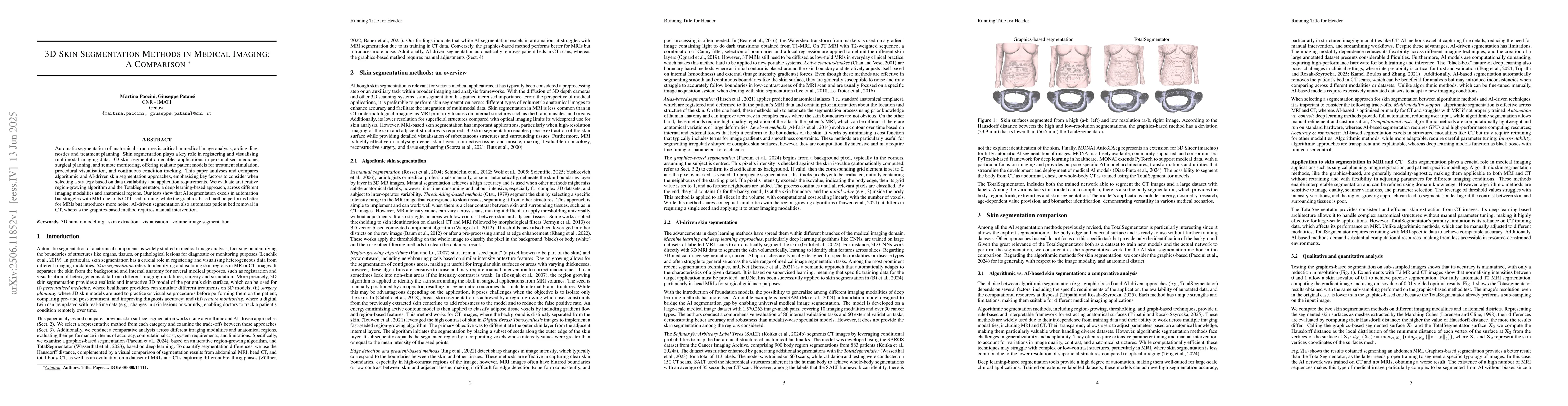
Automatic segmentation of anatomical structures is critical in medical image analysis, aiding diagnostics and treatment planning. Skin segmentation plays a key role in registering and visualising multimodal imaging data. 3D skin segmentation enables applications in personalised medicine, surgical planning, and remote monitoring, offering realistic patient models for treatment simulation, procedural visualisation, and continuous condition tracking. This paper analyses and compares algorithmic and AI-driven skin segmentation approaches, emphasising key factors to consider when selecting a strategy based on data availability and application requirements. We evaluate an iterative region-growing algorithm and the TotalSegmentator, a deep learning-based approach, across different imaging modalities and anatomical regions. Our tests show that AI segmentation excels in automation but struggles with MRI due to its CT-based training, while the graphics-based method performs better for MRIs but introduces more noise. AI-driven segmentation also automates patient bed removal in CT, whereas the graphics-based method requires manual intervention.
AI Key Findings
Get AI-generated insights about this paper's methodology, results, significance, and more — seven facets brought into focus.
Impact
Authors
PDF Preview
Citation Network
Current paper (gray), citations (green), references (blue)
Display is limited for performance on very large graphs.
Discussion 0