A Deep Learning Framework for Nuclear Segmentation and Classification in Histopathological Images
Publication
Metrics
AI Quick Summary
This paper introduces a deep learning framework for nuclear segmentation and classification in histopathological images, utilizing a unified model with three branches: segmentation, HoVer mapping, and classification, to address the challenges of high-level heterogeneity and variations in nuclei. The framework aims to generate nucleus boundaries, calculate pixel distances to their centres, and classify nuclei types.
Paper Preview
Abstract
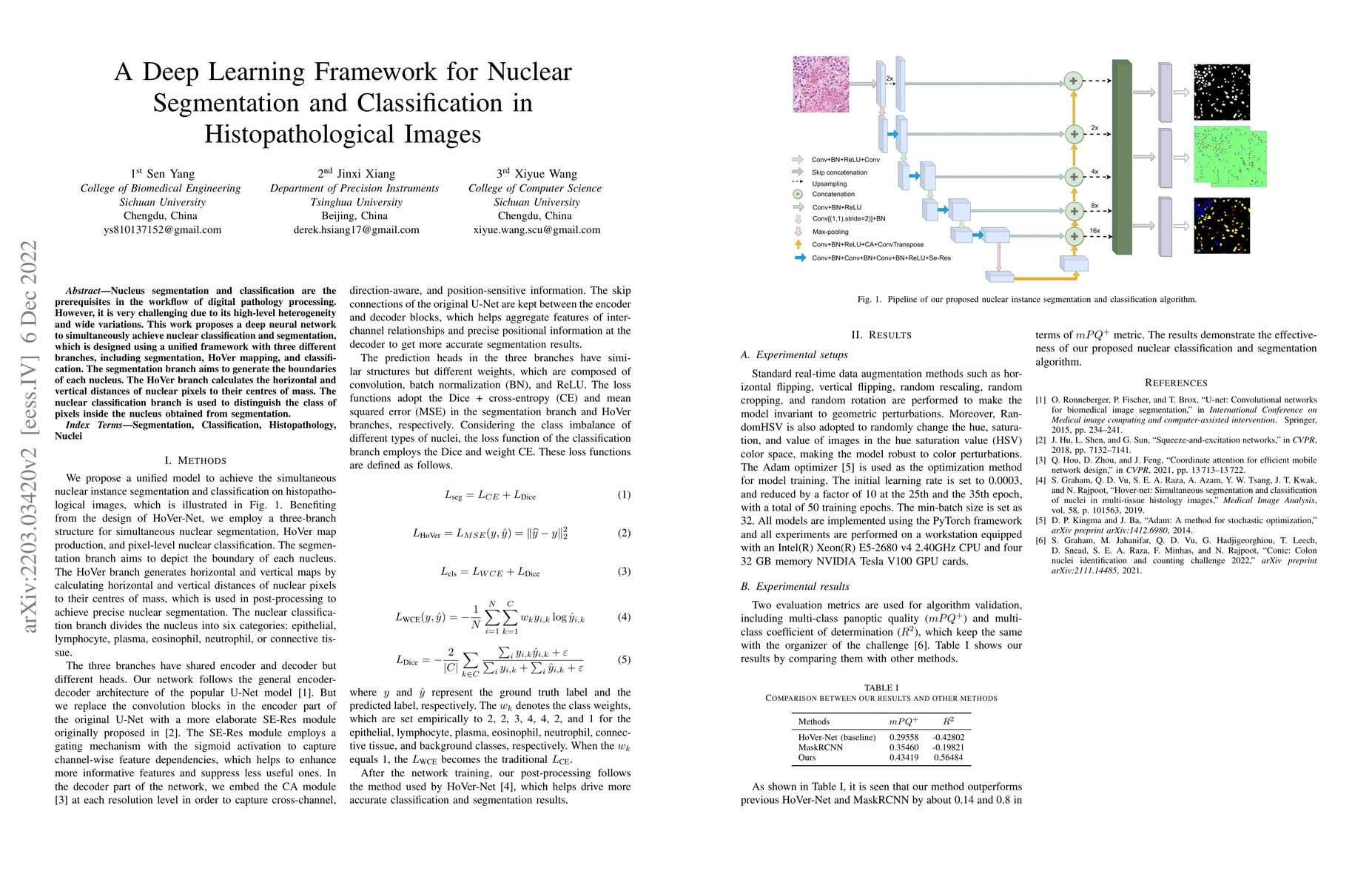
Nucleus segmentation and classification are the prerequisites in the workflow of digital pathology processing. However, it is very challenging due to its high-level heterogeneity and wide variations. This work proposes a deep neural network to simultaneously achieve nuclear classification and segmentation, which is designed using a unified framework with three different branches, including segmentation, HoVer mapping, and classification. The segmentation branch aims to generate the boundaries of each nucleus. The HoVer branch calculates the horizontal and vertical distances of nuclear pixels to their centres of mass. The nuclear classification branch is used to distinguish the class of pixels inside the nucleus obtained from segmentation.
AI Key Findings
Get AI-generated insights about this paper's methodology, results, significance, and more — seven facets brought into focus.
Impact
Paper Details
Authors
PDF Preview
Key Terms
Citation Network
Current paper (gray), citations (green), references (blue)
Display is limited for performance on very large graphs.
Discussion 0