Gland Segmentation in Histopathological Images by Deep Neural Network
Publication
Metrics
AI Quick Summary
This paper proposes a LinkNet-based deep neural network for segmenting gland structures in histopathological images, crucial for cancer diagnosis. The method is shown to be competitive with state-of-the-art techniques using the Warwick-Qu dataset, with performance further enhanced by refining gland edges and hematoxylin components.
Paper Preview
Abstract
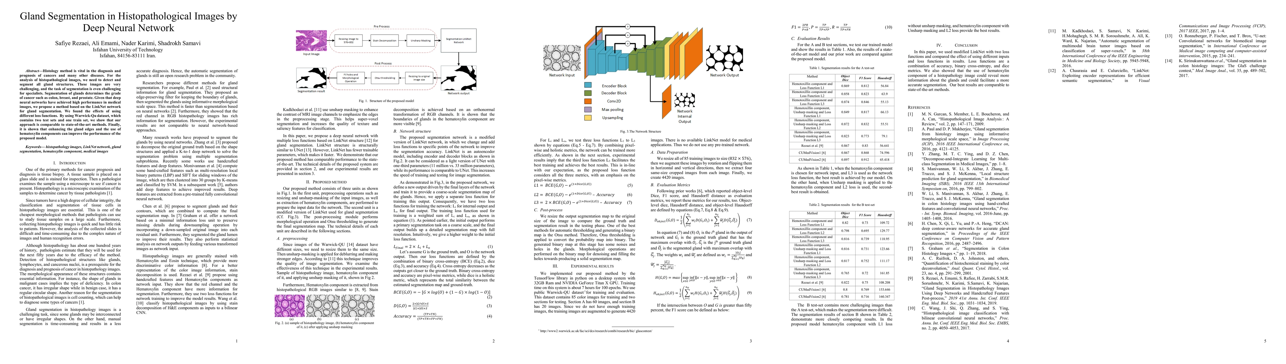
Histology method is vital in the diagnosis and prognosis of cancers and many other diseases. For the analysis of histopathological images, we need to detect and segment all gland structures. These images are very challenging, and the task of segmentation is even challenging for specialists. Segmentation of glands determines the grade of cancer such as colon, breast, and prostate. Given that deep neural networks have achieved high performance in medical images, we propose a method based on the LinkNet network for gland segmentation. We found the effects of using different loss functions. By using Warwick-Qu dataset, which contains two test sets and one train set, we show that our approach is comparable to state-of-the-art methods. Finally, it is shown that enhancing the gland edges and the use of hematoxylin components can improve the performance of the proposed model.
AI Key Findings
Get AI-generated insights about this paper's methodology, results, significance, and more — seven facets brought into focus.
Impact
Paper Details
Authors
PDF Preview
Key Terms
Citation Network
Current paper (gray), citations (green), references (blue)
Display is limited for performance on very large graphs.
Discussion 0