This paper proposes a modified LinkNet deep learning model for segmenting glands in histopathology images, enhanced with handcrafted features like invariant local binary patterns. Experimental results show superior performance compared to state-of-the-art methods, particularly on section B images of the Warwick-QU dataset.
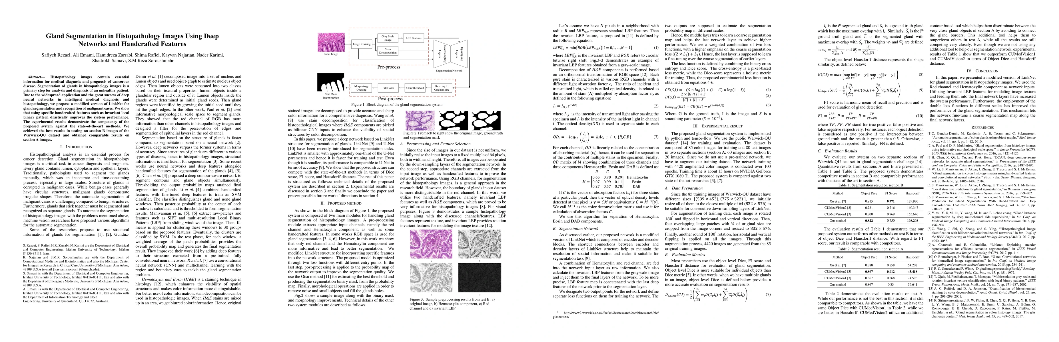
Histopathology images contain essential information for medical diagnosis and prognosis of cancerous disease. Segmentation of glands in histopathology images is a primary step for analysis and diagnosis of an unhealthy patient. Due to the widespread application and the great success of deep neural networks in intelligent medical diagnosis and histopathology, we propose a modified version of LinkNet for gland segmentation and recognition of malignant cases. We show that using specific handcrafted features such as invariant local binary pattern drastically improves the system performance. The experimental results demonstrate the competency of the proposed system against state-of-the-art methods. We achieved the best results in testing on section B images of the Warwick-QU dataset and obtained comparable results on section A images.
Get AI-generated insights about this paper's methodology, results, and significance.
Current paper (gray), citations (green), references (blue)
Title | Authors | Year | Actions |
---|---|---|---|
Gland Segmentation in H&E Histopathological Images using U-Net with Attention Module
|
Z. Yildirim, R. Samet, Emrah Hançer, Nooshin Nemati, Mohamed Traoré Mali
|
2023
|
View Paper |
DARMF-UNet: A dual-branch attention-guided refinement network with multi-scale features fusion U-Net for gland segmentation
|
Junmei Sun, Xin Zhang, Xiumei Li, Ruyu Liu, Tianyang Wang
|
2023
|
View Paper |
Pyramidal position attention model for histopathological image segmentation
|
Z. Bozdağ, M. F. Talu
|
2023
|
View Paper |
Standardized CycleGAN training for unsupervised stain adaptation in invasive carcinoma classification for breast histopathology
|
Nicolas Nerrienet, Rémy Peyret, Marie Sockeel, Stéphane Sockeel
|
2023
|
View Paper |
Region Segmentation of Whole-Slide Images for Analyzing Histological Differentiation of Prostate Adenocarcinoma Using Ensemble EfficientNetB2 U-Net with Transfer Learning Mechanism
|
Kobiljon Ikromjanov, Subrata Bhattacharjee, Rashadul Islam Sumon, Yeong-Byn Hwang, H. Rahman, Myung-Jae Lee, Hee-Cheol Kim, E. Park, N. Cho, Heung-Kook Choi
|
2023
|
View Paper |
Semantic Segmentation in Medical Image Based on Hybrid Dlinknet and Unet
|
Suresh Samudrala, C. Mohan
|
2022
|
View Paper |
DAN-NucNet: A dual attention based framework for nuclei segmentation in cancer histology images under wild clinical conditions
|
Ibtihaj Ahmad, Yong Xia, Hengfei Cui, Z. Islam
|
2022
|
View Paper |
Two Path Gland Segmentation Algorithm of Colon Pathological Image Based on Local Semantic Guidance
|
Songtao Ding, Hongyu Wang, Hu Lu, M. Nappi, Shaohua Wan
|
2022
|
View Paper |
Nuclei and glands instance segmentation in histology images: a narrative review
|
Esha Sadia Nasir, Arshi Parvaiz, M. Fraz
|
2022
|
View Paper |
Gland segmentation in colorectal cancer histopathological images using U-net inspired convolutional network
|
Priyanka Rastogi, K. Khanna, Vijendra Singh
|
2021
|
View Paper |
Automated Gland Detection in Colorectal Histopathological Images
|
Maisun Mohamed Al Zorgani, I. Mehmood, H. Ugail
|
2021
|
View Paper |
Deep Learning Application for Analyzing of Constituents and Their Correlations in the Interpretations of Medical Images
|
Tudor Florin Ursuleanu, Andreea Roxana Luca, L. Gheorghe, Roxana Grigorovici, Stefan Iancu, Maria Hlusneac, C. Preda, A. Grigorovici
|
2021
|
View Paper |
A hybrid deep learning approach for gland segmentation in prostate histopathological images
|
M. Salvi, M. Bosco, L. Molinaro, A. Gambella, M. Papotti, Usha R. Acharya, F. Molinari
|
2021
|
View Paper |
The impact of pre- and post-image processing techniques on deep learning frameworks: A comprehensive review for digital pathology image analysis
|
M. Salvi, U. Acharya, F. Molinari, K. Meiburger
|
2020
|
View Paper |
Brain Tumor Classification Using Medial Residual Encoder Layers
|
Zahra Sobhaninia, N. Karimi, P. Khadivi, R. Roshandel, S. Samavi
|
2020
|
View Paper |
Brain Tumor Classification Using Medial Residual Encoder Layers
|
Zahra Sobhaninia, N. Karimi, P. Khadivi, Roshank Roshandel, S. Samavi
|
2020
|
View Paper |
Eigenloss: Combined PCA-Based Loss Function for Polyp Segmentation
|
L. F. Sánchez-Peralta, A. Picón, J. Antequera-Barroso, J. Ortega-Morán, F. Sánchez-Margallo, J. B. Pagador
|
2020
|
View Paper |
Multi-Task Deep Learning for Image Segmentation Using Recursive Approximation Tasks
|
Rihuan Ke, Aurélie Bugeau, N. Papadakis, Mark Kirkland, P. Schuetz, C. Schönlieb
|
2020
|
View Paper |
A survey on incorporating domain knowledge into deep learning for medical image analysis
|
Xiaozhen Xie, J. Niu, Xuefeng Liu, Zhengsu Chen, Shaojie Tang, Shui Yu
|
2020
|
View Paper |
A Survey on Domain Knowledge Powered Deep Learning for Medical Image Analysis
|
Xiaozhen Xie, J. Niu, Xuefeng Liu, Zhengsu Chen, Shaojie Tang
|
2020
|
View Paper |
Gland Segmentation in Histopathological Images by Deep Neural Network
|
Safiyeh Rezaei, Ali Emami, N. Karimi, S. Samavi
|
2019
|
View Paper |
Automatic Gland Segmentation for Detection of CRC Using Enhanced SegNet Neural Network
|
Mohan Mahanty, D. Bhattacharyya, Divya Midhunchakkaravarthy
|
2021
|
View Paper |
Comments (0)