A generalizable approach based on U-Net model for automatic Intra retinal cyst segmentation in SD-OCT images
Publication
Metrics
AI Quick Summary
This paper proposes a U-Net-based approach for the automatic segmentation of intra retinal cysts in SD-OCT images, addressing challenges faced by previous techniques. The method includes data adjustment and prior information embedding, followed by a connection module in the U-Net for effective information transfer, achieving mean Dice values of 0.78 and 0.81 on two public datasets.
Paper Preview
Abstract
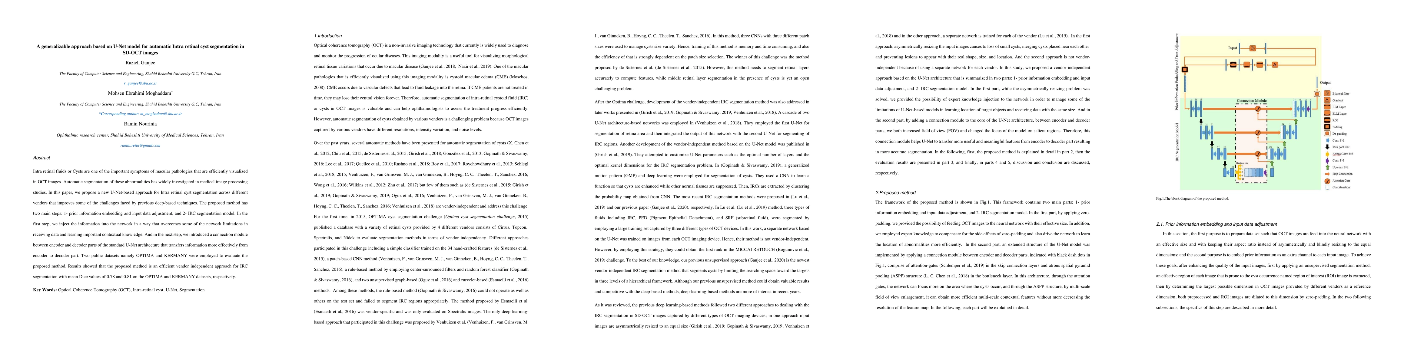
Intra retinal fluids or Cysts are one of the important symptoms of macular pathologies that are efficiently visualized in OCT images. Automatic segmentation of these abnormalities has been widely investigated in medical image processing studies. In this paper, we propose a new U-Net-based approach for Intra retinal cyst segmentation across different vendors that improves some of the challenges faced by previous deep-based techniques. The proposed method has two main steps: 1- prior information embedding and input data adjustment, and 2- IRC segmentation model. In the first step, we inject the information into the network in a way that overcomes some of the network limitations in receiving data and learning important contextual knowledge. And in the next step, we introduced a connection module between encoder and decoder parts of the standard U-Net architecture that transfers information more effectively from the encoder to the decoder part. Two public datasets namely OPTIMA and KERMANY were employed to evaluate the proposed method. Results showed that the proposed method is an efficient vendor-independent approach for IRC segmentation with mean Dice values of 0.78 and 0.81 on the OPTIMA and KERMANY datasets, respectively.
AI Key Findings
Get AI-generated insights about this paper's methodology, results, significance, and more — seven facets brought into focus.
Impact
Paper Details
Authors
PDF Preview
Key Terms
Citation Network
Current paper (gray), citations (green), references (blue)
Display is limited for performance on very large graphs.
Discussion 0