Quick Summary:

This paper proposes an automated method for cyst segmentation in OCT retinal images using domain knowledge and ensemble learning with Random Forests. The biologically inspired approach achieves a mean dice coefficient of 0.3893, demonstrating potential for robust cyst detection and segmentation in 3D OCT volumes.

Summary

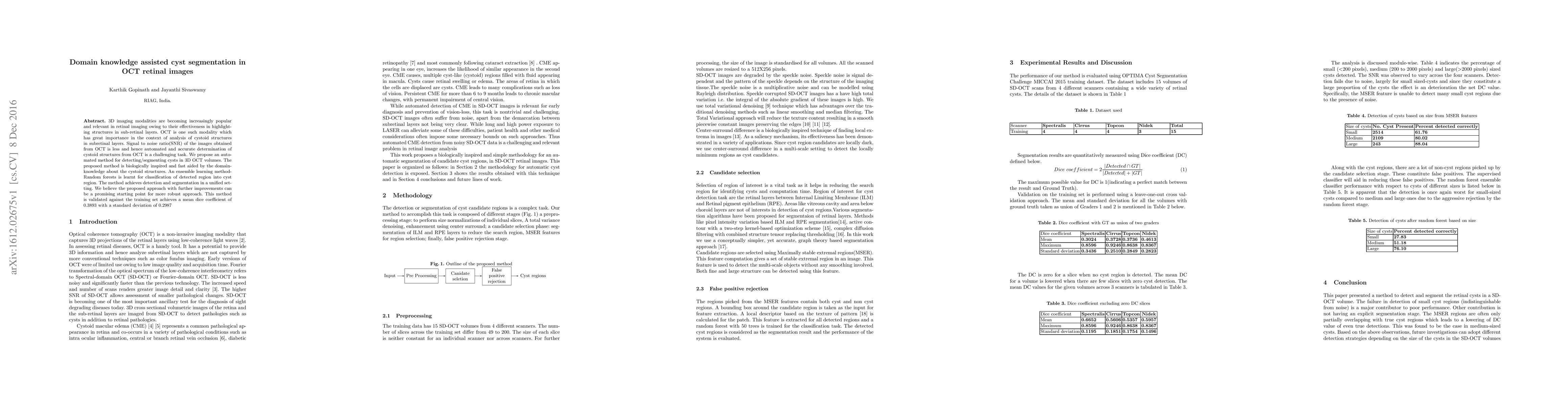
3D imaging modalities are becoming increasingly popular and relevant in retinal imaging owing to their effectiveness in highlighting structures in sub-retinal layers. OCT is one such modality which has great importance in the context of analysis of cystoid structures in subretinal layers. Signal to noise ratio(SNR) of the images obtained from OCT is less and hence automated and accurate determination of cystoid structures from OCT is a challenging task. We propose an automated method for detecting/segmenting cysts in 3D OCT volumes. The proposed method is biologically inspired and fast aided by the domain knowledge about the cystoid structures. An ensemble learning methodRandom forests is learnt for classification of detected region into cyst region. The method achieves detection and segmentation in a unified setting. We believe the proposed approach with further improvements can be a promising starting point for more robust approach. This method is validated against the training set achieves a mean dice coefficient of 0.3893 with a standard deviation of 0.2987

AI Key Findings

Get AI-generated insights about this paper's methodology, results, and significance.

Paper Details

PDF Preview

Download PDF

Key Terms

structures (0.217)
automated (0.213)
layers (0.186)
imaging (0.179)
achieves (0.171)
region (0.155)
volumes (0.149)
method (0.149)
aided (0.147)
snr (0.146)
modality (0.145)
method achieves (0.145)
starting point (0.145)
owing (0.142)
highlighting (0.141)
noise ratio (0.140)
believe (0.139)
validated (0.137)
signal noise (0.135)
modalities (0.134)

Citation Network

Current paper (gray), citations (green), references (blue)

Current Paper
Citations
References
Click to view
Title Authors Year Actions
A generalizable approach based on the U‐Net model for automatic intraretinal cyst segmentation in SD‐OCT images
Razieh Ganjee, Mohsen Ebrahimi Moghaddam, R. Nourinia
2023
View Paper
A Review of Machine Learning Algorithms for Retinal Cyst Segmentation on Optical Coherence Tomography
Xing Wei, R. Sui
2023
View Paper
A generalizable approach based on U-Net model for automatic Intra retinal cyst segmentation in SD-OCT images
Razieh Ganjee, Mohsen Ebrahimi Moghaddam, R. Nourinia
2022
View Paper
Deep learning-based classification and segmentation of retinal cavitations on optical coherence tomography images of macular telangiectasia type 2
Jessica Loo, Cindy X. Cai, J. Choong, E. Chew, M. Friedlander, G. Jaffe, Sina Farsiu
2020
View Paper
An unsupervised hierarchical approach for automatic intra-retinal cyst segmentation in spectral-domain optical coherence tomography images.
Razieh Ganjee, M. Ebrahimi Moghaddam, R. Nourinia
2020
View Paper
Depthwise Separable Convolutional Neural Network Model for Intra-Retinal Cyst Segmentation
Girish G. N., B. Saikumar, Sohini Roychowdhury, Abhishek R. Kothari, Jeny Rajan
2019
View Paper
Segmentation of Intra-Retinal Cysts From Optical Coherence Tomography Images Using a Fully Convolutional Neural Network Model
G. Girish, Bibhash Thakur, S. Chowdhury, Abhishek R. Kothari, Jeny Rajan
2019
View Paper
Classification of Retinal Cysts on SD-OCT Images Using Stacked Auto-Encoder
K. Alsaih, T. Tang, F. Mériaudeau, G. Lemaître, M. Rastgoo, D. Sidibé
2018
View Paper
Segmentation of Retinal Cysts From Optical Coherence Tomography Volumes Via Selective Enhancement
Karthik Gopinath, J. Sivaswamy
2017
View Paper
CYST SEGMENTATION ON OPTICAL COHERENCE TOMOGRAPHY IMAGE USING FCNN
K.SHEIKDAVOOD, R. Kiruthiga, V.SOWMIYAPRIYA, S.VIJAYA, Ug Student
2021
View Paper
A design for an automated Optical Coherence Tomography analysis system
J. Sivaswamy, Christian Desrosiers, Karthik Gopinath
2018
View Paper

Comments (0)

This website uses cookies to ensure you get the best experience. Learn more