Summary
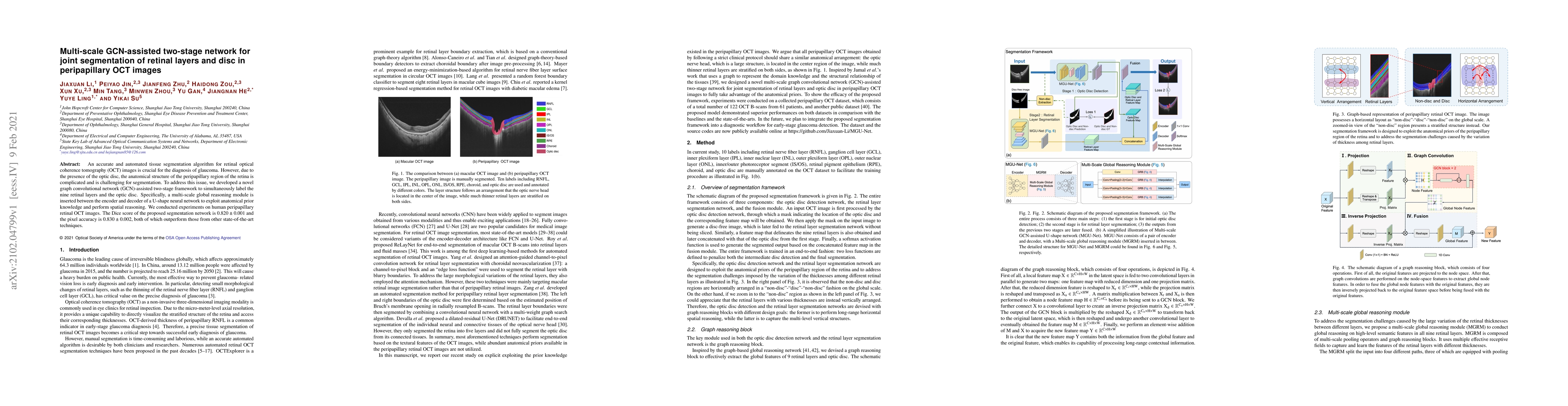
An accurate and automated tissue segmentation algorithm for retinal optical coherence tomography (OCT) images is crucial for the diagnosis of glaucoma. However, due to the presence of the optic disc, the anatomical structure of the peripapillary region of the retina is complicated and is challenging for segmentation. To address this issue, we developed a novel graph convolutional network (GCN)-assisted two-stage framework to simultaneously label the nine retinal layers and the optic disc. Specifically, a multi-scale global reasoning module is inserted between the encoder and decoder of a U-shape neural network to exploit anatomical prior knowledge and perform spatial reasoning. We conducted experiments on human peripapillary retinal OCT images. The Dice score of the proposed segmentation network is 0.820$\pm$0.001 and the pixel accuracy is 0.830$\pm$0.002, both of which outperform those from other state-of-the-art techniques.
AI Key Findings
Get AI-generated insights about this paper's methodology, results, and significance.
Paper Details
PDF Preview
Key Terms
Citation Network
Current paper (gray), citations (green), references (blue)
Display is limited for performance on very large graphs.
Similar Papers
Found 4 papersReliable Joint Segmentation of Retinal Edema Lesions in OCT Images
Yong Liu, Meng Wang, Kai Yu et al.
Multi-Scale Convolutional Neural Network for Automated AMD Classification using Retinal OCT Images
Saman Sotoudeh-Paima, Hamid Soltanian-Zadeh, Ata Jodeiri et al.
| Title | Authors | Year | Actions |
|---|
Comments (0)