Summary

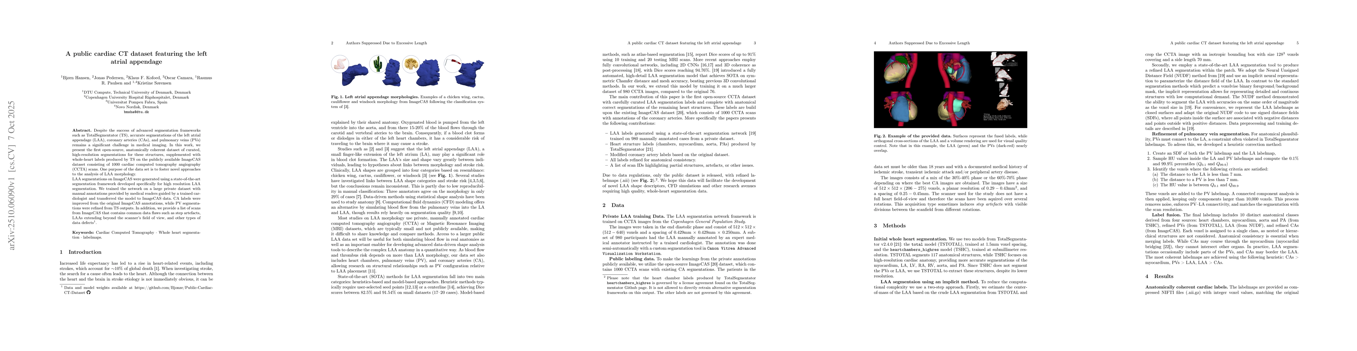
Despite the success of advanced segmentation frameworks such as TotalSegmentator (TS), accurate segmentations of the left atrial appendage (LAA), coronary arteries (CAs), and pulmonary veins (PVs) remain a significant challenge in medical imaging. In this work, we present the first open-source, anatomically coherent dataset of curated, high-resolution segmentations for these structures, supplemented with whole-heart labels produced by TS on the publicly available ImageCAS dataset consisting of 1000 cardiac computed tomography angiography (CCTA) scans. One purpose of the data set is to foster novel approaches to the analysis of LAA morphology. LAA segmentations on ImageCAS were generated using a state-of-the-art segmentation framework developed specifically for high resolution LAA segmentation. We trained the network on a large private dataset with manual annotations provided by medical readers guided by a trained cardiologist and transferred the model to ImageCAS data. CA labels were improved from the original ImageCAS annotations, while PV segmentations were refined from TS outputs. In addition, we provide a list of scans from ImageCAS that contains common data flaws such as step artefacts, LAAs extending beyond the scanner's field of view, and other types of data defects.

AI Key Findings

Generated Oct 09, 2025

Methodology

The study introduces a public cardiac CT dataset featuring the left atrial appendage (LAA), combining high-resolution segmentations from TotalSegmentator (TS) and a state-of-the-art LAA segmentation framework. The dataset includes refined pulmonary vein (PV) segmentations, coronary artery (CA) labels, and anatomical consistency checks. Data preprocessing, label fusion, and quality control are performed to ensure anatomical coherence.

Key Results

  • The proposed dataset provides high-resolution, anatomically coherent segmentations for LAA, PVs, and CAs, enabling detailed morphological analysis of the LAA.
  • LAA segmentations using the NUDF method achieve smooth and accurate representations, capturing critical anatomical features like trabeculation and lobes, which are essential for understanding blood flow and stroke risk.
  • The dataset includes 1000 CCTA scans with curated labels, along with a list of scans containing data flaws such as step artefacts and LAA out-of-field-of-view issues.

Significance

This research addresses the challenge of accurate LAA segmentation, which is critical for stroke risk assessment in atrial fibrillation patients. The dataset provides a valuable resource for studying anatomical variability and improving diagnostic tools.

Technical Contribution

The paper introduces a novel approach combining TotalSegmentator outputs, neural distance field-based LAA segmentation, and label refinement techniques to generate high-quality, anatomically consistent label maps for cardiac structures.

Novelty

This work presents the first open-source, high-resolution dataset featuring detailed LAA segmentations, along with refined PV and CA labels, enabling robust analysis of complex cardiac anatomies and morphology.

Limitations

  • The dataset lacks patient metadata such as age, sex, and disease status, limiting comprehensive clinical analysis.
  • The dataset includes a mix of end-diastolic (ED) and end-systolic (ES) scans without clear phase labeling, which may affect volume estimation and analysis.

Future Work

  • Developing methods to integrate patient metadata for more comprehensive stroke risk analysis.
  • Exploring advanced segmentation techniques to handle complex anatomical structures and improve accuracy in challenging regions like the LAA ostium.

Paper Details

PDF Preview

Download PDF

Comments (0)

This website uses cookies to ensure you get the best experience. Learn more