Summary
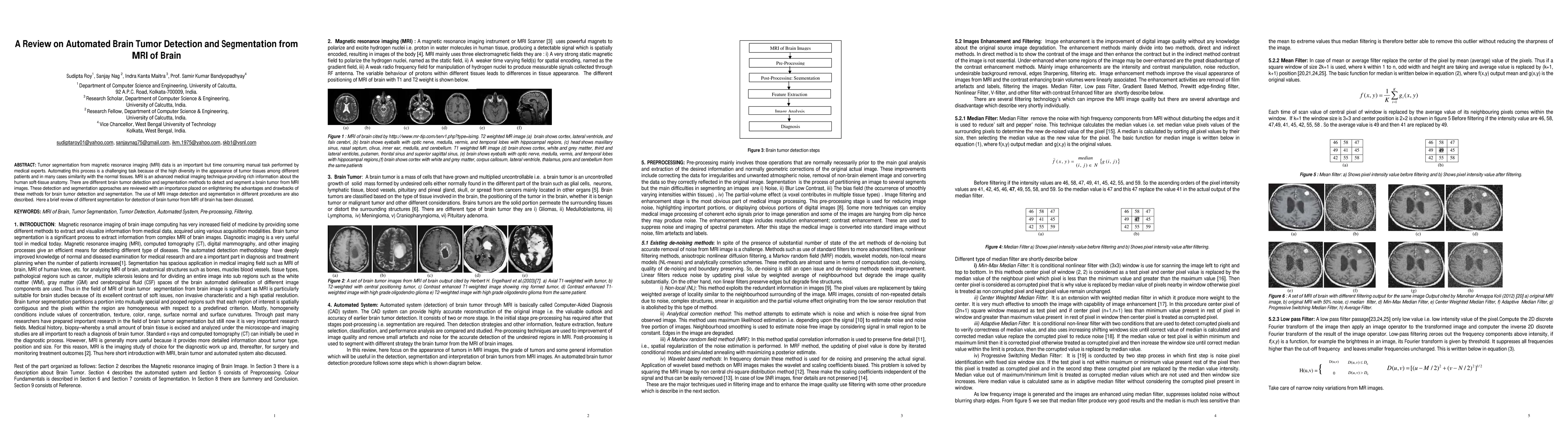
Tumor segmentation from magnetic resonance imaging (MRI) data is an important but time consuming manual task performed by medical experts. Automating this process is a challenging task because of the high diversity in the appearance of tumor tissues among different patients and in many cases similarity with the normal tissues. MRI is an advanced medical imaging technique providing rich information about the human soft-tissue anatomy. There are different brain tumor detection and segmentation methods to detect and segment a brain tumor from MRI images. These detection and segmentation approaches are reviewed with an importance placed on enlightening the advantages and drawbacks of these methods for brain tumor detection and segmentation. The use of MRI image detection and segmentation in different procedures are also described. Here a brief review of different segmentation for detection of brain tumor from MRI of brain has been discussed.
AI Key Findings
Get AI-generated insights about this paper's methodology, results, and significance.
Paper Details
PDF Preview
Key Terms
Citation Network
Current paper (gray), citations (green), references (blue)
Display is limited for performance on very large graphs.
Similar Papers
Found 4 papersFully Automated Tumor Segmentation for Brain MRI data using Multiplanner UNet
Erik B Dam, Mathias Perslev, Sumit Pandey et al.
Brain Tumor Segmentation from MRI Images using Deep Learning Techniques
Mayank Dixit, Ayan Gupta, Vipul Kumar Mishra et al.
| Title | Authors | Year | Actions |
|---|
Comments (0)