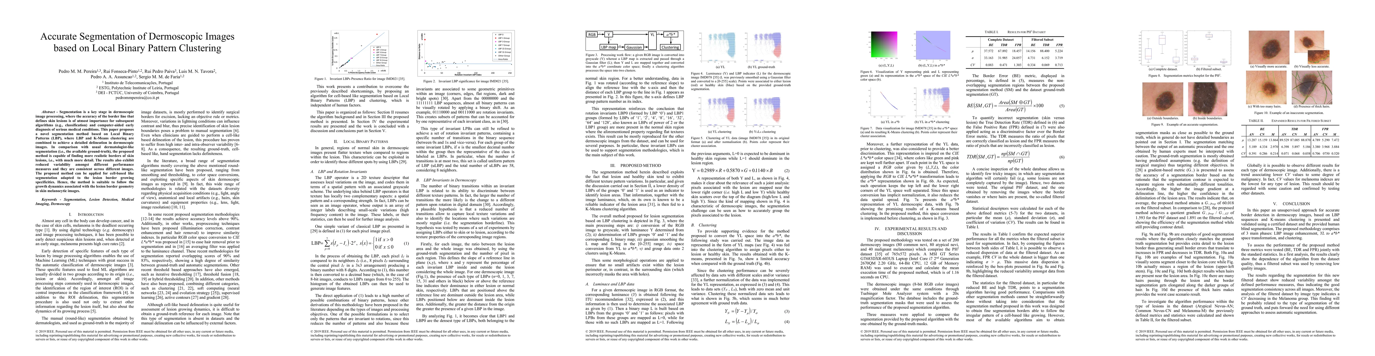
ArxivLens

Quick Summary:

This paper introduces a novel segmentation method for dermoscopic images using Local Binary Patterns (LBP) and K-Means clustering, achieving more detailed and realistic delineation of skin lesion borders compared to traditional methods. The method demonstrates consistent performance across different images and is suitable for tracking lesion border growth dynamics.

arXiv Id: 1902.06347
Comments: submitted to MIPRO DC 2019
Date Published: 2019-02-21
Date Updated: 2019-02-21

Categories:

Key Terms (TF-IDF):

skin (0.332)
segmentation (0.289)
images (0.199)
proposed method (0.185)
method (0.150)
aided (0.149)
diagnosis (0.142)
method applied (0.140)
like (0.139)
adapted (0.136)
defines (0.135)
ground truth (0.133)
subsequent (0.132)
truth (0.132)
method based (0.127)
paper proposes (0.125)
medical (0.123)
variability (0.123)
usual (0.122)
growing (0.121)
License: http://arxiv.org/licenses/nonexclusive-distrib/1.0/

Summary:

Segmentation is a key stage in dermoscopic image processing, where the accuracy of the border line that defines skin lesions is of utmost importance for subsequent algorithms (e.g., classification) and computer-aided early diagnosis of serious medical conditions. This paper proposes a novel segmentation method based on Local Binary Patterns (LBP), where LBP and K-Means clustering are combined to achieve a detailed delineation in dermoscopic images. In comparison with usual dermatologist-like segmentation (i.e., the available ground-truth), the proposed method is capable of finding more realistic borders of skin lesions, i.e., with much more detail. The results also exhibit reduced variability amongst different performance measures and they are consistent across different images. The proposed method can be applied for cell-based like segmentation adapted to the lesion border growing specificities. Hence, the method is suitable to follow the growth dynamics associated with the lesion border geometry in skin melanocytic images.

Citation & Reference Network Visualization

This graph shows how this paper is connected to other research. The current paper is shown in dark gray, papers that cite it are in green, and papers it references are in blue. You can:

Current Paper
Papers citing this work (Citations)
Papers cited by this work (References)

• Hover over any node to see the paper title

• Click a node to view that paper's details

• Drag nodes to rearrange the graph

• Use the zoom controls (bottom right) to zoom in/out

Citations and References

Title Authors Year Actions
CMTT-JTracker: a fully test-time adaptive framework serving automated cell lineage construction
Liuyin Chen, Sanyuan Fu, Zijun Zhang
2024
View Paper
Image Segmentation Metrics in Skin Lesion: Accuracy, Sensitivity, Specificity, Dice Coefficient, Jaccard Index, and Matthews Correlation Coefficient
A. W. Setiawan
2020
View Paper
Dermoscopic skin lesion image segmentation based on Local Binary Pattern Clustering: Comparative study
Pedro M. M. Pereira, R. Fonseca-Pinto, Rui Pedro Paiva, P. Assunção, Luís M. N. Tavora, Lucas A. Thomaz, S. Faria
2020
View Paper
Skin lesion classification enhancement using border-line features - The melanoma vs nevus problem
Pedro M. M. Pereira, R. Fonseca-Pinto, Rui Pedro Paiva, P. Assunção, Luís M. N. Tavora, Lucas A. Thomaz, S. Faria
2020
View Paper
A Content-Based Approach to Medical Image Retrieval
A. K, N. K., Rukmani Devi D.
2021
View Paper
Biomedical Signal Processing and Control
Pedro M. M. Pereira, R. Fonseca-Pinto, Pedro Paiva, Pedro A.A. Assuncao b, Luís M. N. Tavora, Lucas A. Thomaz, S. M. Faria
2020
View Paper
Joint Segmentation and Diagnosis of Skin Lesions
Francisco CastelBranco da Silveira, Sena Esteves
2020
View Paper

Comments (0)

This website uses cookies to ensure you get the best experience. Learn more