Saliency-based segmentation of dermoscopic images using color information
Publication
Metrics
AI Quick Summary
This paper proposes a novel method for skin lesion segmentation in dermoscopic images by integrating color information with saliency-based segmentation, achieving higher accuracy compared to existing saliency-only methods. The method includes pre-processing steps to enhance image quality and a binarization process inspired by human visual perception.
Paper Preview
Abstract
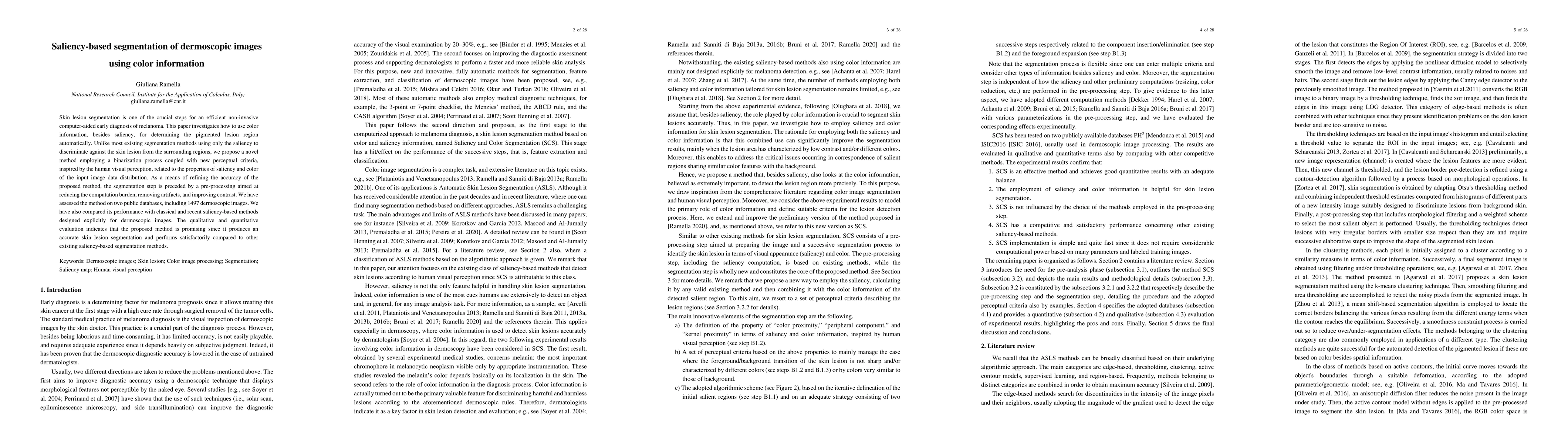
Skin lesion segmentation is one of the crucial steps for an efficient non-invasive computer-aided early diagnosis of melanoma. This paper investigates how color information, besides saliency, can be used to determine the pigmented lesion region automatically. Unlike most existing segmentation methods using only the saliency in order to discriminate against the skin lesion from the surrounding regions, we propose a novel method employing a binarization process coupled with new perceptual criteria, inspired by the human visual perception, related to the properties of saliency and color of the input image data distribution. As a means of refining the accuracy of the proposed method, the segmentation step is preceded by a pre-processing aimed at reducing the computation burden, removing artifacts, and improving contrast. We have assessed the method on two public databases, including 1497 dermoscopic images. We have also compared its performance with classical and recent saliency-based methods designed explicitly for dermoscopic images. The qualitative and quantitative evaluation indicates that the proposed method is promising since it produces an accurate skin lesion segmentation and performs satisfactorily compared to other existing saliency-based segmentation methods.
AI Key Findings
Get AI-generated insights about this paper's methodology, results, significance, and more — seven facets brought into focus.
Impact
Paper Details
Authors
PDF Preview
Key Terms
Citation Network
Current paper (gray), citations (green), references (blue)
Display is limited for performance on very large graphs.
Discussion 0