Publication
Metrics
AI Quick Summary
This paper presents a machine learning super-resolution algorithm to enhance low-field MRI (LF-MRI) images, synthesizing 1 mm isotropic scans from T1-weighted and T2-weighted LF-MRI sequences. The results show that automated segmentation tools perform well on these enhanced images, correlating highly with high-field MRI measurements, thus improving the diagnostic potential of LF-MRI.
Paper Preview
Abstract
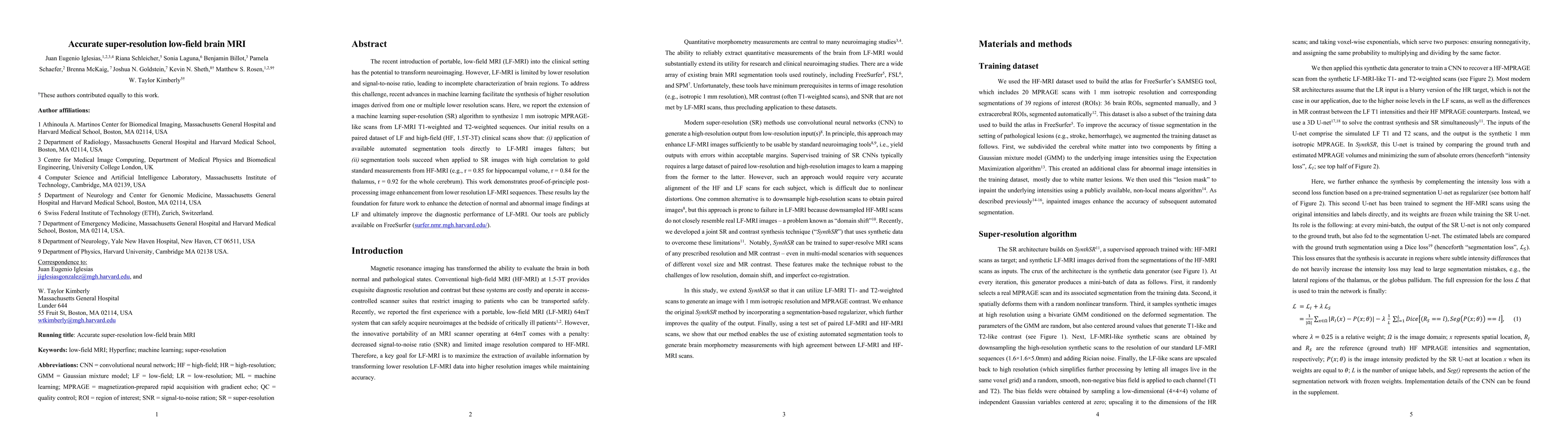
The recent introduction of portable, low-field MRI (LF-MRI) into the clinical setting has the potential to transform neuroimaging. However, LF-MRI is limited by lower resolution and signal-to-noise ratio, leading to incomplete characterization of brain regions. To address this challenge, recent advances in machine learning facilitate the synthesis of higher resolution images derived from one or multiple lower resolution scans. Here, we report the extension of a machine learning super-resolution (SR) algorithm to synthesize 1 mm isotropic MPRAGE-like scans from LF-MRI T1-weighted and T2-weighted sequences. Our initial results on a paired dataset of LF and high-field (HF, 1.5T-3T) clinical scans show that: (i) application of available automated segmentation tools directly to LF-MRI images falters; but (ii) segmentation tools succeed when applied to SR images with high correlation to gold standard measurements from HF-MRI (e.g., r = 0.85 for hippocampal volume, r = 0.84 for the thalamus, r = 0.92 for the whole cerebrum). This work demonstrates proof-of-principle post-processing image enhancement from lower resolution LF-MRI sequences. These results lay the foundation for future work to enhance the detection of normal and abnormal image findings at LF and ultimately improve the diagnostic performance of LF-MRI. Our tools are publicly available on FreeSurfer (surfer.nmr.mgh.harvard.edu/).
AI Key Findings
Get AI-generated insights about this paper's methodology, results, significance, and more — seven facets brought into focus.
Impact
Paper Details
Authors
PDF Preview
Key Terms
Citation Network
Current paper (gray), citations (green), references (blue)
Display is limited for performance on very large graphs.
Discussion 0