Academic Profile
Statistics
Similar Authors
Papers on arXiv
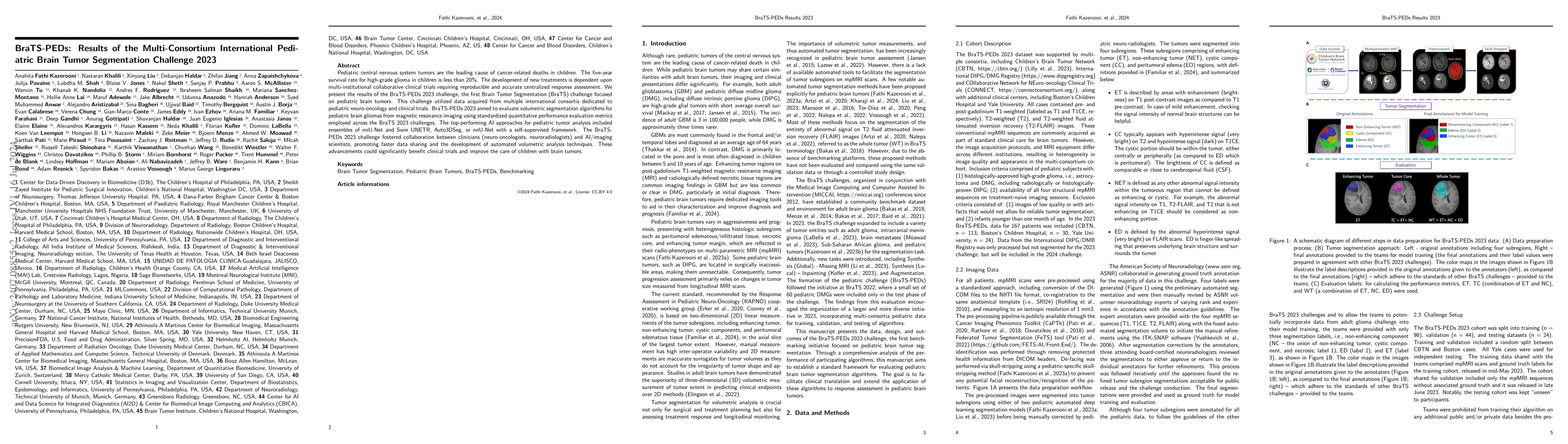
Pediatric central nervous system tumors are the leading cause of cancer-related deaths in children. The five-year survival rate for high-grade glioma in children is less than 20%. The development of n...
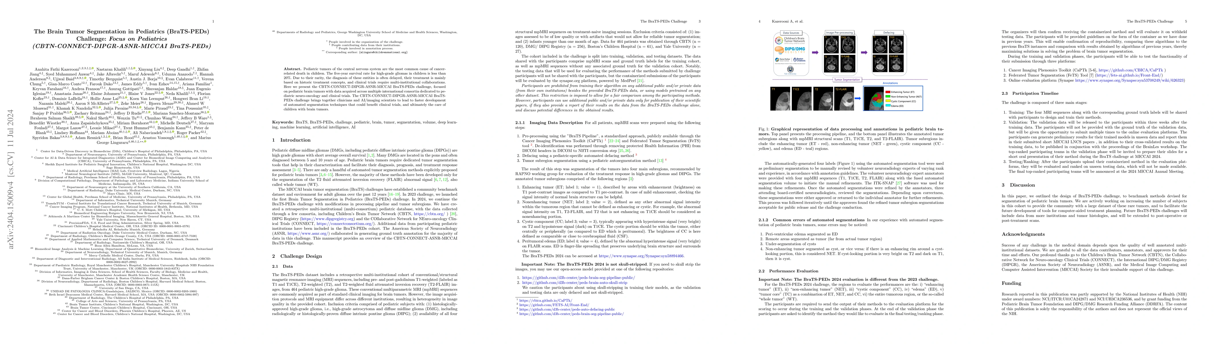
Pediatric tumors of the central nervous system are the most common cause of cancer-related death in children. The five-year survival rate for high-grade gliomas in children is less than 20%. Due to th...
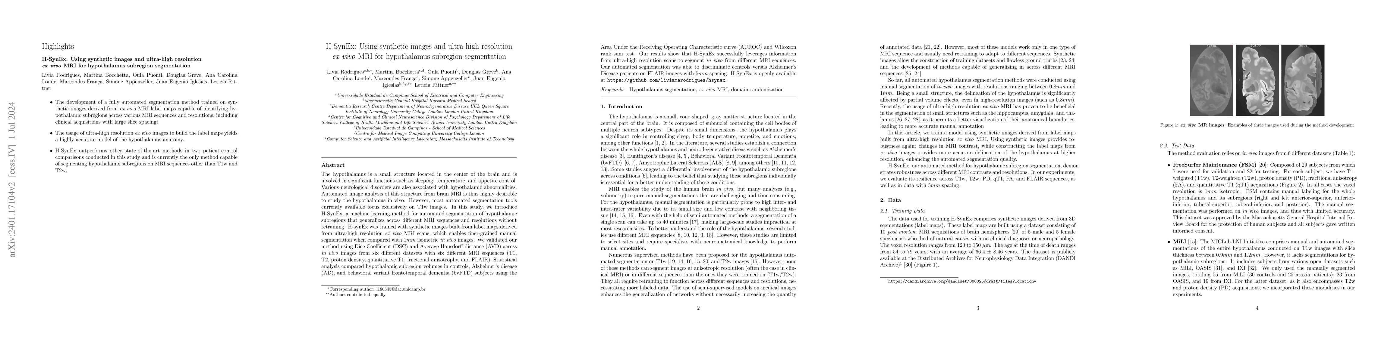
The hypothalamus is a small structure located in the center of the brain and is involved in significant functions such as sleeping, temperature, and appetite control. Various neurological disorders ar...
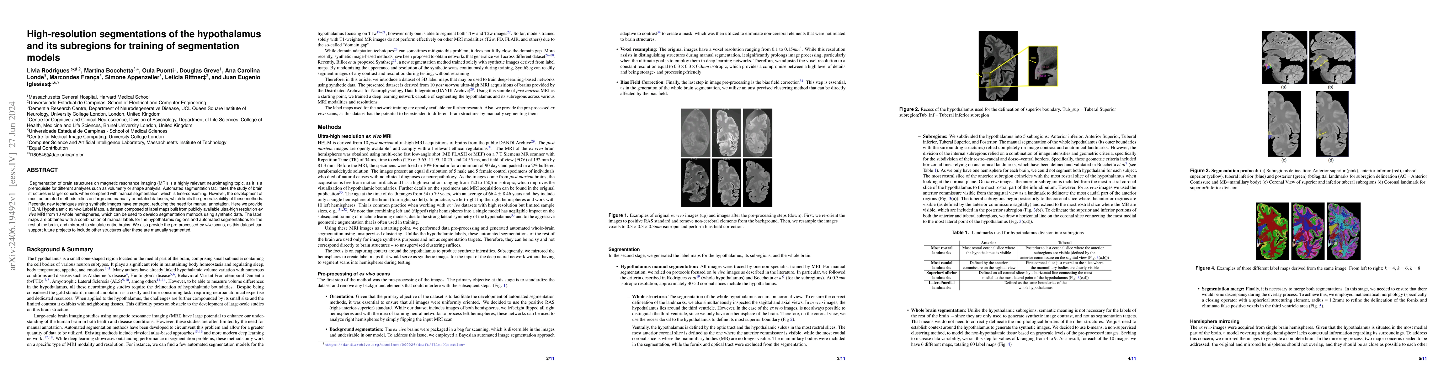
Segmentation of brain structures on magnetic resonance imaging (MRI) is a highly relevant neuroimaging topic, as it is a prerequisite for different analyses such as volumetry or shape analysis. Automa...
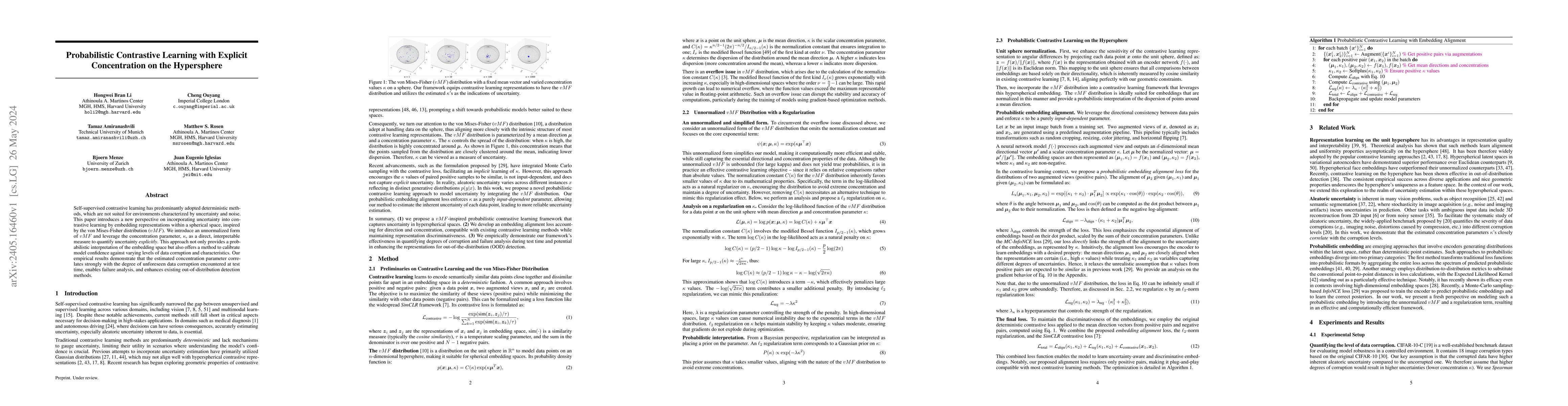
Self-supervised contrastive learning has predominantly adopted deterministic methods, which are not suited for environments characterized by uncertainty and noise. This paper introduces a new perspe...
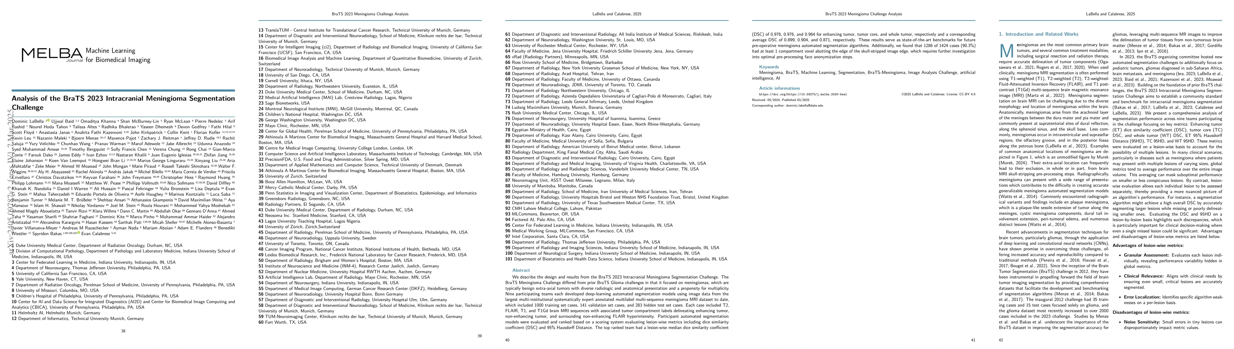
We describe the design and results from the BraTS 2023 Intracranial Meningioma Segmentation Challenge. The BraTS Meningioma Challenge differed from prior BraTS Glioma challenges in that it focused o...
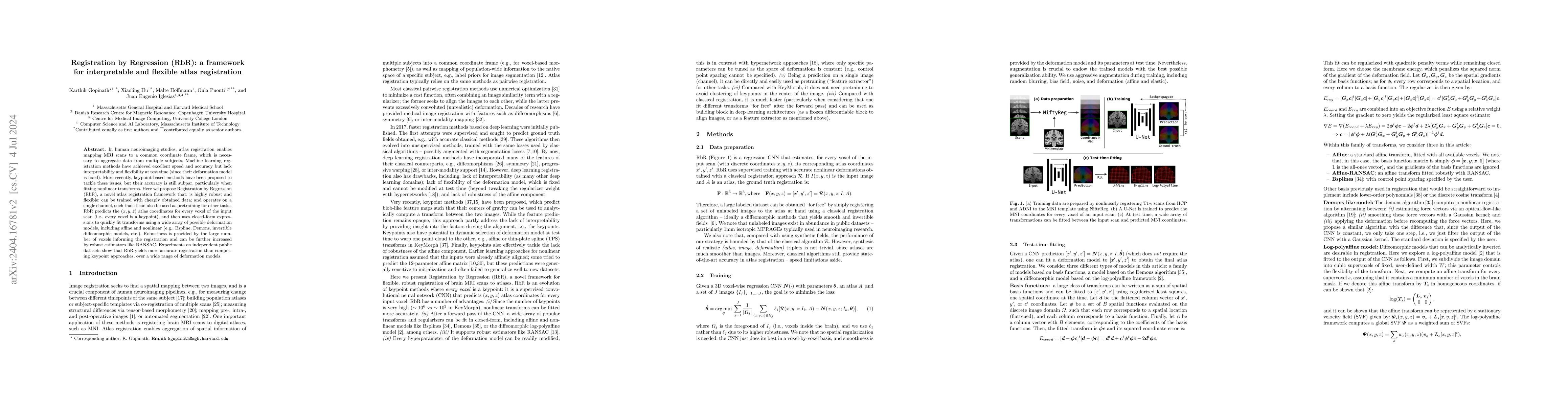
In human neuroimaging studies, atlas registration enables mapping MRI scans to a common coordinate frame, which is necessary to aggregate data from multiple subjects. Machine learning registration m...
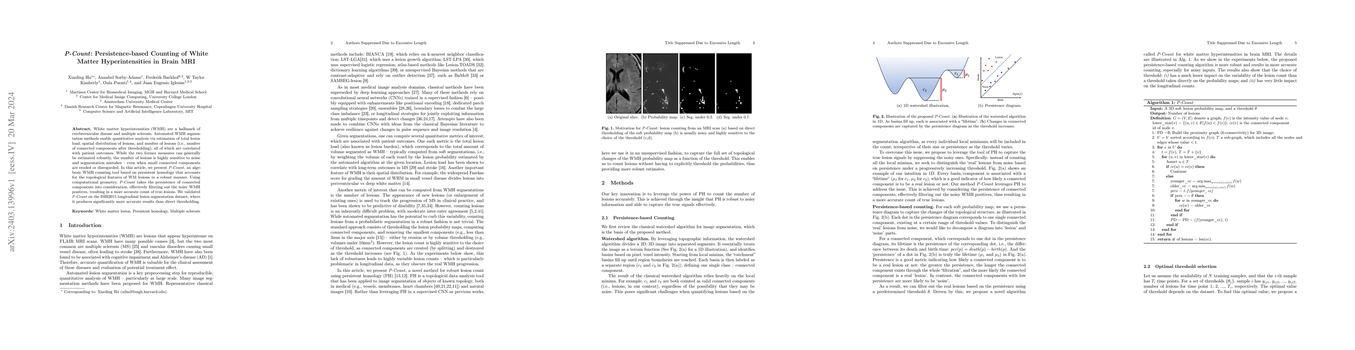
White matter hyperintensities (WMH) are a hallmark of cerebrovascular disease and multiple sclerosis. Automated WMH segmentation methods enable quantitative analysis via estimation of total lesion l...
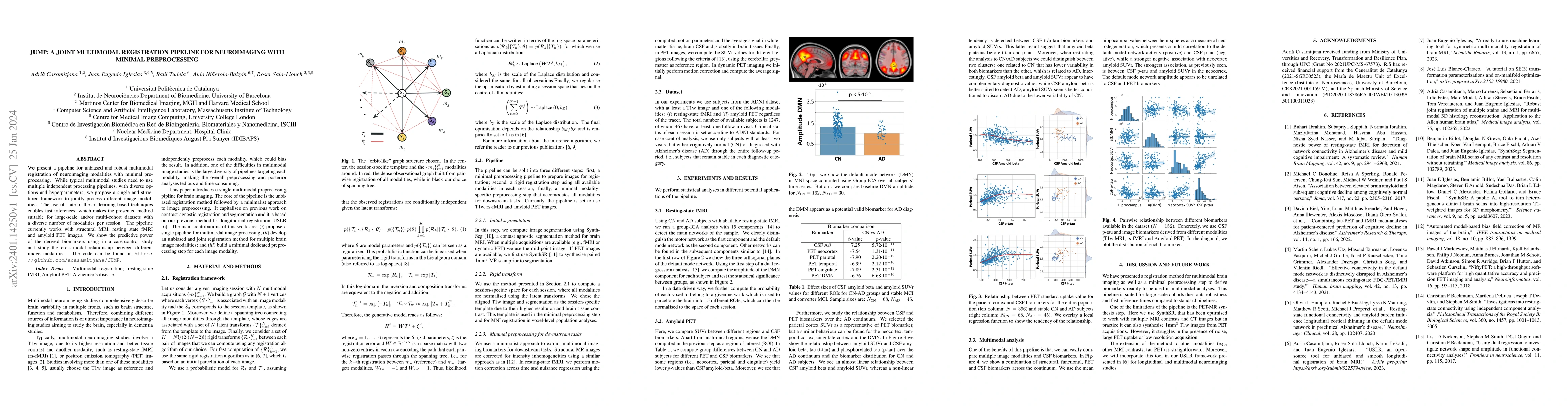
We present a pipeline for unbiased and robust multimodal registration of neuroimaging modalities with minimal pre-processing. While typical multimodal studies need to use multiple independent proces...
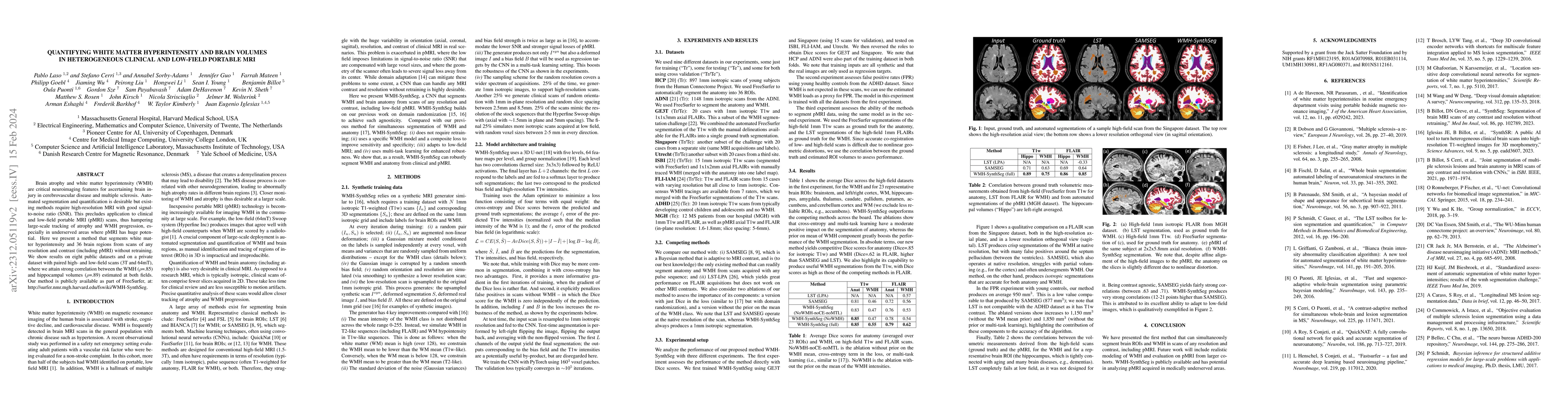
Brain atrophy and white matter hyperintensity (WMH) are critical neuroimaging features for ascertaining brain injury in cerebrovascular disease and multiple sclerosis. Automated segmentation and qua...
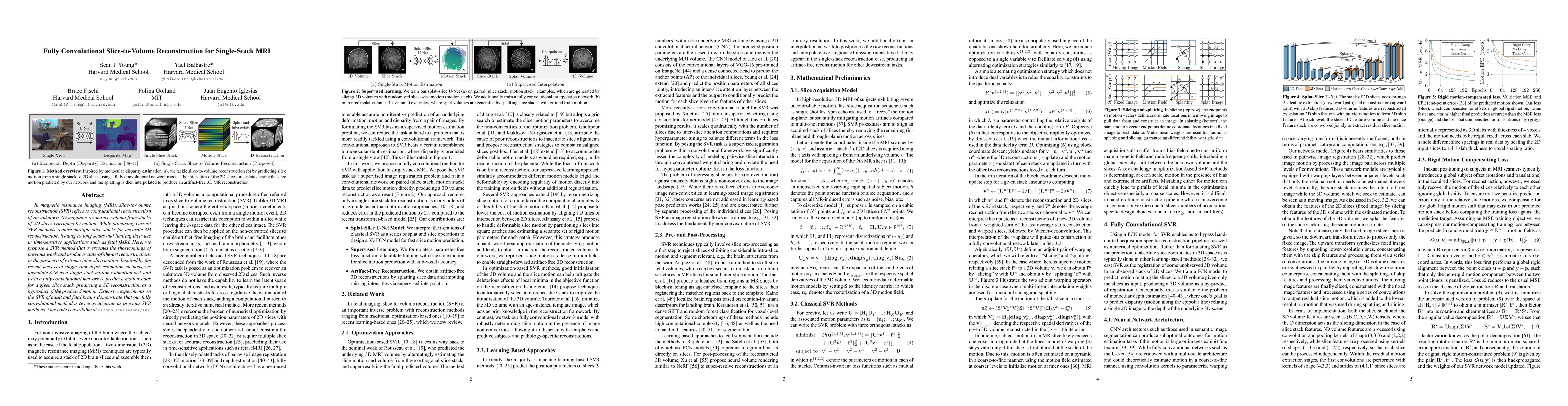
In magnetic resonance imaging (MRI), slice-to-volume reconstruction (SVR) refers to computational reconstruction of an unknown 3D magnetic resonance volume from stacks of 2D slices corrupted by moti...
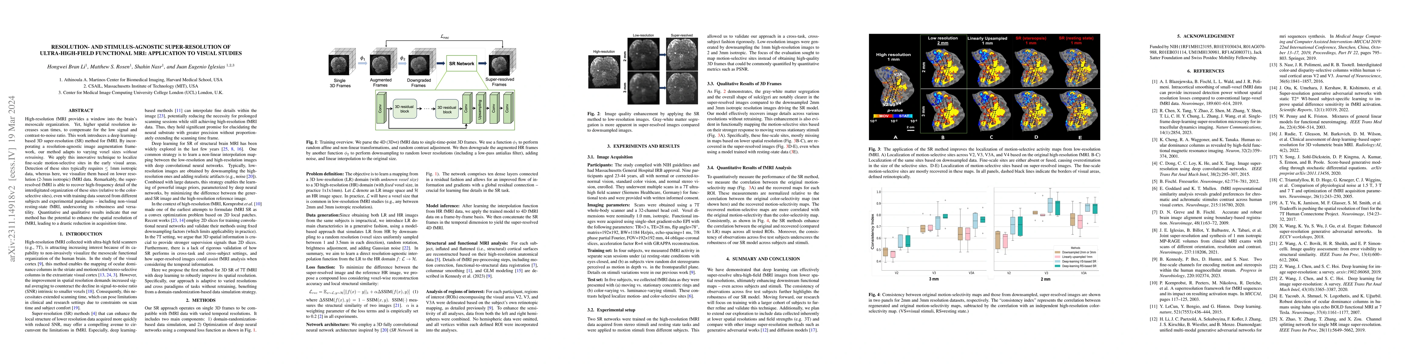
High-resolution fMRI provides a window into the brain's mesoscale organization. Yet, higher spatial resolution increases scan times, to compensate for the low signal and contrast-to-noise ratio. Thi...
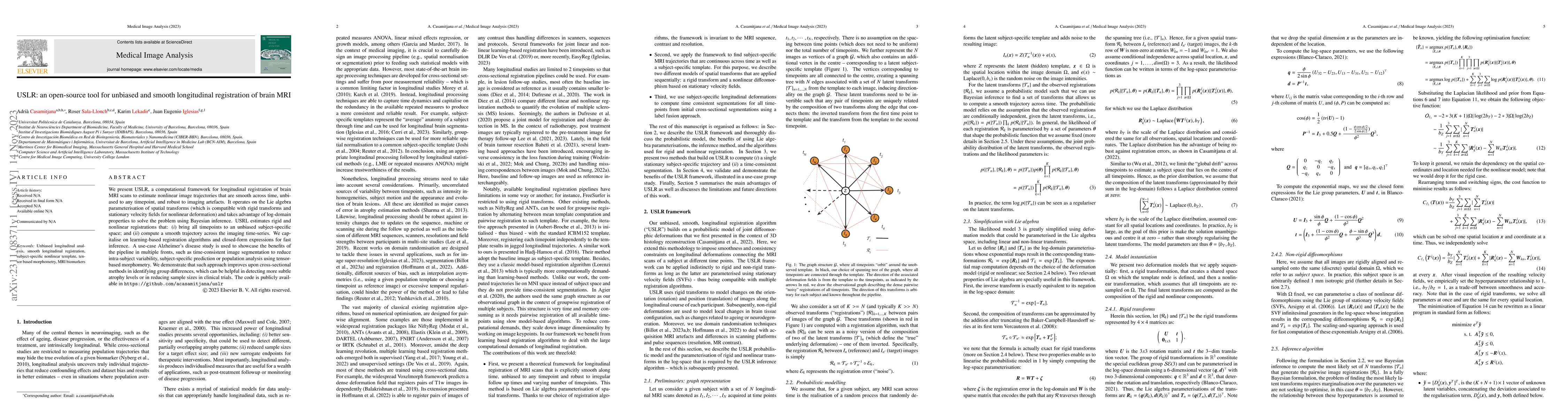
We present USLR, a computational framework for longitudinal registration of brain MRI scans to estimate nonlinear image trajectories that are smooth across time, unbiased to any timepoint, and robus...
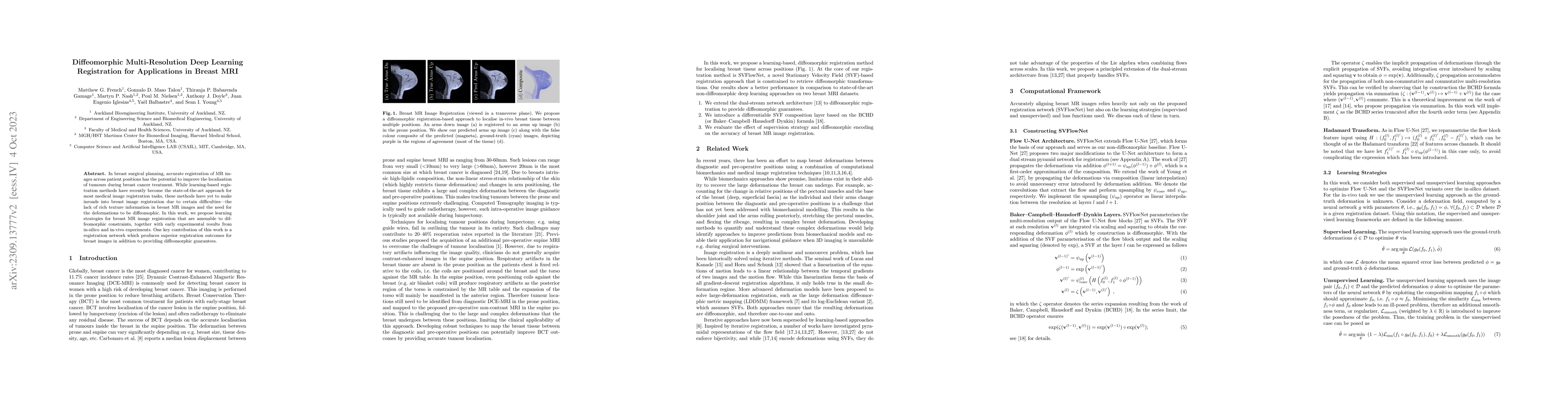
In breast surgical planning, accurate registration of MR images across patient positions has the potential to improve the localisation of tumours during breast cancer treatment. While learning-based...
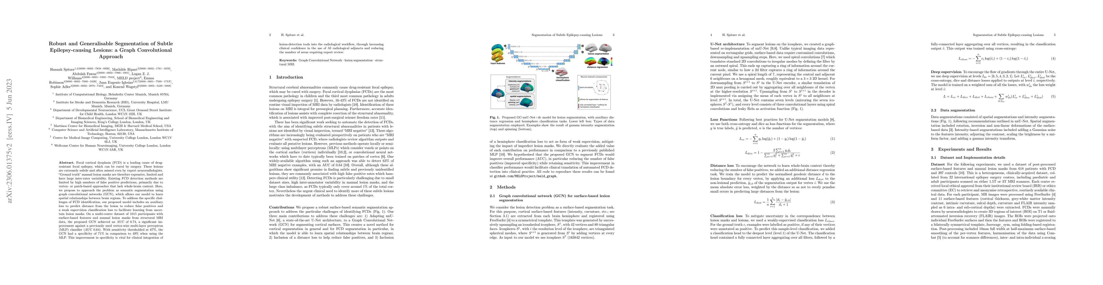
Focal cortical dysplasia (FCD) is a leading cause of drug-resistant focal epilepsy, which can be cured by surgery. These lesions are extremely subtle and often missed even by expert neuroradiologist...
The translation of AI-generated brain metastases (BM) segmentation into clinical practice relies heavily on diverse, high-quality annotated medical imaging datasets. The BraTS-METS 2023 challenge ha...
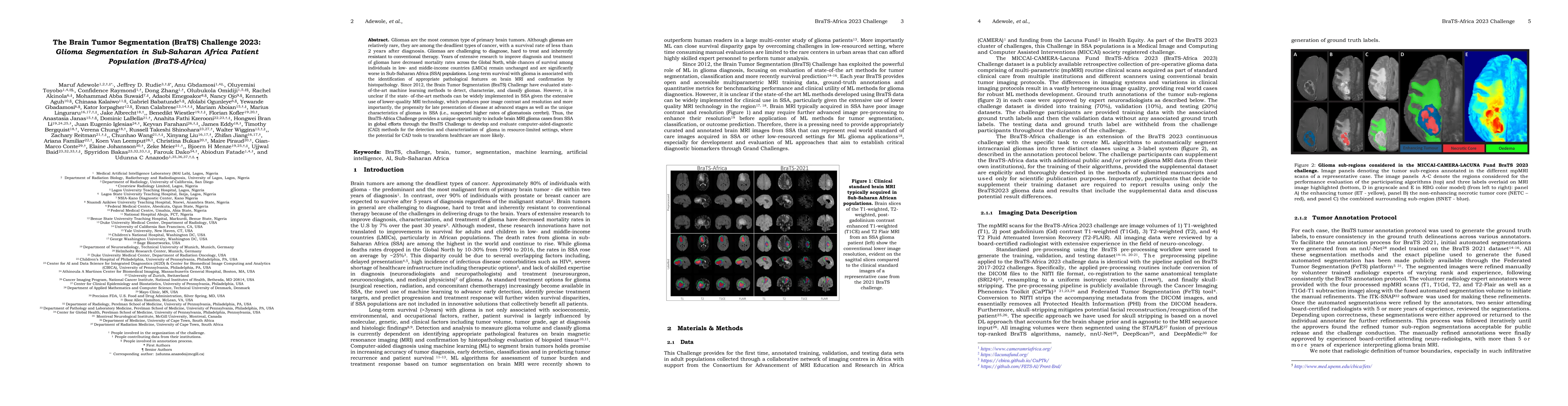
Gliomas are the most common type of primary brain tumors. Although gliomas are relatively rare, they are among the deadliest types of cancer, with a survival rate of less than 2 years after diagnosi...
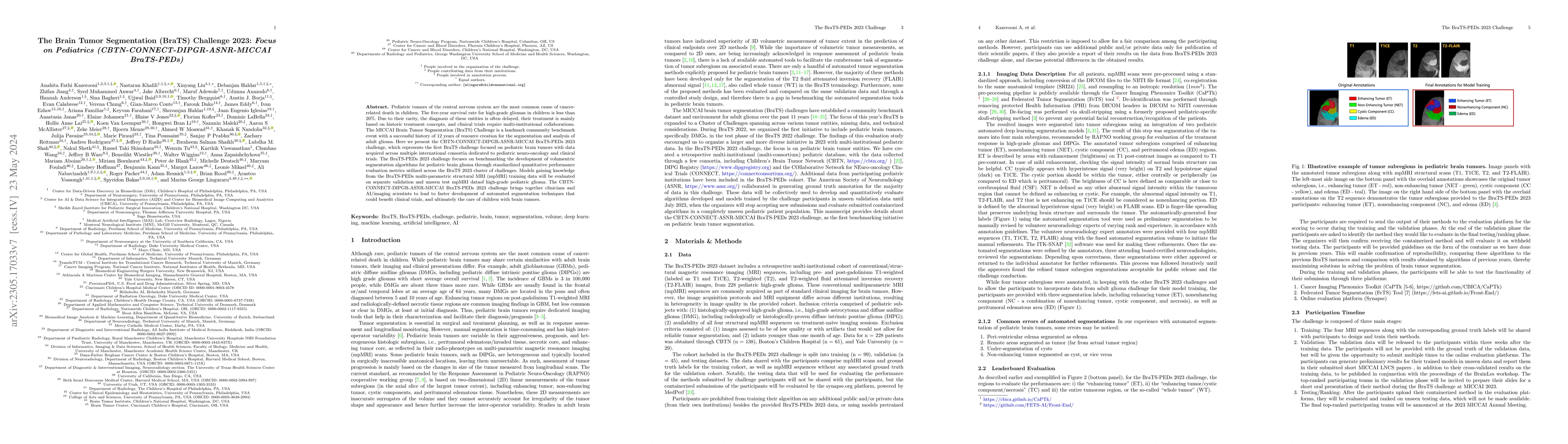
Pediatric tumors of the central nervous system are the most common cause of cancer-related death in children. The five-year survival rate for high-grade gliomas in children is less than 20\%. Due to...
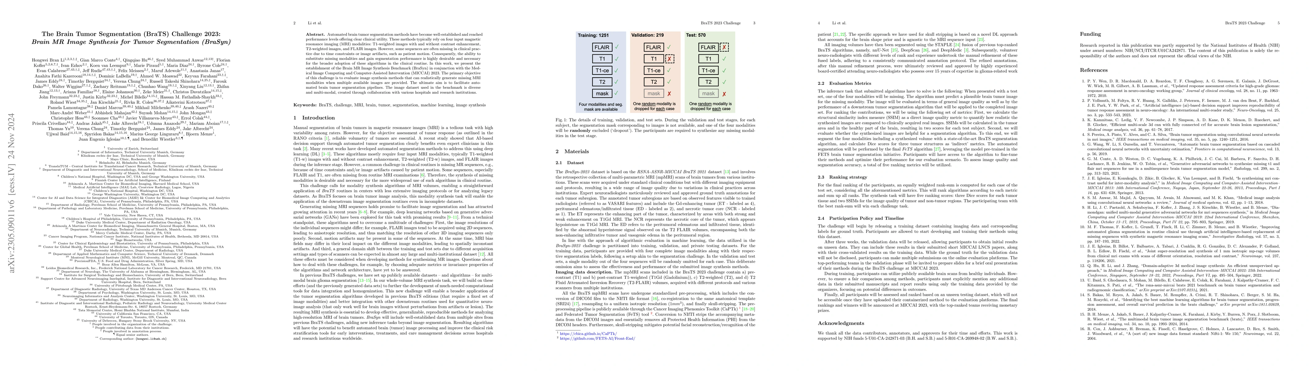
Automated brain tumor segmentation methods have become well-established and reached performance levels offering clear clinical utility. These methods typically rely on four input magnetic resonance ...
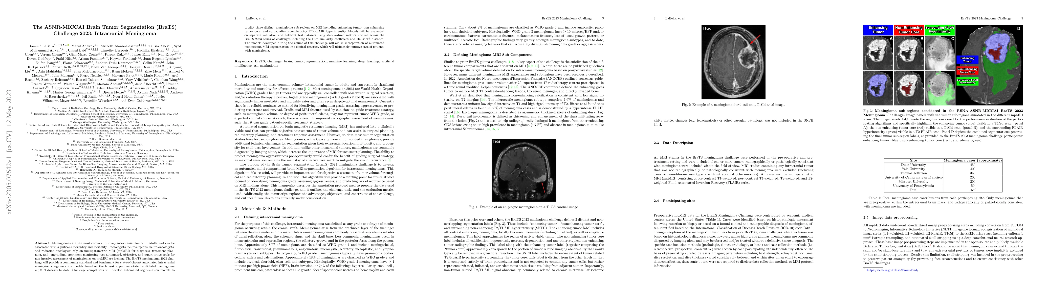
Meningiomas are the most common primary intracranial tumor in adults and can be associated with significant morbidity and mortality. Radiologists, neurosurgeons, neuro-oncologists, and radiation onc...
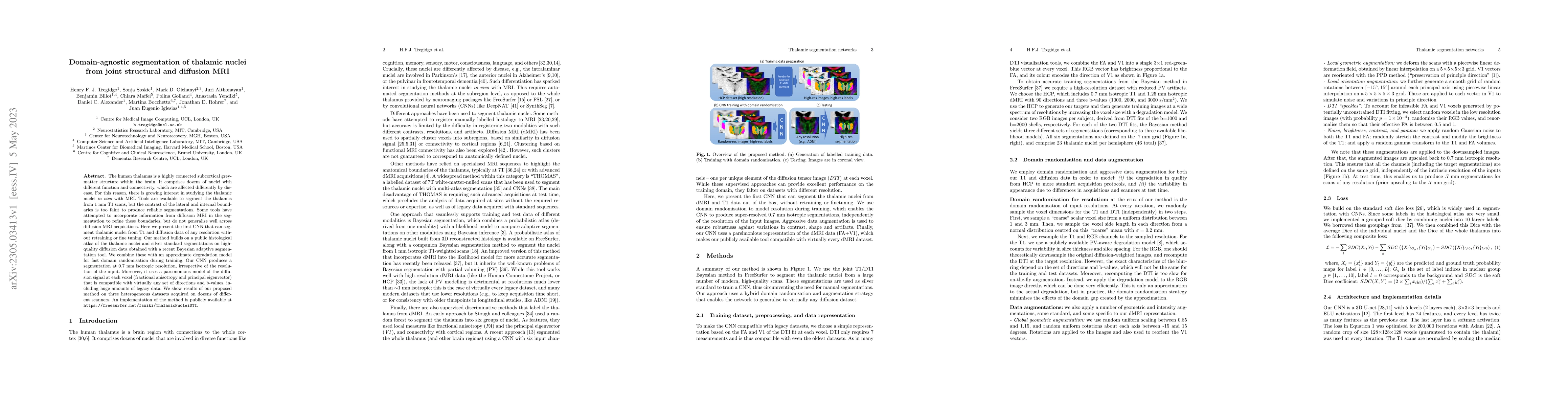
The human thalamus is a highly connected subcortical grey-matter structure within the brain. It comprises dozens of nuclei with different function and connectivity, which are affected differently by...
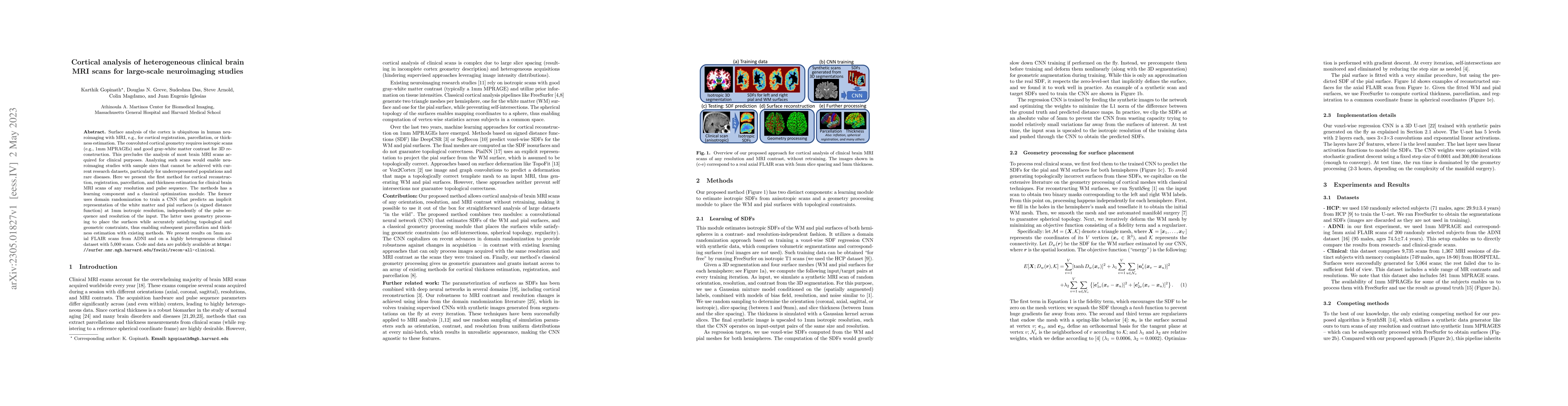
Surface analysis of the cortex is ubiquitous in human neuroimaging with MRI, e.g., for cortical registration, parcellation, or thickness estimation. The convoluted cortical geometry requires isotrop...
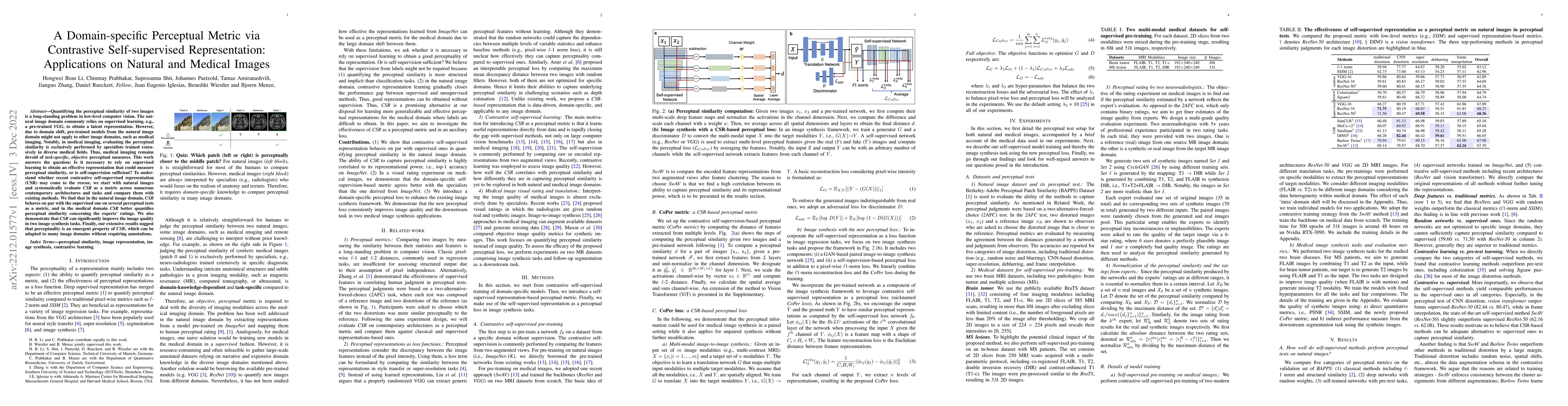
Quantifying the perceptual similarity of two images is a long-standing problem in low-level computer vision. The natural image domain commonly relies on supervised learning, e.g., a pre-trained VGG,...
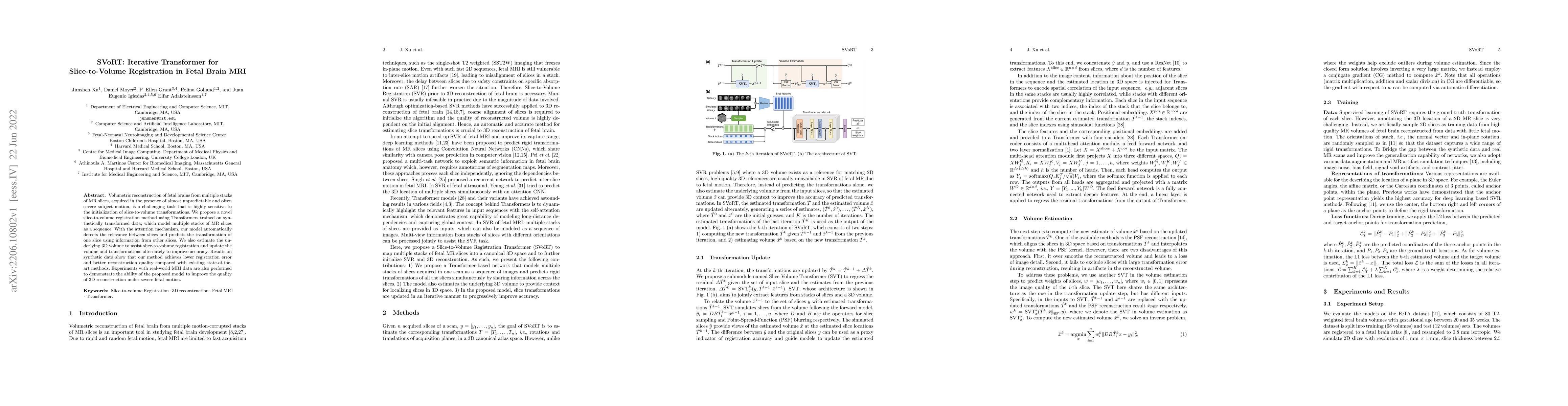
Volumetric reconstruction of fetal brains from multiple stacks of MR slices, acquired in the presence of almost unpredictable and often severe subject motion, is a challenging task that is highly se...
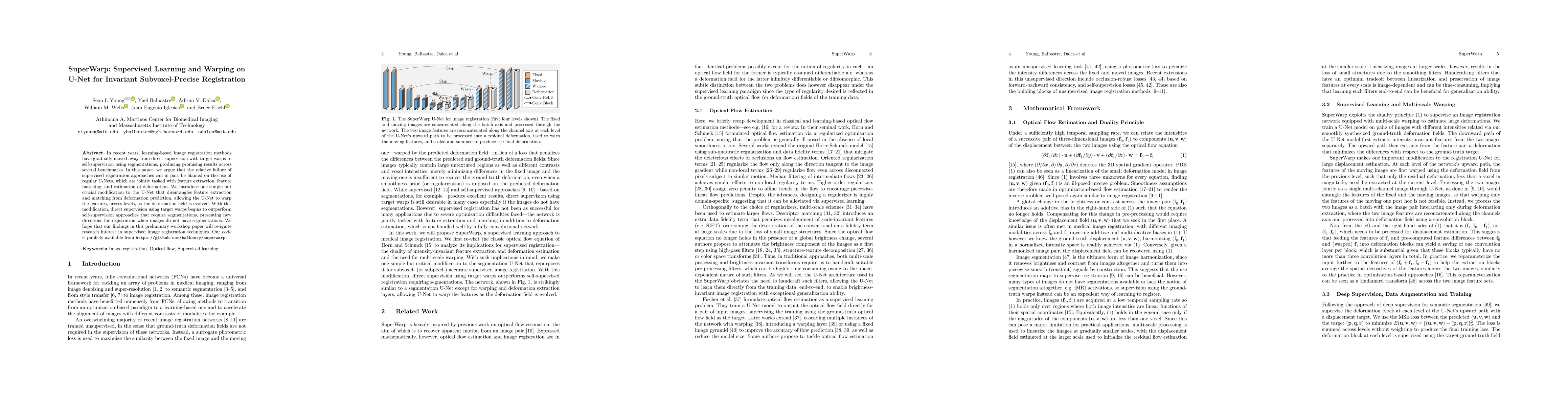
In recent years, learning-based image registration methods have gradually moved away from direct supervision with target warps to instead use self-supervision, with excellent results in several regi...
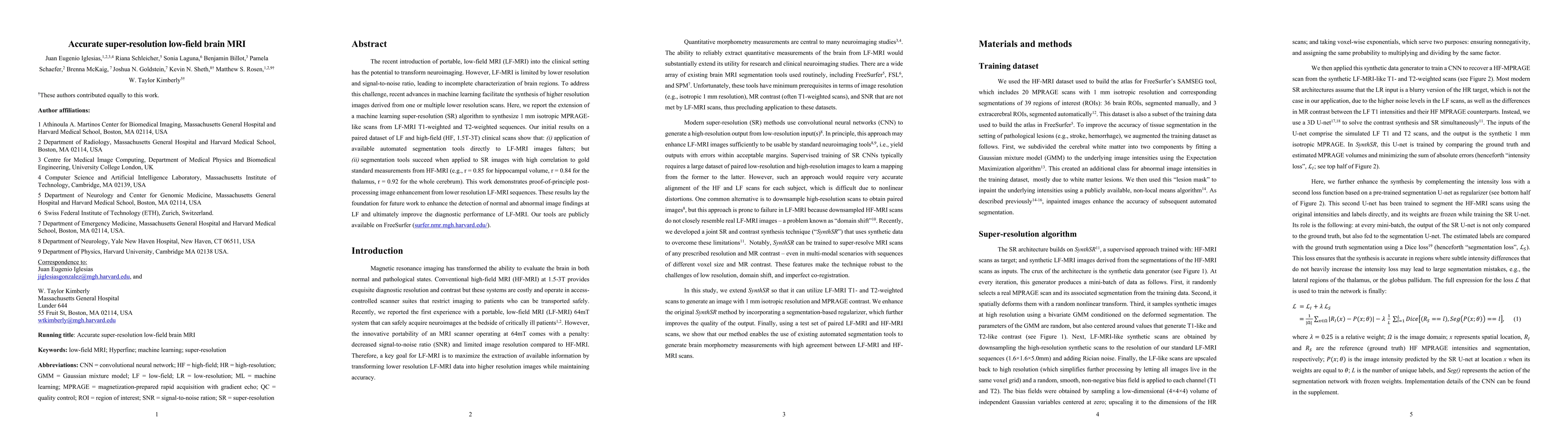
The recent introduction of portable, low-field MRI (LF-MRI) into the clinical setting has the potential to transform neuroimaging. However, LF-MRI is limited by lower resolution and signal-to-noise ...
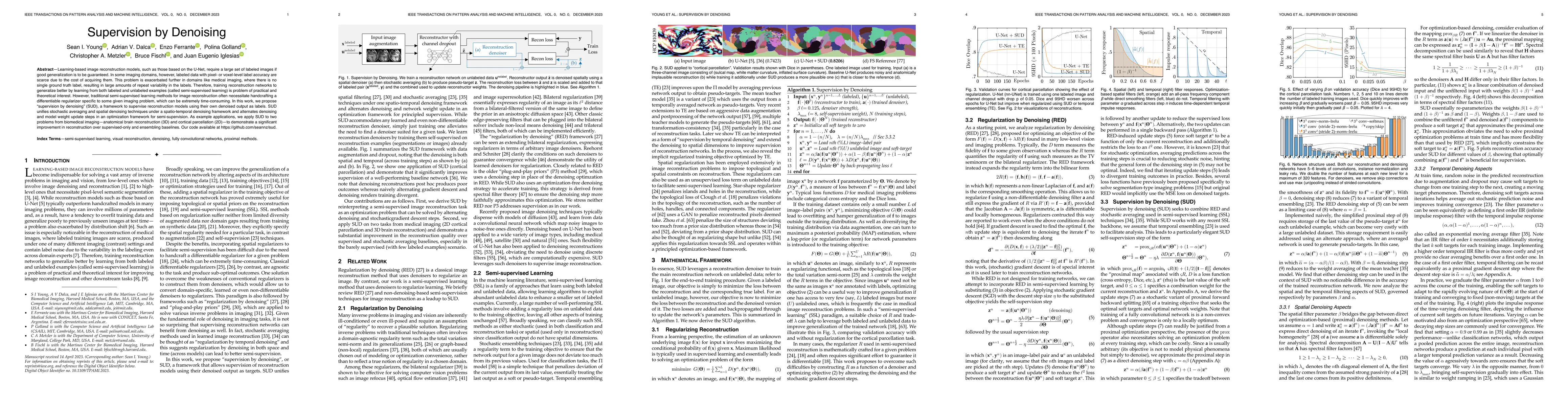
Learning-based image reconstruction models, such as those based on the U-Net, require a large set of labeled images if good generalization is to be guaranteed. In some imaging domains, however, labe...
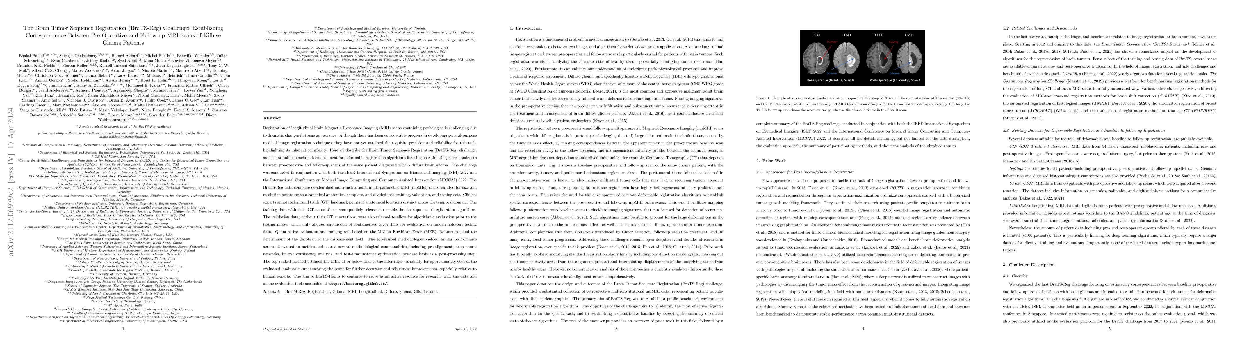
Registration of longitudinal brain MRI scans containing pathologies is challenging due to dramatic changes in tissue appearance. Although there has been progress in developing general-purpose medica...
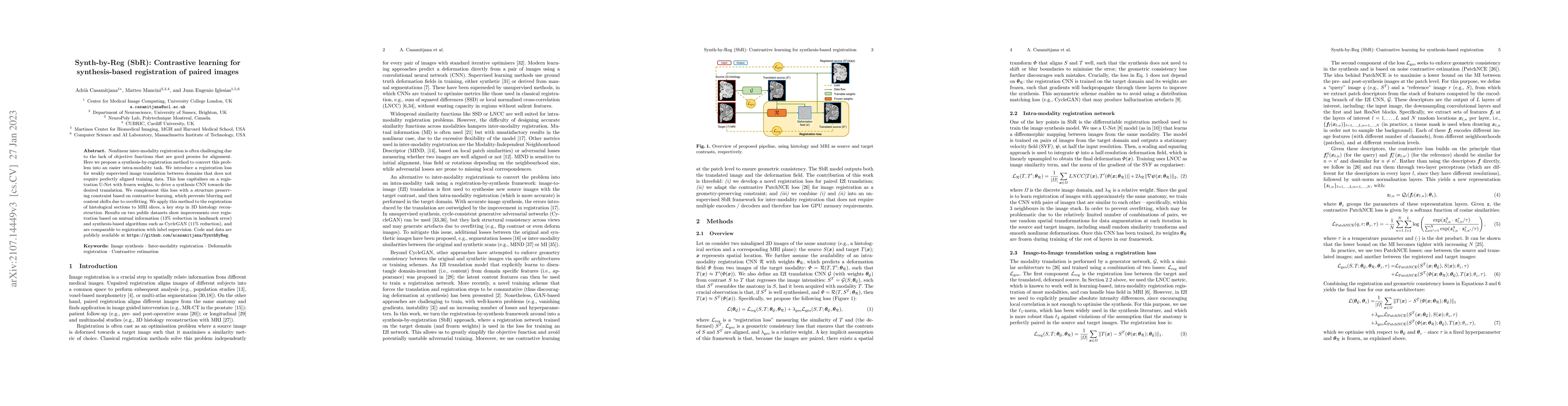
Nonlinear inter-modality registration is often challenging due to the lack of objective functions that are good proxies for alignment. Here we propose a synthesis-by-registration method to convert t...
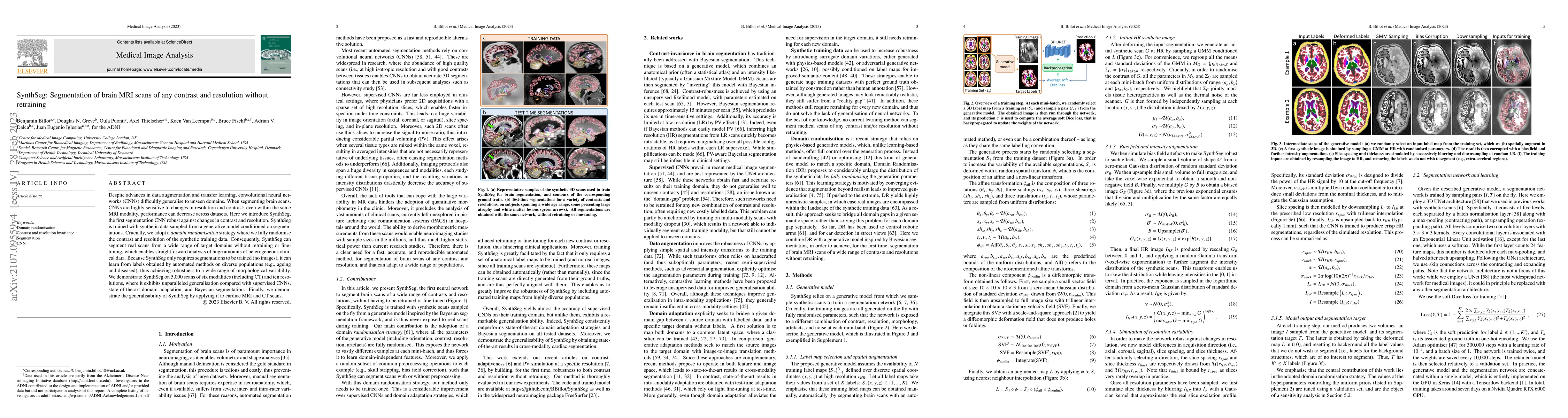
Despite advances in data augmentation and transfer learning, convolutional neural networks (CNNs) difficultly generalise to unseen domains. When segmenting brain scans, CNNs are highly sensitive to ...
Short inversion time inversion recovery (STIR) MRI is widely used in clinical practice to identify and quantify inflammation in axial spondyloarthritis. However, assessment of STIR images is limited...
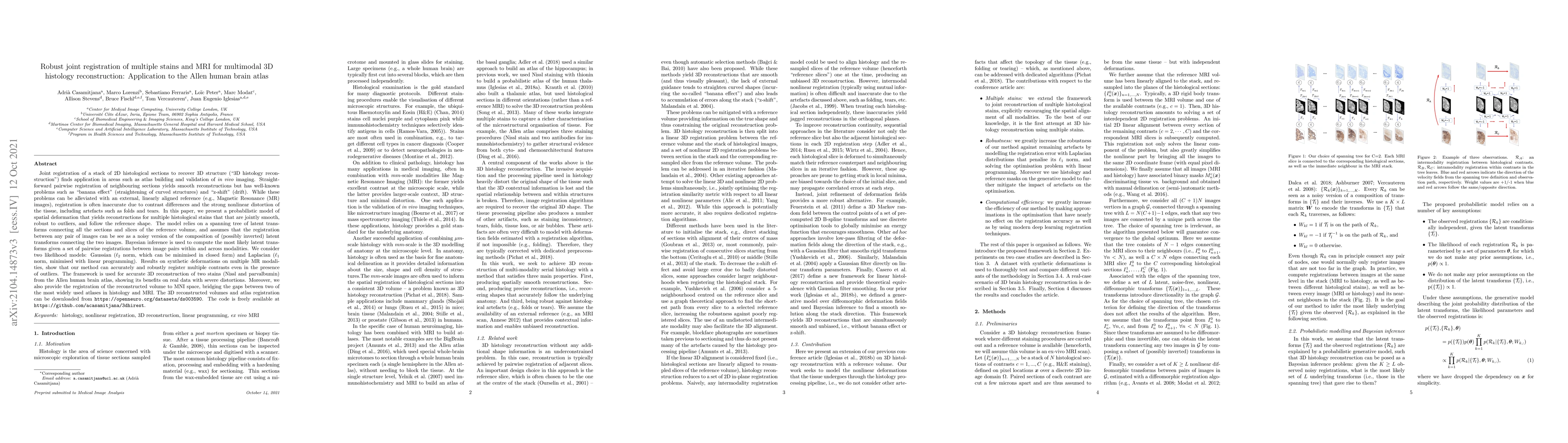
Joint registration of a stack of 2D histological sections to recover 3D structure (``3D histology reconstruction'') finds application in areas such as atlas building and validation of \emph{in vivo}...
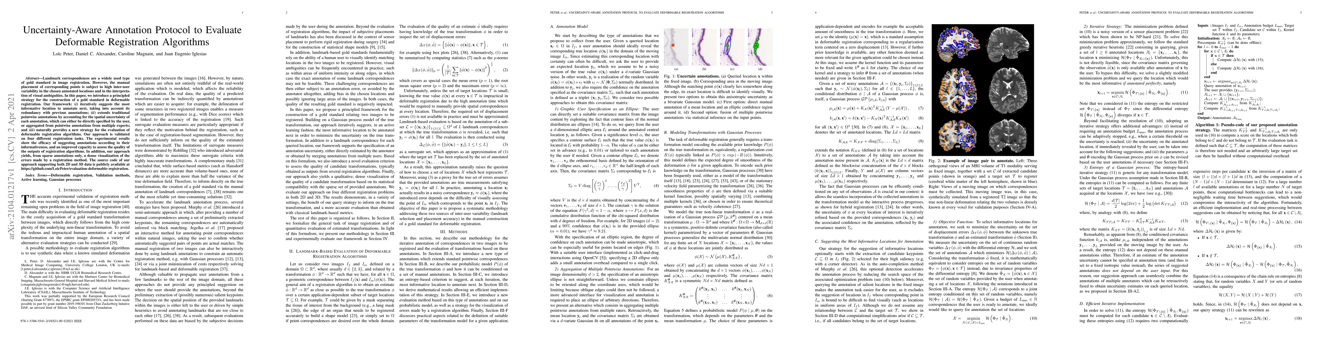
Landmark correspondences are a widely used type of gold standard in image registration. However, the manual placement of corresponding points is subject to high inter-user variability in the chosen ...
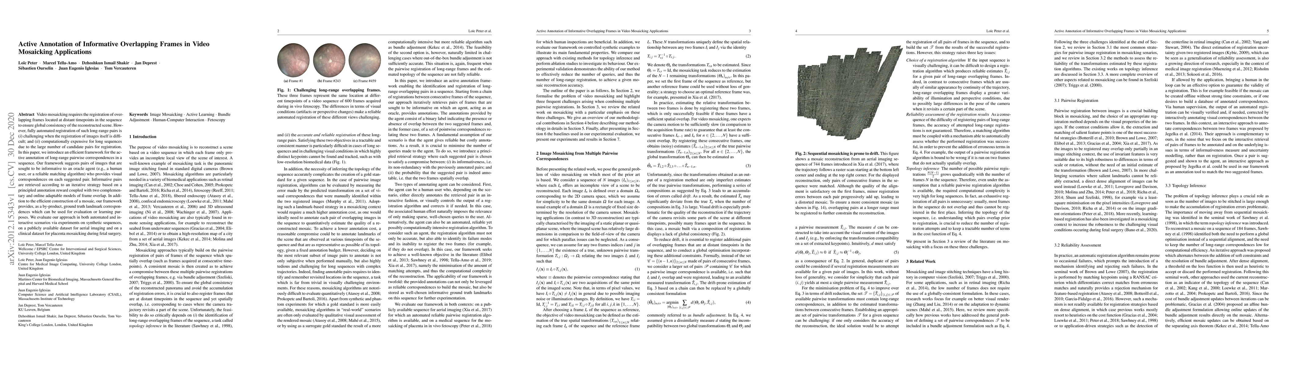
Video mosaicking requires the registration of overlapping frames located at distant timepoints in the sequence to ensure global consistency of the reconstructed scene. However, fully automated regis...
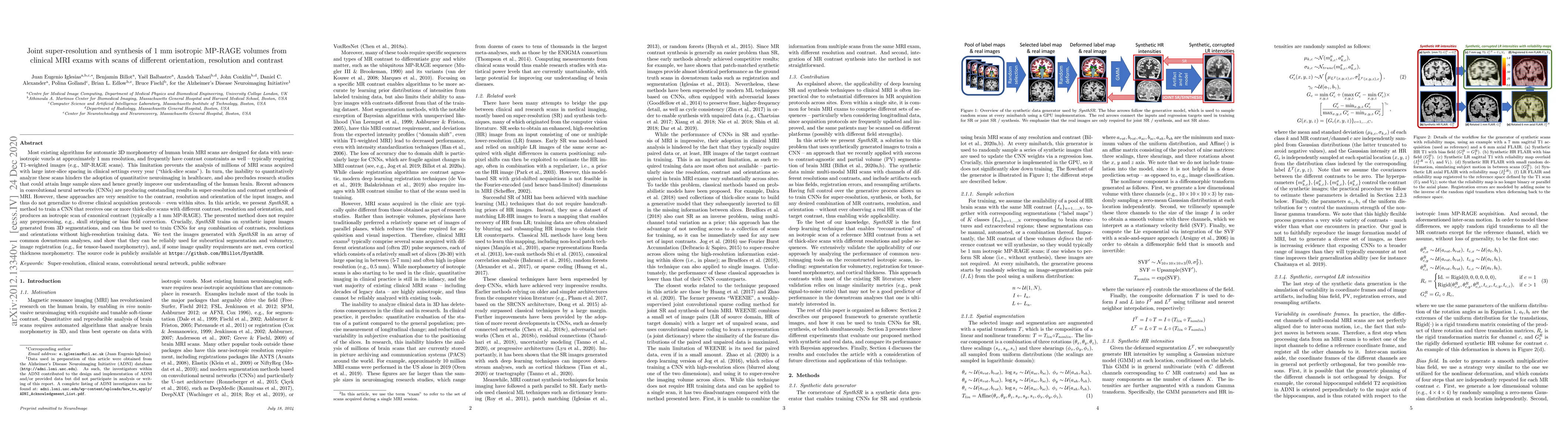
Most existing algorithms for automatic 3D morphometry of human brain MRI scans are designed for data with near-isotropic voxels at approximately 1 mm resolution, and frequently have contrast constra...
DeepReg (https://github.com/DeepRegNet/DeepReg) is a community-supported open-source toolkit for research and education in medical image registration using deep learning.
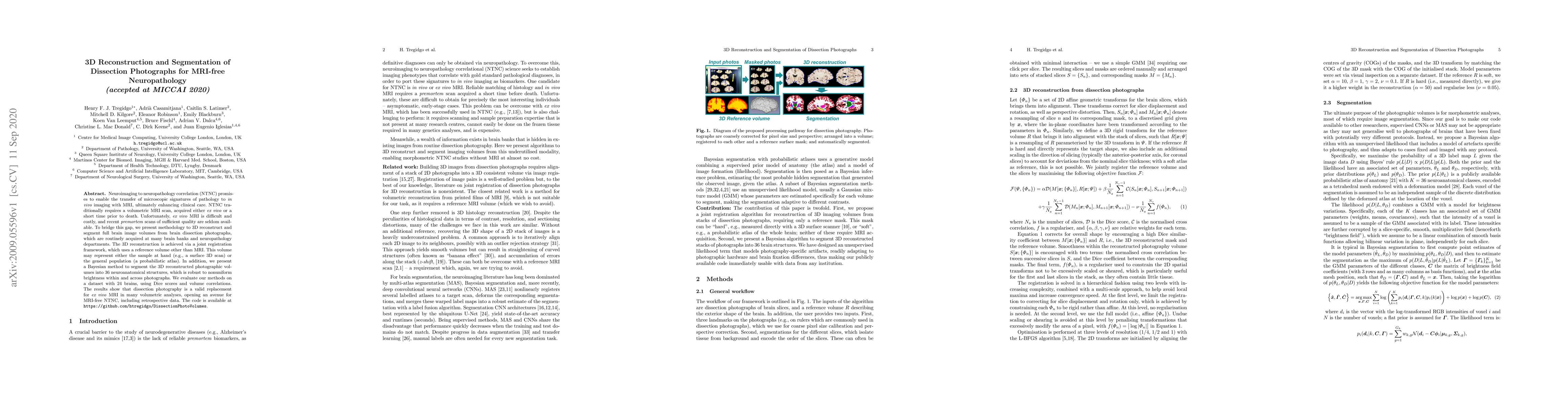
Neuroimaging to neuropathology correlation (NTNC) promises to enable the transfer of microscopic signatures of pathology to in vivo imaging with MRI, ultimately enhancing clinical care. NTNC traditi...
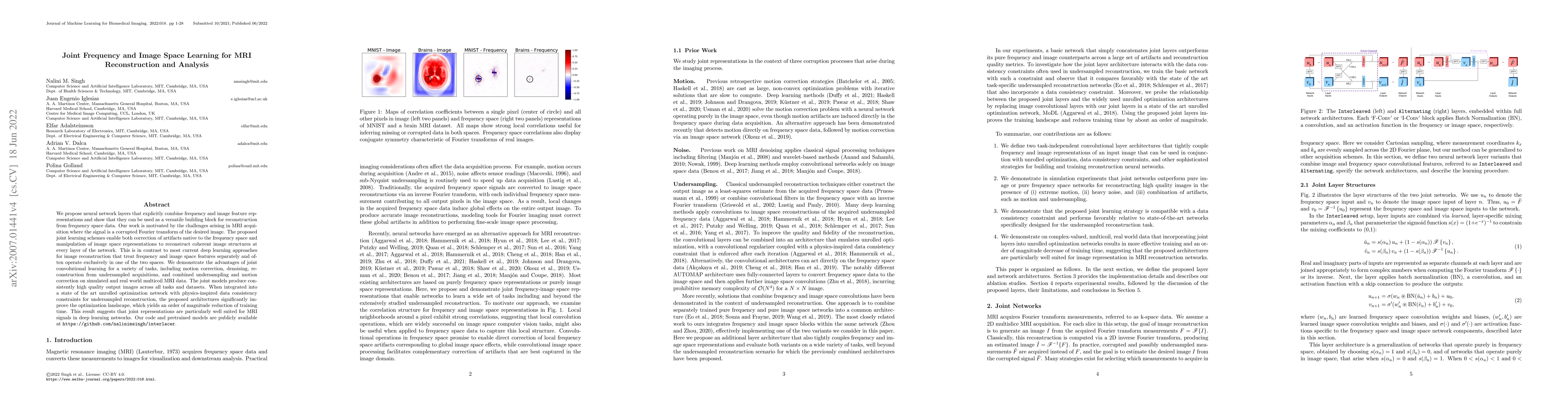
We propose neural network layers that explicitly combine frequency and image feature representations and show that they can be used as a versatile building block for reconstruction from frequency sp...
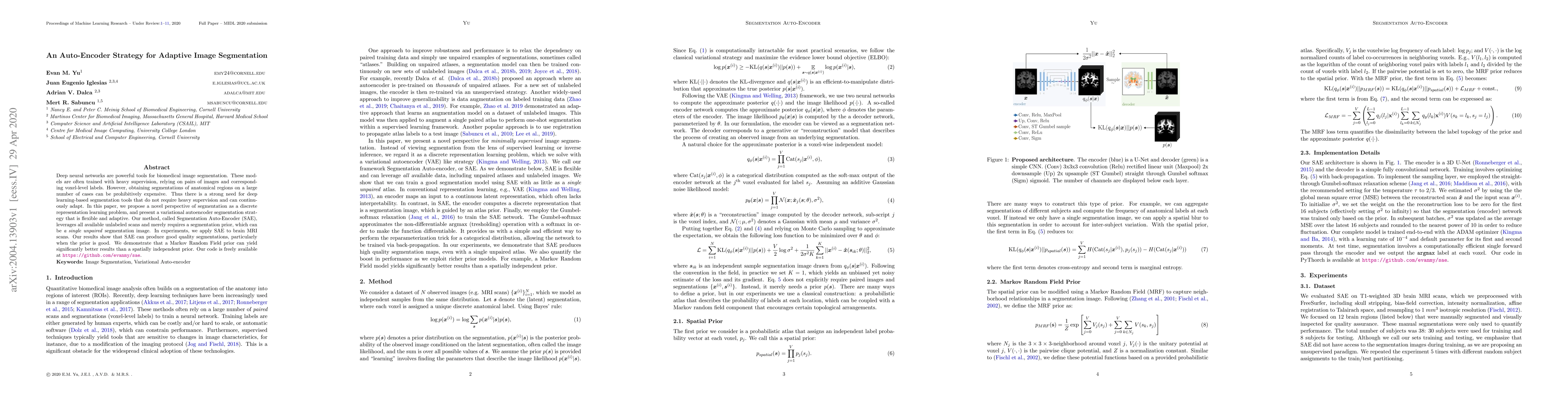
Deep neural networks are powerful tools for biomedical image segmentation. These models are often trained with heavy supervision, relying on pairs of images and corresponding voxel-level labels. How...
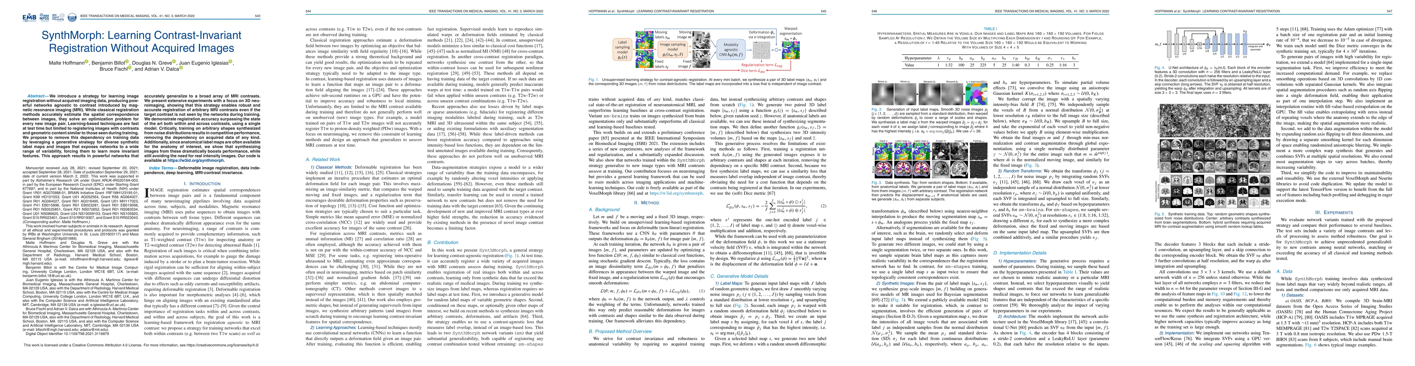
We introduce a strategy for learning image registration without acquired imaging data, producing powerful networks agnostic to contrast introduced by magnetic resonance imaging (MRI). While classica...
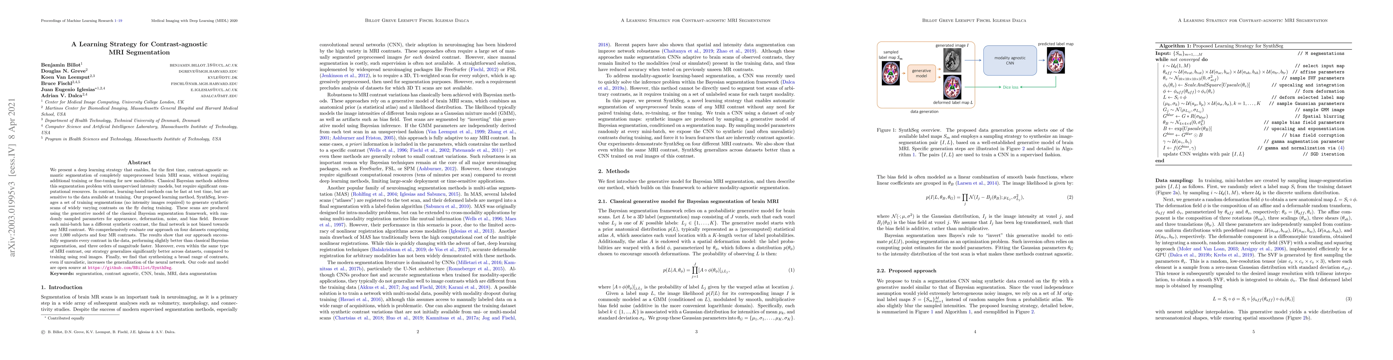
We present a deep learning strategy that enables, for the first time, contrast-agnostic semantic segmentation of completely unpreprocessed brain MRI scans, without requiring additional training or f...
The development of automated tools for brain morphometric analysis in infants has lagged significantly behind analogous tools for adults. This gap reflects the greater challenges in this domain due ...
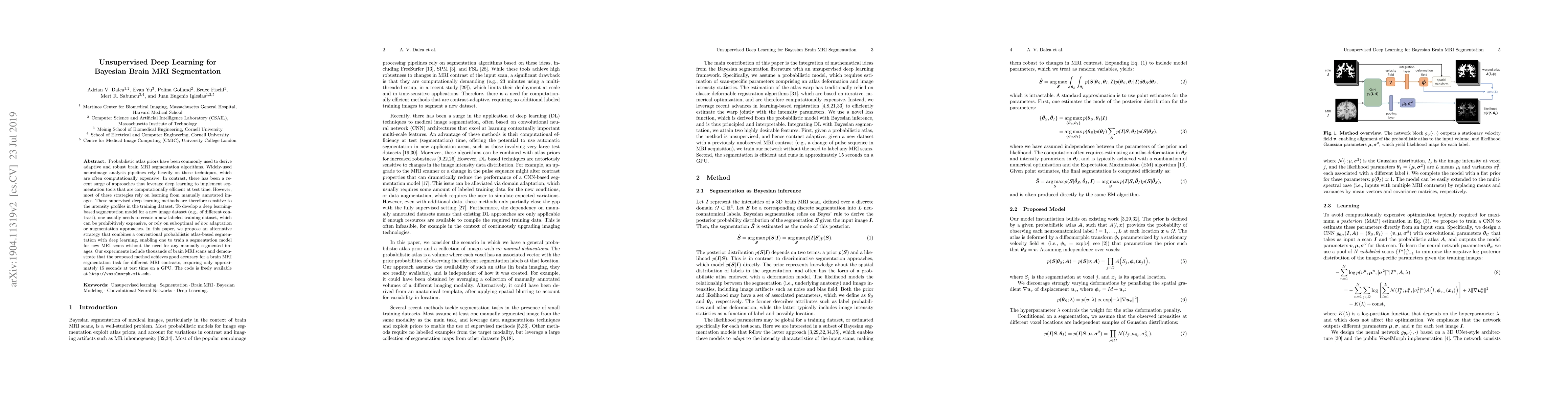
Probabilistic atlas priors have been commonly used to derive adaptive and robust brain MRI segmentation algorithms. Widely-used neuroimage analysis pipelines rely heavily on these techniques, which ...
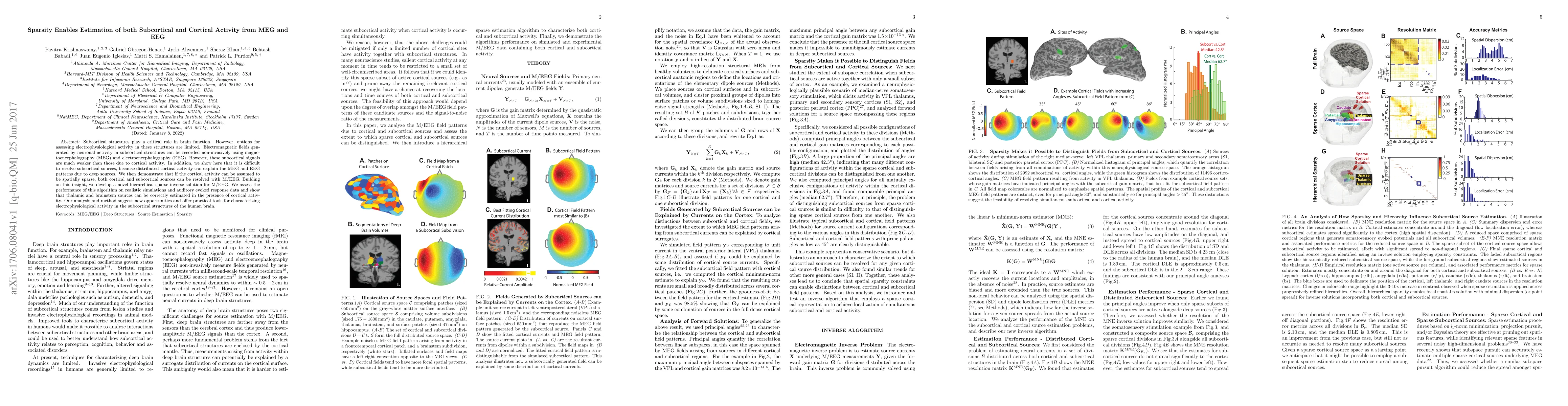
Subcortical structures play a critical role in brain function. However, options for assessing electrophysiological activity in these structures are limited. Electromagnetic fields generated by neuro...
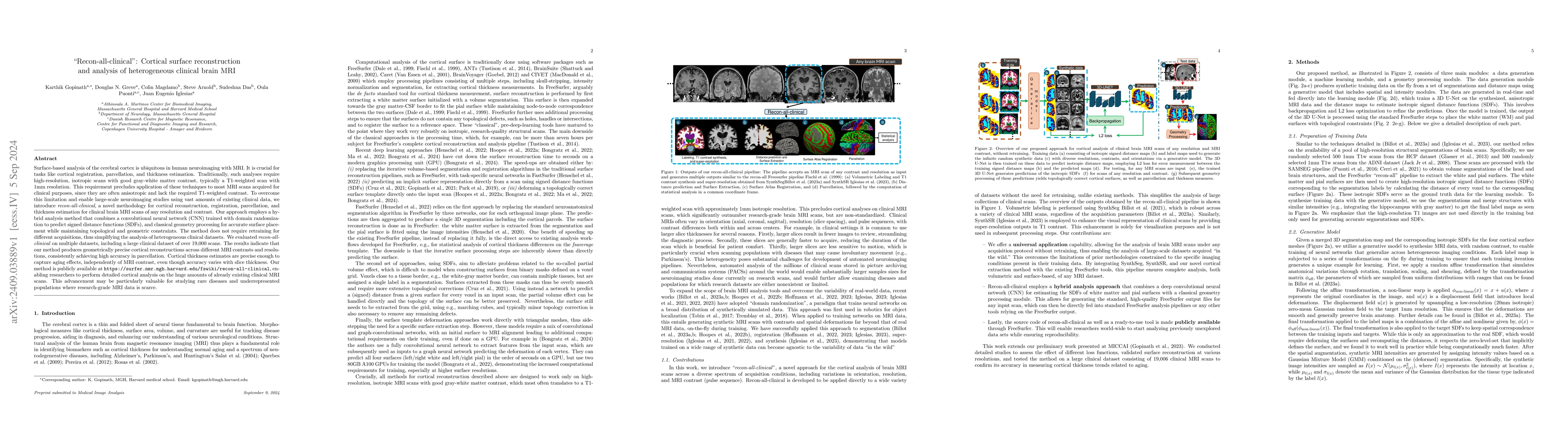
Surface-based analysis of the cerebral cortex is ubiquitous in human neuroimaging with MRI. It is crucial for cortical registration, parcellation, and thickness estimation. Traditionally, these analys...
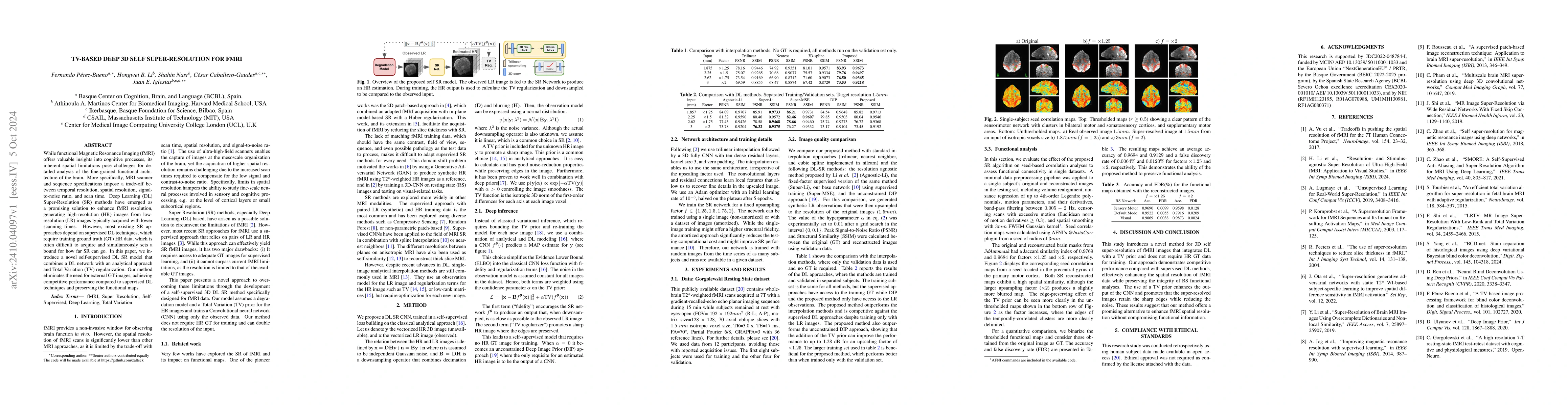
While functional Magnetic Resonance Imaging (fMRI) offers valuable insights into cognitive processes, its inherent spatial limitations pose challenges for detailed analysis of the fine-grained functio...
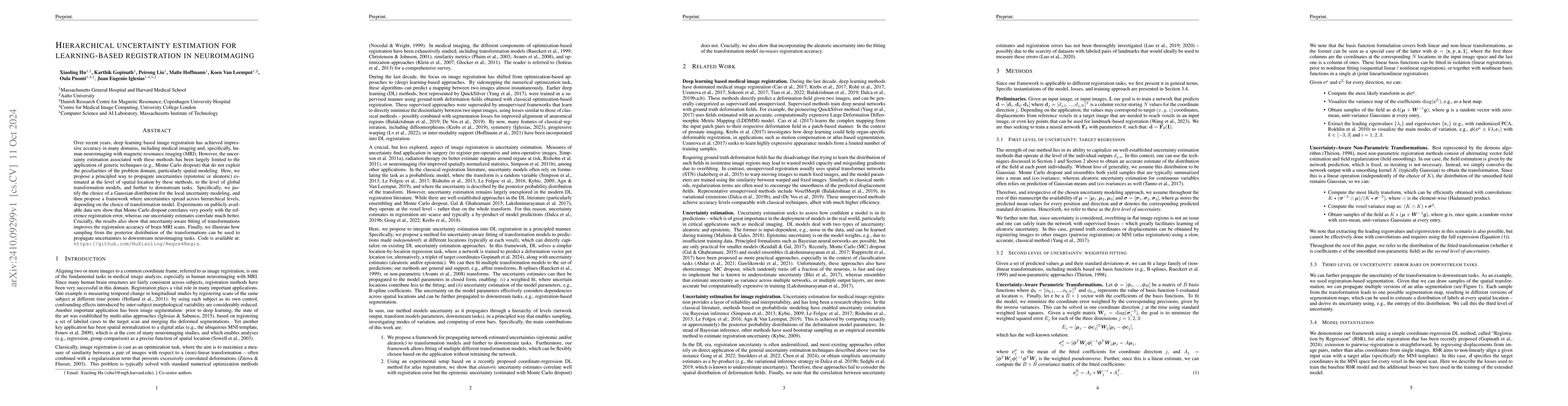
Over recent years, deep learning based image registration has achieved impressive accuracy in many domains, including medical imaging and, specifically, human neuroimaging with magnetic resonance imag...
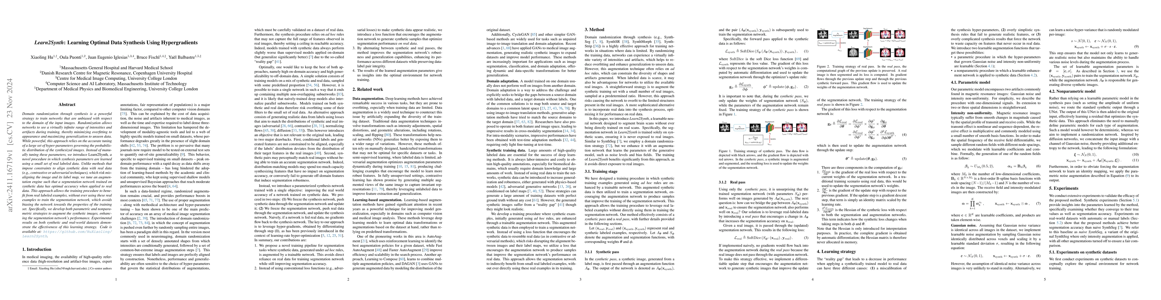
Domain randomization through synthesis is a powerful strategy to train networks that are unbiased with respect to the domain of the input images. Randomization allows networks to see a virtually infin...
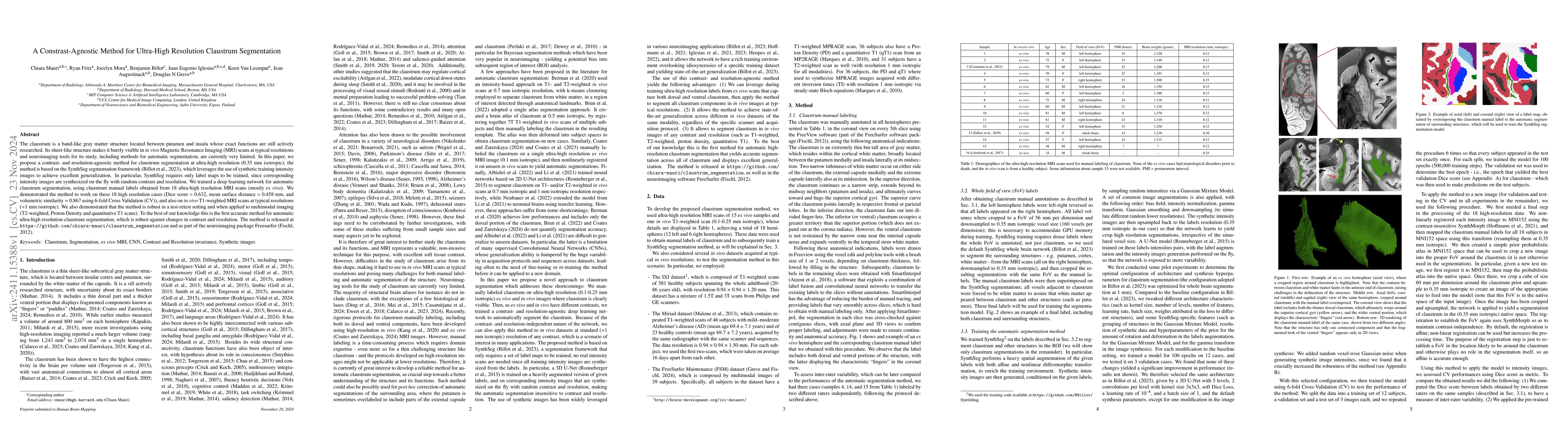
The claustrum is a band-like gray matter structure located between putamen and insula whose exact functions are still actively researched. Its sheet-like structure makes it barely visible in in vivo M...
Correlation of neuropathology with MRI has the potential to transfer microscopic signatures of pathology to invivo scans. Recently, a classical registration method has been proposed, to build these co...
Despite continuous advancements in cancer treatment, brain metastatic disease remains a significant complication of primary cancer and is associated with an unfavorable prognosis. One approach for imp...
Surface-based cortical analysis is valuable for a variety of neuroimaging tasks, such as spatial normalization, parcellation, and gray matter (GM) thickness estimation. However, most tools for estimat...
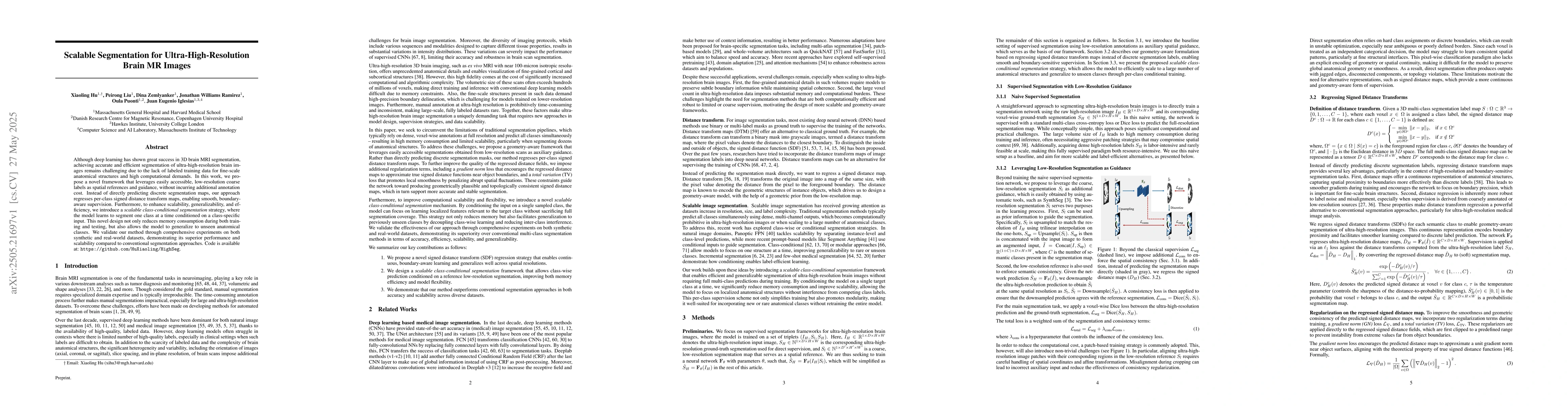
Although deep learning has shown great success in 3D brain MRI segmentation, achieving accurate and efficient segmentation of ultra-high-resolution brain images remains challenging due to the lack of ...
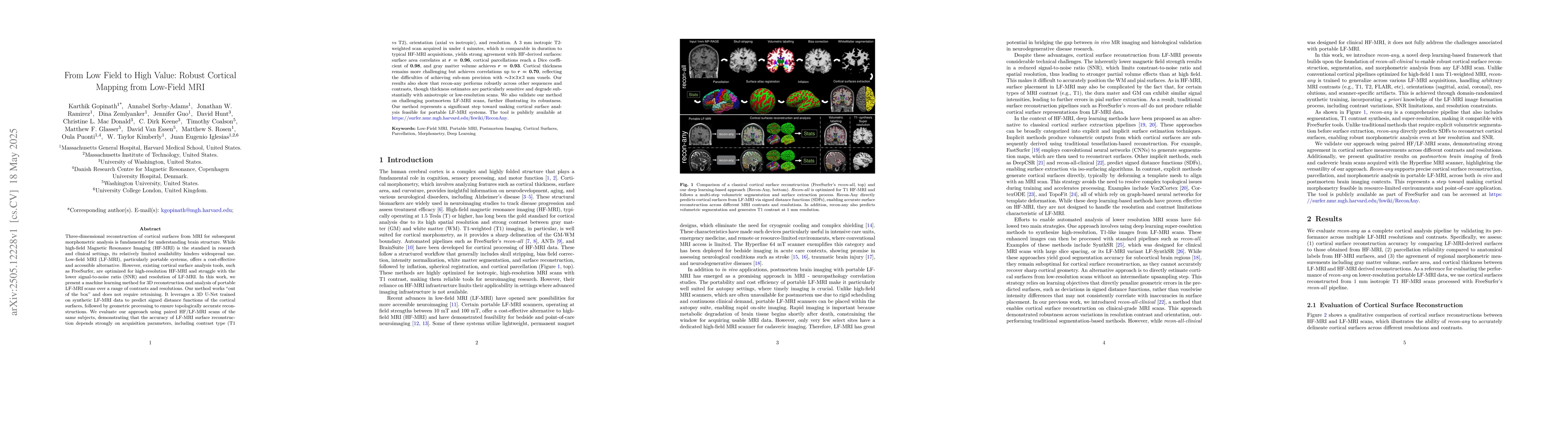
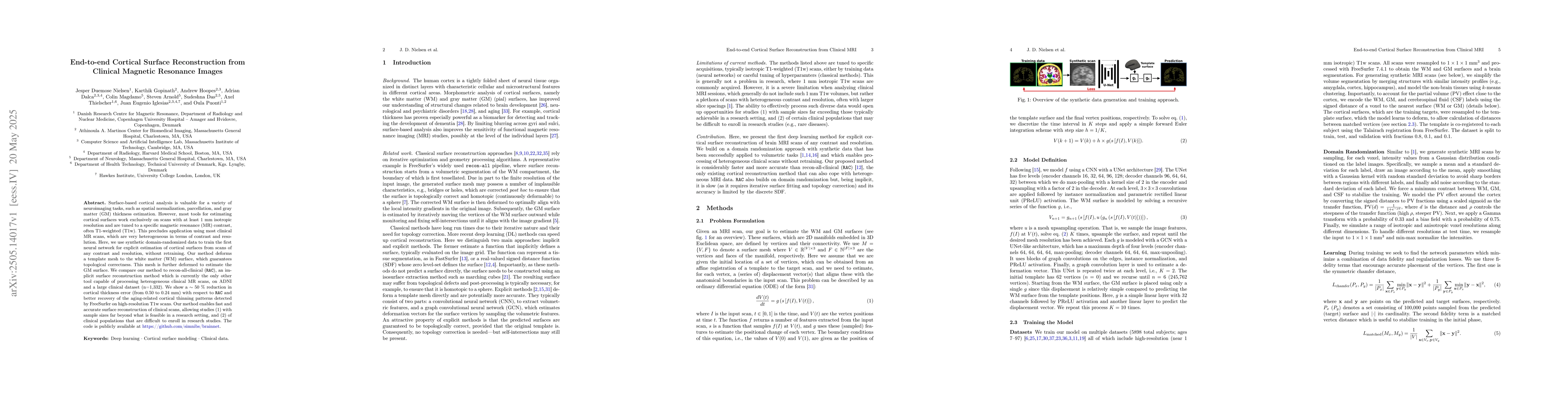
Three-dimensional reconstruction of cortical surfaces from MRI for morphometric analysis is fundamental for understanding brain structure. While high-field MRI (HF-MRI) is standard in research and cli...
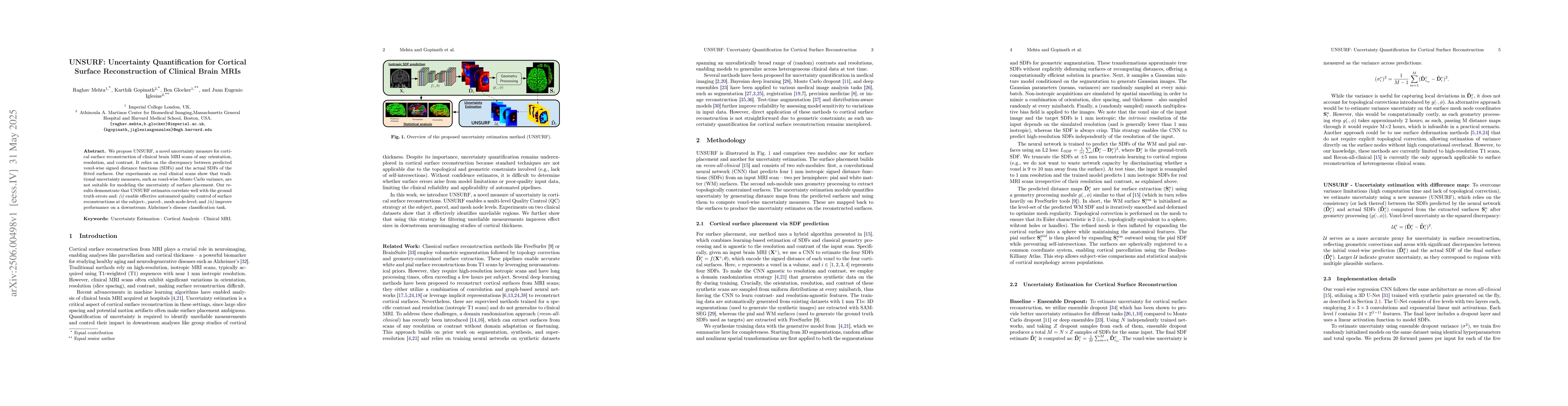
We propose UNSURF, a novel uncertainty measure for cortical surface reconstruction of clinical brain MRI scans of any orientation, resolution, and contrast. It relies on the discrepancy between predic...
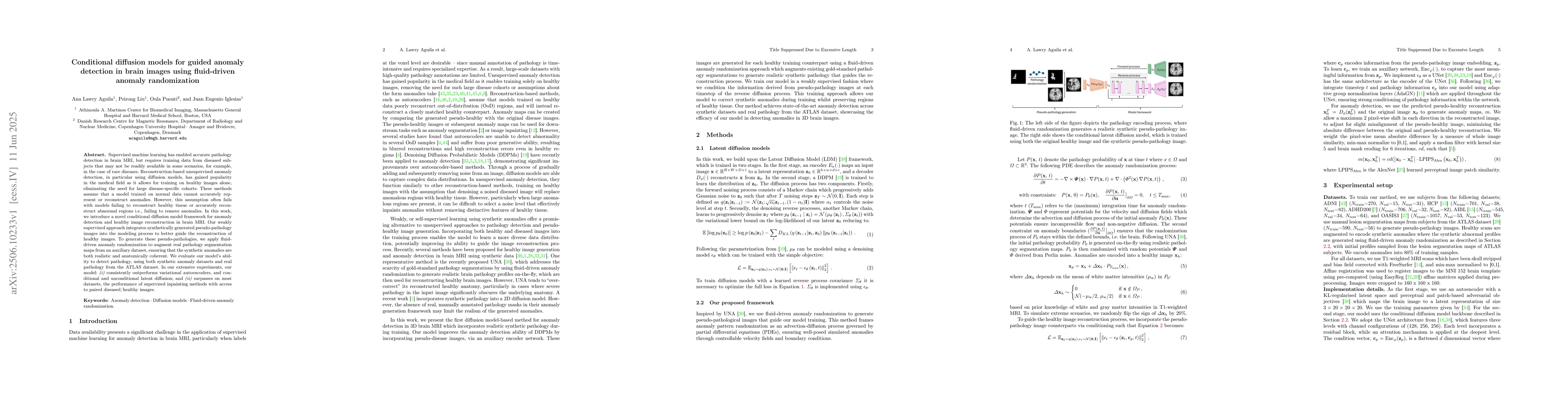
Supervised machine learning has enabled accurate pathology detection in brain MRI, but requires training data from diseased subjects that may not be readily available in some scenarios, for example, i...
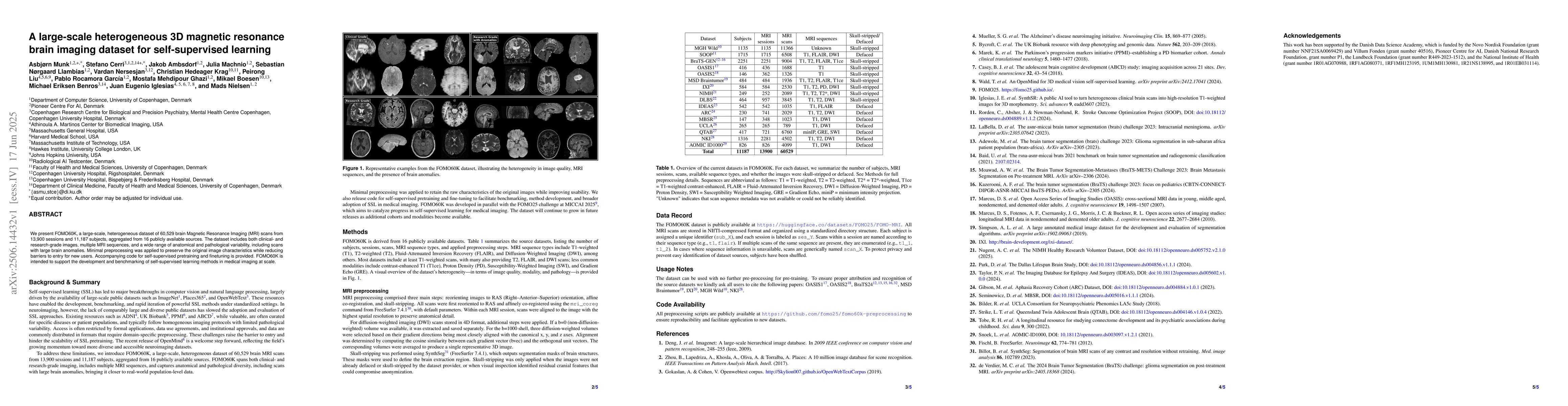
We present FOMO60K, a large-scale, heterogeneous dataset of 60,529 brain Magnetic Resonance Imaging (MRI) scans from 13,900 sessions and 11,187 subjects, aggregated from 16 publicly available sources....
The Brain Tumor Segmentation (BraTS) cluster of challenges has significantly advanced brain tumor image analysis by providing large, curated datasets and addressing clinically relevant tasks. However,...
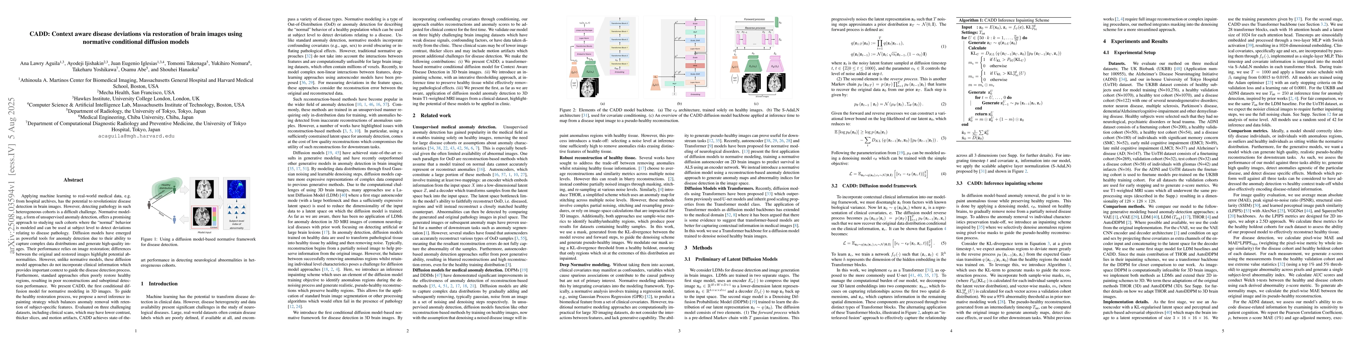
Applying machine learning to real-world medical data, e.g. from hospital archives, has the potential to revolutionize disease detection in brain images. However, detecting pathology in such heterogene...
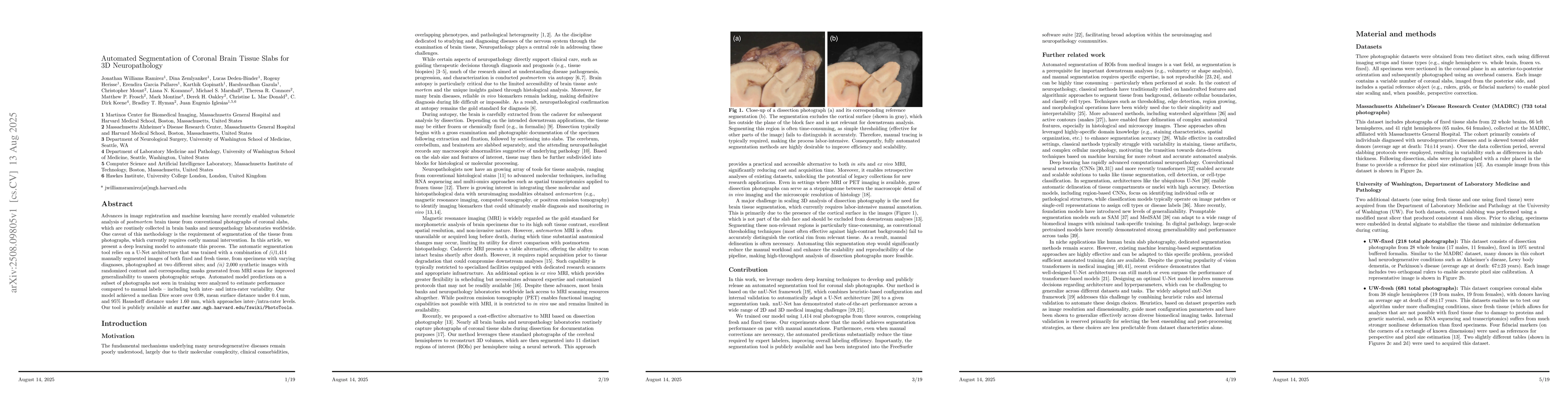
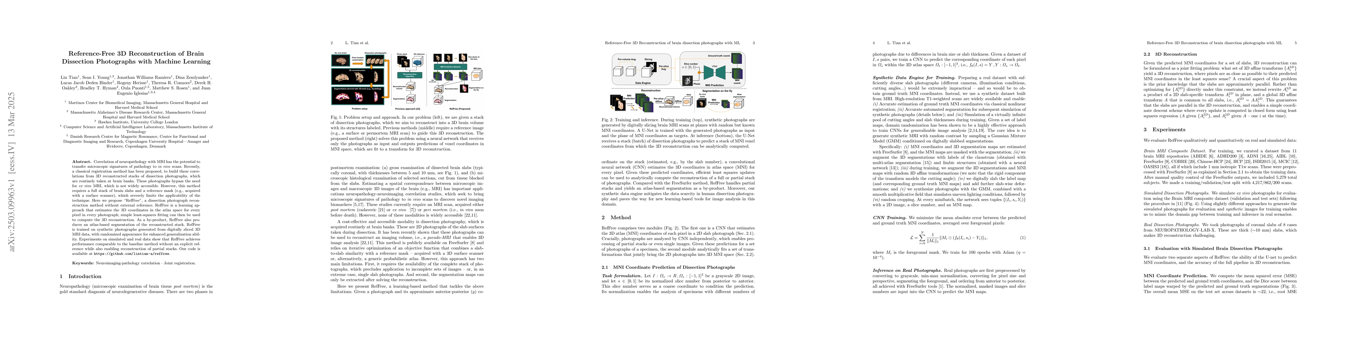
Advances in image registration and machine learning have recently enabled volumetric analysis of \emph{postmortem} brain tissue from conventional photographs of coronal slabs, which are routinely coll...
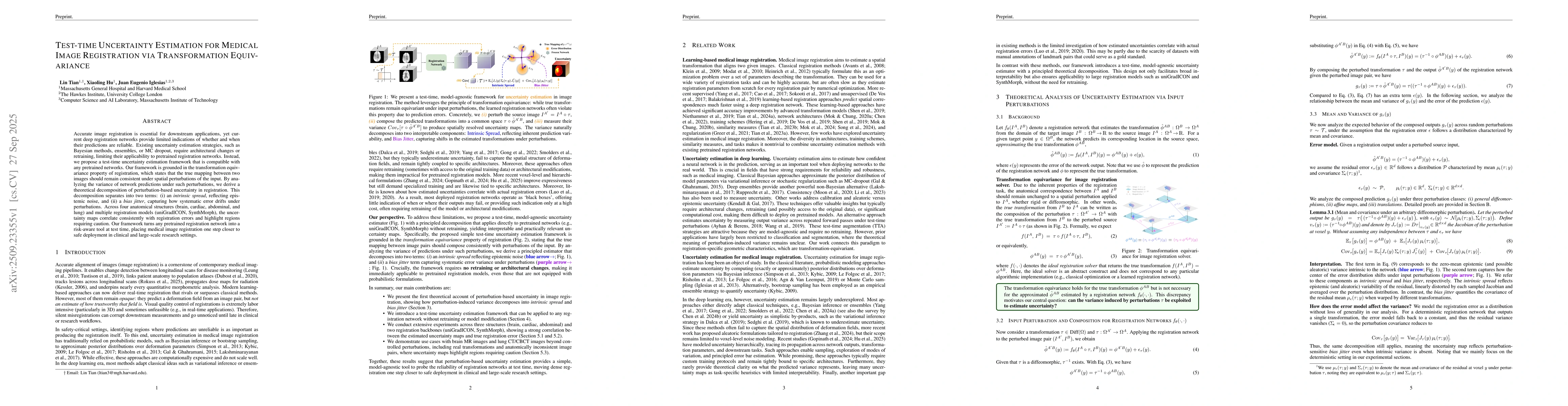
Accurate image registration is essential for downstream applications, yet current deep registration networks provide limited indications of whether and when their predictions are reliable. Existing un...
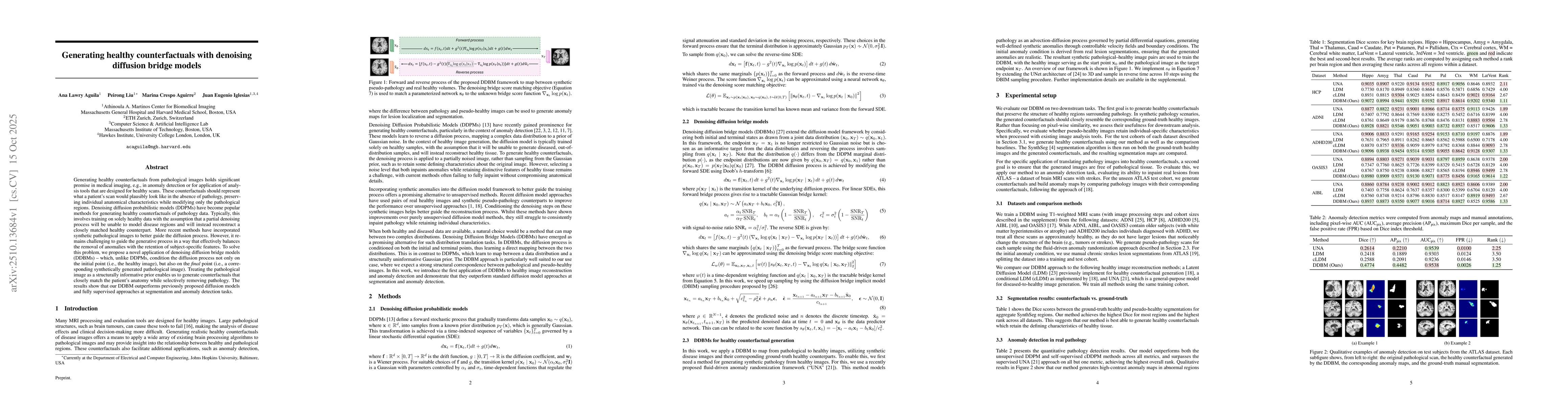
Generating healthy counterfactuals from pathological images holds significant promise in medical imaging, e.g., in anomaly detection or for application of analysis tools that are designed for healthy ...
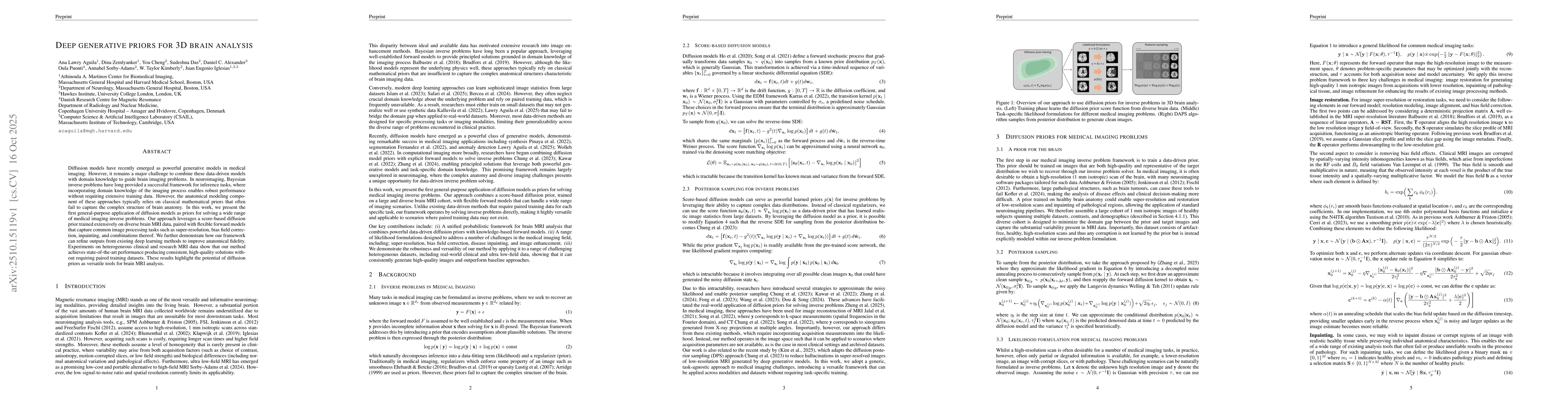
Diffusion models have recently emerged as powerful generative models in medical imaging. However, it remains a major challenge to combine these data-driven models with domain knowledge to guide brain ...
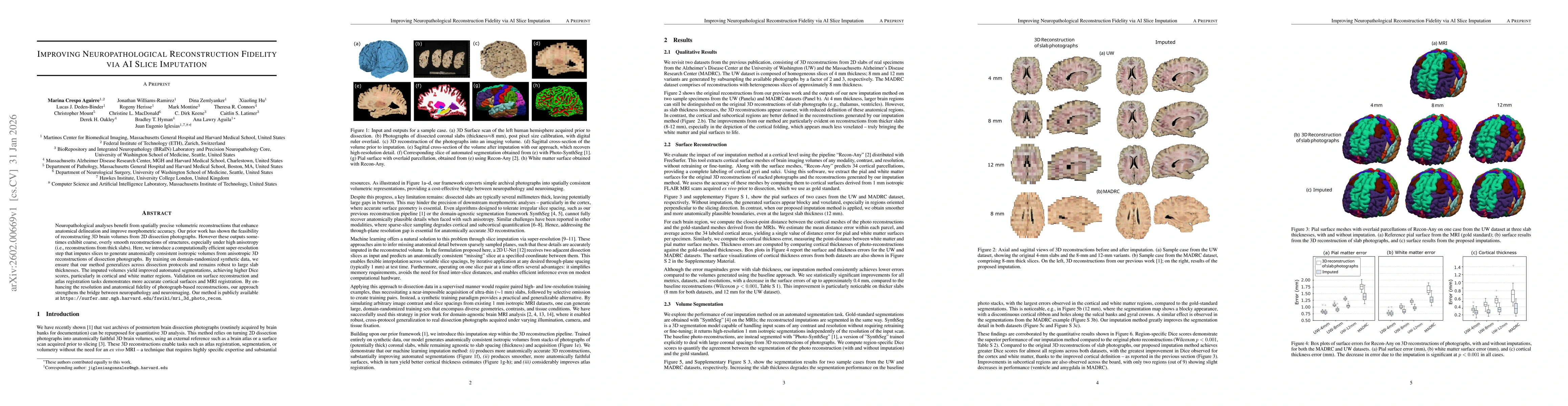
Neuropathological analyses benefit from spatially precise volumetric reconstructions that enhance anatomical delineation and improve morphometric accuracy. Our prior work has shown the feasibility of ...
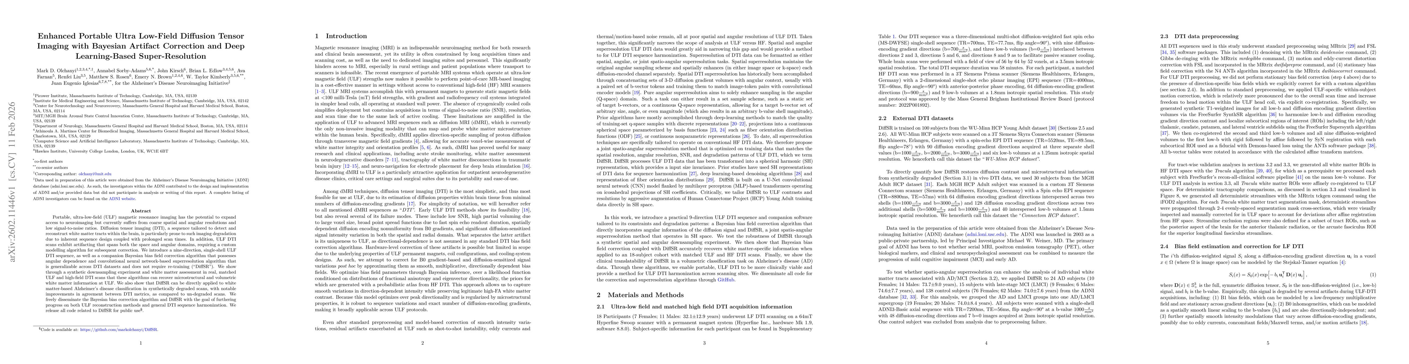
Portable, ultra-low-field (ULF) magnetic resonance imaging has the potential to expand access to neuroimaging but currently suffers from coarse spatial and angular resolutions and low signal-to-noise ...
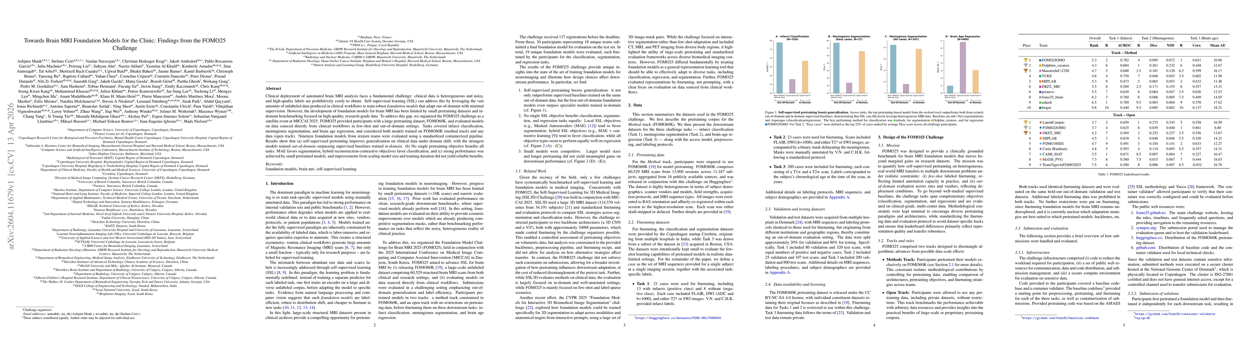
Clinical deployment of automated brain MRI analysis faces a fundamental challenge: clinical data is heterogeneous and noisy, and high-quality labels are prohibitively costly to obtain. Self-supervised...