Publication
Metrics
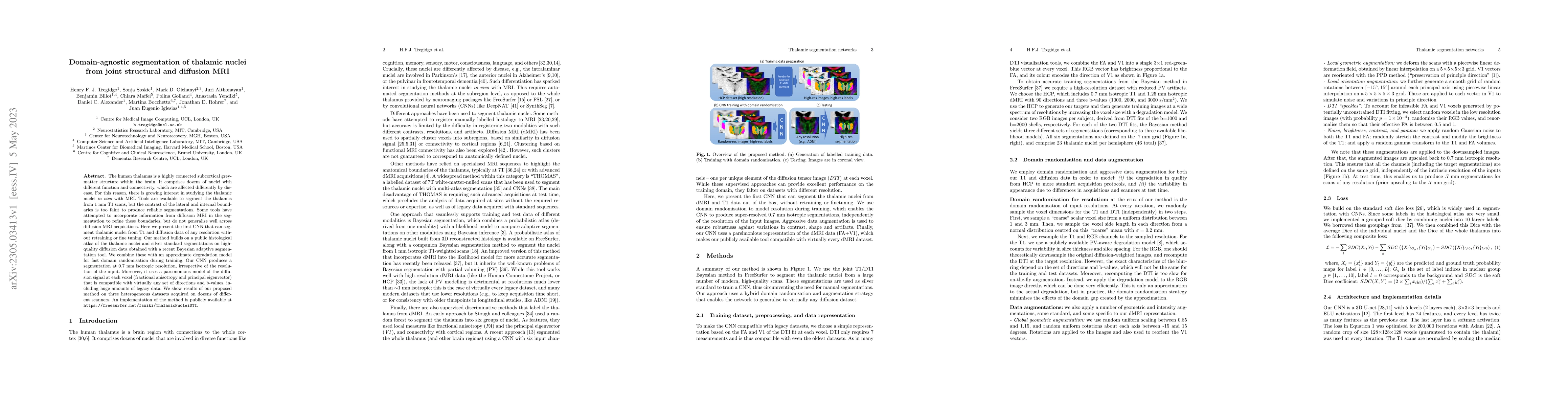
AI Quick Summary
This paper introduces a domain-agnostic CNN that segments thalamic nuclei from both structural T1 and diffusion MRI data of any resolution without retraining. The method leverages a public histological atlas and high-quality diffusion data to achieve reliable segmentations at 0.7 mm isotropic resolution across heterogeneous datasets from various scanners.
Paper Preview
Abstract
The human thalamus is a highly connected subcortical grey-matter structure within the brain. It comprises dozens of nuclei with different function and connectivity, which are affected differently by disease. For this reason, there is growing interest in studying the thalamic nuclei in vivo with MRI. Tools are available to segment the thalamus from 1 mm T1 scans, but the contrast of the lateral and internal boundaries is too faint to produce reliable segmentations. Some tools have attempted to incorporate information from diffusion MRI in the segmentation to refine these boundaries, but do not generalise well across diffusion MRI acquisitions. Here we present the first CNN that can segment thalamic nuclei from T1 and diffusion data of any resolution without retraining or fine tuning. Our method builds on a public histological atlas of the thalamic nuclei and silver standard segmentations on high-quality diffusion data obtained with a recent Bayesian adaptive segmentation tool. We combine these with an approximate degradation model for fast domain randomisation during training. Our CNN produces a segmentation at 0.7 mm isotropic resolution, irrespective of the resolution of the input. Moreover, it uses a parsimonious model of the diffusion signal at each voxel (fractional anisotropy and principal eigenvector) that is compatible with virtually any set of directions and b-values, including huge amounts of legacy data. We show results of our proposed method on three heterogeneous datasets acquired on dozens of different scanners. An implementation of the method is publicly available at https://freesurfer.net/fswiki/ThalamicNucleiDTI.
AI Key Findings
Get AI-generated insights about this paper's methodology, results, significance, and more — seven facets brought into focus.
Impact
Paper Details
Authors
PDF Preview
Key Terms
Citation Network
Current paper (gray), citations (green), references (blue)
Display is limited for performance on very large graphs.
Discussion 0