ArxivLens

Publication

Published: Mar 13, 2025
Categories: eess.IV, cs.CV

Metrics

Source: ArXiv

AI Quick Summary

This paper introduces RefFree, a machine learning method for reconstructing 3D brain dissection photographs without requiring external reference masks, enabling the reconstruction of partial stacks. RefFree estimates 3D coordinates directly from photographs and achieves performance comparable to traditional methods, facilitating neuropathology correlations with MRI.

Quick Answers

What is "Reference-Free 3D Reconstruction of Brain Dissection Photographs with Machine Learning" about?

This paper introduces RefFree, a machine learning method for reconstructing 3D brain dissection photographs without requiring external reference masks, enabling the reconstruction of partial stacks. RefFree estimates 3D coordinates directly from photographs and achieves performance comparable to traditional methods, facilitating neuropathology correlations with MRI.

What methodology did the authors use?

The research proposes RefFree, a reference-free 3D reconstruction method for dissection photographs, trained on synthetic data. It uses a CNN to predict MNI coordinates from photographs, followed by least-squares fitting for 3D reconstruction. The method is trained on synthetic photographs generated from digitally sliced 3D MRI data with randomized appearance. More in Methodology →

What are the key results?

RefFree achieves comparable performance to the baseline method without an explicit reference. — The method can reconstruct partial stacks of photographs. More in Key Results →

Why is this work significant?

This research offers a valuable alternative to existing reference-based methods in FreeSurfer for full-stack reconstruction, addressing scenarios where reference data are unavailable. It provides a versatile tool for postmortem brain examination, potentially enhancing the correlation of neuropathology with in vivo scans. More in Significance →

What are the main limitations?

RefFree currently relies on slice indices and cannot account for non-rigid deformations during reconstruction. — The method assumes slabs are approximately parallel, which might not always be the case in real-world scenarios. More in Limitations →

Paper Preview

Abstract

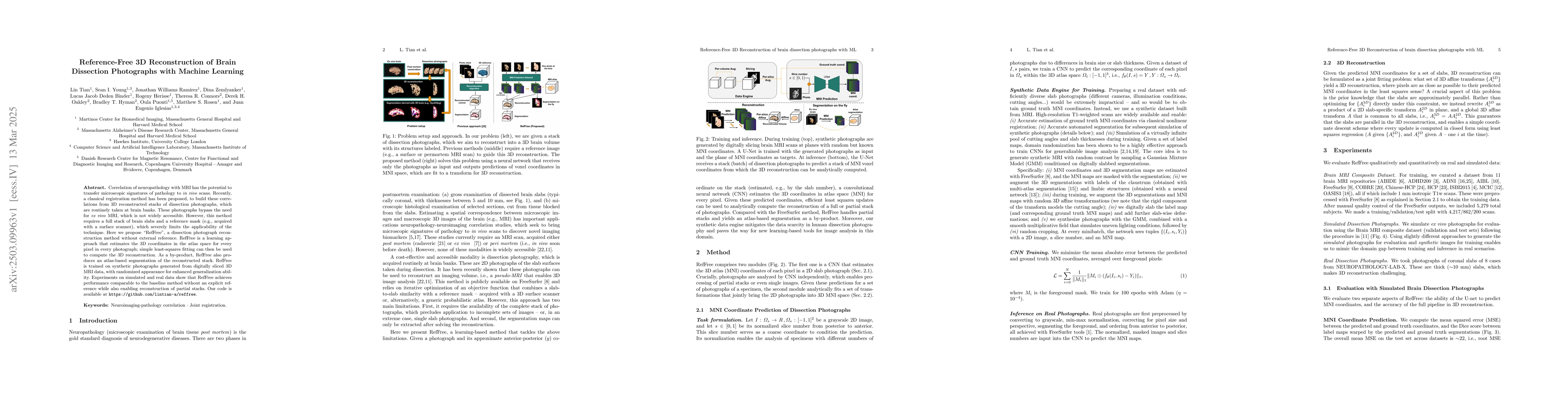
Correlation of neuropathology with MRI has the potential to transfer microscopic signatures of pathology to invivo scans. Recently, a classical registration method has been proposed, to build these correlations from 3D reconstructed stacks of dissection photographs, which are routinely taken at brain banks. These photographs bypass the need for exvivo MRI, which is not widely accessible. However, this method requires a full stack of brain slabs and a reference mask (e.g., acquired with a surface scanner), which severely limits the applicability of the technique. Here we propose RefFree, a dissection photograph reconstruction method without external reference. RefFree is a learning approach that estimates the 3D coordinates in the atlas space for every pixel in every photograph; simple least-squares fitting can then be used to compute the 3D reconstruction. As a by-product, RefFree also produces an atlas-based segmentation of the reconstructed stack. RefFree is trained on synthetic photographs generated from digitally sliced 3D MRI data, with randomized appearance for enhanced generalization ability. Experiments on simulated and real data show that RefFree achieves performance comparable to the baseline method without an explicit reference while also enabling reconstruction of partial stacks. Our code is available at https://github.com/lintian-a/reffree.

Key Findings, in focus

Seven facets of this paper, analysed and brought into focus by AI.

AI-generated · verify against source Generated Jun 10, 2025
In focus · one-line synthesis

This research offers a valuable alternative to existing reference-based methods in FreeSurfer for full-stack reconstruction, addressing scenarios where reference data are unavailable.

01
MethodologyHow they did it

The research proposes RefFree, a reference-free 3D reconstruction method for dissection photographs, trained on synthetic data. It uses a CNN to predict MNI coordinates from photographs, followed by least-squares fitting for 3D reconstruction. The method is trained on synthetic photographs generated from digitally sliced 3D MRI data with randomized appearance.

02
ResultsWhat they found
  • RefFree achieves comparable performance to the baseline method without an explicit reference.
  • The method can reconstruct partial stacks of photographs.
  • Experiments on simulated and real data demonstrate its effectiveness.
  • The approach also produces an atlas-based segmentation of the reconstructed stack as a by-product.
  • RefFree outperforms existing reference-based methods in scenarios where baseline methods fall short, such as with partial stacks.
03
SignificanceWhy it matters

This research offers a valuable alternative to existing reference-based methods in FreeSurfer for full-stack reconstruction, addressing scenarios where reference data are unavailable. It provides a versatile tool for postmortem brain examination, potentially enhancing the correlation of neuropathology with in vivo scans.

04
Technical ContributionsWhat's new technically

The main technical contribution is a learning-based approach for 3D reconstruction of brain dissection photographs without an external reference, using synthetic data generated from digitally sliced 3D MRI data.

05
NoveltyWhat sets it apart

This work is novel as it introduces a reference-free method for 3D reconstruction of brain dissection photographs, trained on synthetic data, which outperforms existing reference-based methods in handling partial stacks and varying appearances, thus expanding the applicability of the technique in real-world neuropathology studies.

06
LimitationsWhat to be cautious about
  • RefFree currently relies on slice indices and cannot account for non-rigid deformations during reconstruction.
  • The method assumes slabs are approximately parallel, which might not always be the case in real-world scenarios.
07
Future WorkWhere it goes next
  • Explore eliminating dependency on slice indices.
  • Model nonlinear deformations crucial for fresh tissue cutting.
  • Improve simulation of dissection photography using 3D models of slices and photorealistic rendering.
  • Further validate the method on downstream applications.

Impact

25
References

Paper Details

Paper ID: 2503.09963
Categories:
eess.IV cs.CV

How to Cite This Paper

Reference manager: RIS EndNote

Citation Network

Current paper (gray), citations (green), references (blue)

Display is limited for performance on very large graphs.

Current Paper
Citations
References
Click to view


Discussion 0

This website uses cookies to ensure you get the best experience. Learn more