Publication
Metrics
AI Quick Summary
This paper proposes an automatic 3D U-Net convolutional neural network for segmenting acute ischemic stroke lesions in non-contrast CT brain scans. The model, enhanced with squeeze-and-excitation blocks and residual connections, achieved an average Dice coefficient of 0.628, demonstrating effective segmentation performance.
Paper Preview
Abstract
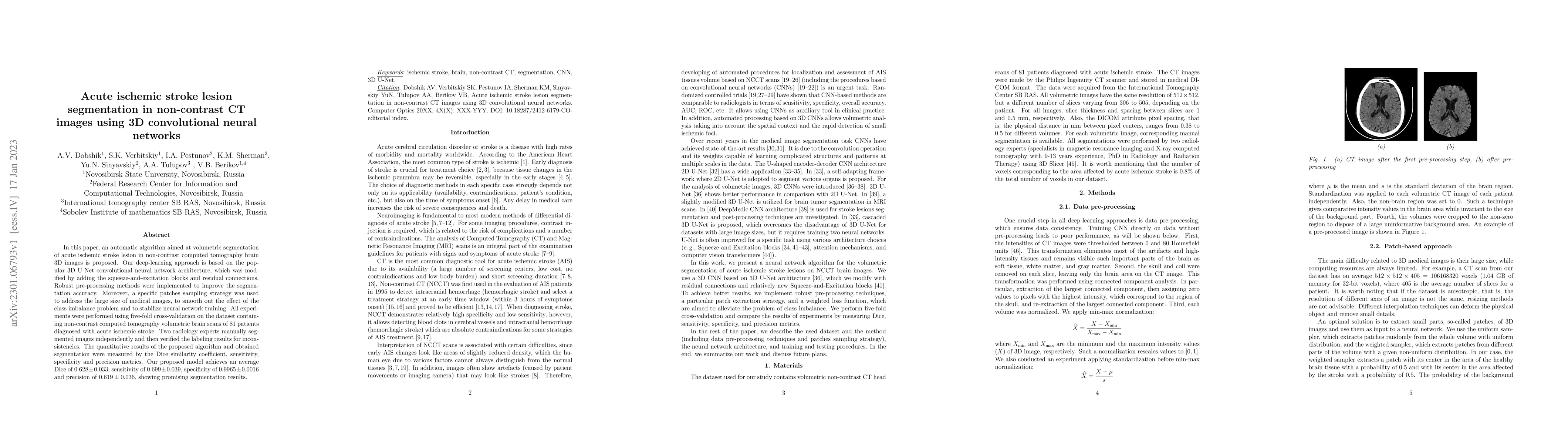
In this paper, an automatic algorithm aimed at volumetric segmentation of acute ischemic stroke lesion in non-contrast computed tomography brain 3D images is proposed. Our deep-learning approach is based on the popular 3D U-Net convolutional neural network architecture, which was modified by adding the squeeze-and-excitation blocks and residual connections. Robust pre-processing methods were implemented to improve the segmentation accuracy. Moreover, a specific patches sampling strategy was used to address the large size of medical images, to smooth out the effect of the class imbalance problem and to stabilize neural network training. All experiments were performed using five-fold cross-validation on the dataset containing non-contrast computed tomography volumetric brain scans of 81 patients diagnosed with acute ischemic stroke. Two radiology experts manually segmented images independently and then verified the labeling results for inconsistencies. The quantitative results of the proposed algorithm and obtained segmentation were measured by the Dice similarity coefficient, sensitivity, specificity and precision metrics. Our proposed model achieves an average Dice of $0.628\pm0.033$, sensitivity of $0.699\pm0.039$, specificity of $0.9965\pm0.0016$ and precision of $0.619\pm0.036$, showing promising segmentation results.
AI Key Findings
Get AI-generated insights about this paper's methodology, results, significance, and more — seven facets brought into focus.
Impact
Paper Details
Authors
PDF Preview
Key Terms
Citation Network
Current paper (gray), citations (green), references (blue)
Display is limited for performance on very large graphs.
Discussion 0