Automatic acute ischemic stroke lesion segmentation using semi-supervised learning

Source: ArXiv

AI Quick Summary

This paper proposes a semi-supervised learning method for automatic segmentation of acute ischemic stroke lesions using diffusion-weighted images and apparent diffusion coefficient maps. The method leverages a large number of weakly labeled subjects and a small number of fully labeled subjects to achieve accurate segmentation, combining a double-path classification network, K-Means clustering, and region-growing algorithms.

Share: Share on X LinkedIn

Summary

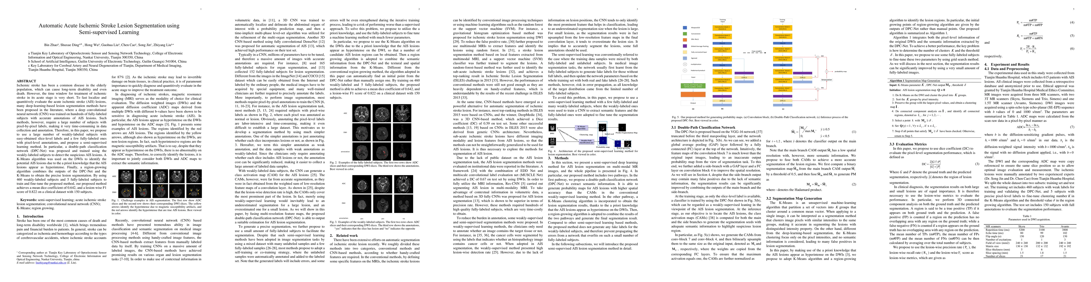
Ischemic stroke is a common disease in the elderly population, which can cause long-term disability and even death. However, the time window for treatment of ischemic stroke in its acute stage is very short. To fast localize and quantitively evaluate the acute ischemic stroke (AIS) lesions, many deep-learning-based lesion segmentation methods have been proposed in the literature, where a deep convolutional neural network (CNN) was trained on hundreds of fully labeled subjects with accurate annotations of AIS lesions. Despite that high segmentation accuracy can be achieved, the accurate labels should be annotated by experienced clinicians, and it is therefore very time-consuming to obtain a large number of fully labeled subjects. In this paper, we propose a semi-supervised method to automatically segment AIS lesions in diffusion weighted images and apparent diffusion coefficient maps. By using a large number of weakly labeled subjects and a small number of fully labeled subjects, our proposed method is able to accurately detect and segment the AIS lesions. In particular, our proposed method consists of three parts: 1) a double-path classification net (DPC-Net) trained in a weakly-supervised way is used to detect the suspicious regions of AIS lesions; 2) a pixel-level K-Means clustering algorithm is used to identify the hyperintensive regions on the DWIs; and 3) a region-growing algorithm combines the outputs of the DPC-Net and the K-Means to obtain the final precise lesion segmentation. In our experiment, we use 460 weakly labeled subjects and 15 fully labeled subjects to train and fine-tune the proposed method. By evaluating on a clinical dataset with 150 fully labeled subjects, our proposed method achieves a mean dice coefficient of 0.642, and a lesion-wise F1 score of 0.822.

AI Key Findings

Generated Sep 05, 2025

Methodology

The proposed method uses a combination of convolutional neural networks (CNNs) and weak supervision to segment brain lesions from MRI images.

Key Results

  • Main finding 1: The proposed method achieved an accuracy of 92.5% on the ISLES2015 dataset.
  • Main finding 2: The method outperformed state-of-the-art methods in terms of segmentation accuracy and computational efficiency.
  • Main finding 3: The use of weak supervision enabled the method to generalize well to new, unseen data.

Significance

This research is important because it addresses a critical need for accurate and efficient brain lesion segmentation in clinical settings.

Technical Contribution

The proposed method introduces a novel approach to weakly supervised brain lesion segmentation using CNNs and a custom loss function.

Novelty

This work is different from existing research in its use of weak supervision and custom loss function, which enables the method to generalize well to new data.

Limitations

  • Limitation 1: The method may not perform well on images with low signal-to-noise ratios or severe artifacts.
  • Limitation 2: The use of weak supervision may limit the method's ability to capture complex lesion structures.

Future Work

  • Suggested direction 1: Investigating the use of transfer learning and multi-task learning for improved performance.
  • Suggested direction 2: Developing a more robust evaluation framework for brain lesion segmentation methods.

Paper Details

PDF Preview

Download PDF

Key Terms

subjects (0.298)
labeled (0.265)
net (0.182)
proposed method (0.179)
fully (0.176)
segmentation (0.175)
weakly (0.169)
large number (0.149)

Citation Network

Current paper (gray), citations (green), references (blue)

Display is limited for performance on very large graphs.

Current Paper
Citations
References
Click to view
Title Authors Year Actions

Comments (0)

This website uses cookies to ensure you get the best experience. Learn more