Automatic Skin Lesion Segmentation using Semi-supervised Learning Technique
Publication
Metrics
AI Quick Summary
This paper proposes an automatic skin lesion segmentation technique using semi-supervised learning for dermoscopic images to aid in early skin cancer diagnosis. The method involves preprocessing to remove noise and semi-supervised segmentation using K-means clustering and color feature analysis, validated using images from the ISIC 2017 challenge.
Paper Preview
Abstract
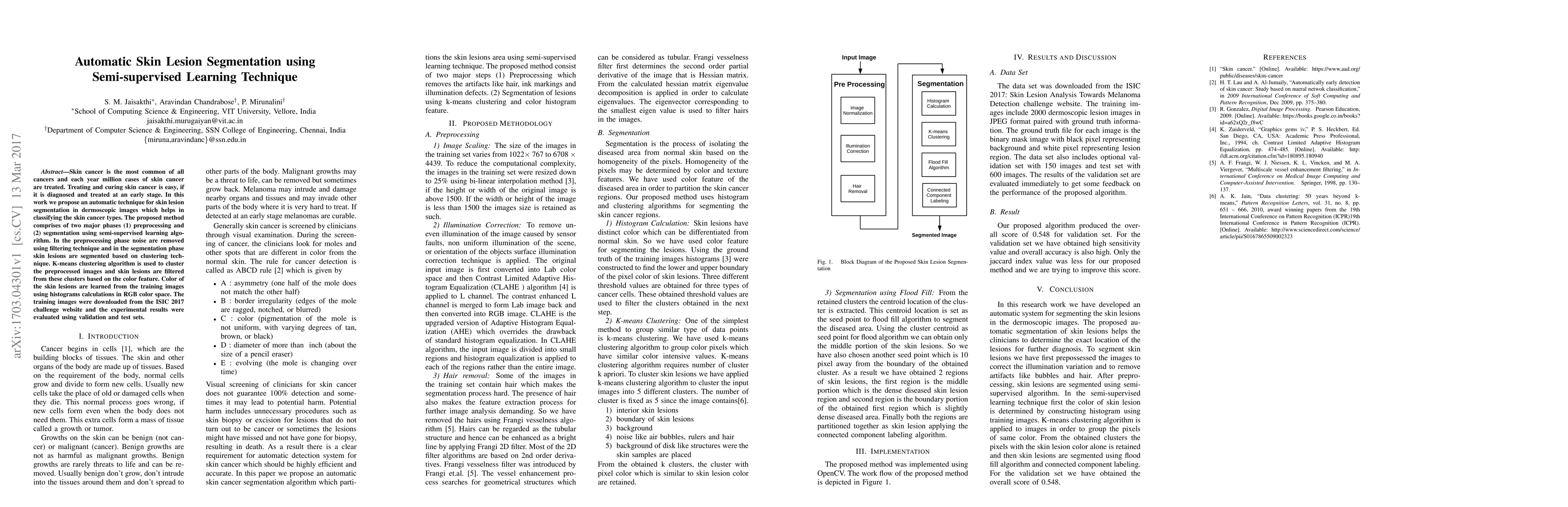
Skin cancer is the most common of all cancers and each year million cases of skin cancer are treated. Treating and curing skin cancer is easy, if it is diagnosed and treated at an early stage. In this work we propose an automatic technique for skin lesion segmentation in dermoscopic images which helps in classifying the skin cancer types. The proposed method comprises of two major phases (1) preprocessing and (2) segmentation using semi-supervised learning algorithm. In the preprocessing phase noise are removed using filtering technique and in the segmentation phase skin lesions are segmented based on clustering technique. K-means clustering algorithm is used to cluster the preprocessed images and skin lesions are filtered from these clusters based on the color feature. Color of the skin lesions are learned from the training images using histograms calculations in RGB color space. The training images were downloaded from the ISIC 2017 challenge website and the experimental results were evaluated using validation and test sets.
AI Key Findings
Get AI-generated insights about this paper's methodology, results, significance, and more — seven facets brought into focus.
Impact
Paper Details
PDF Preview
Key Terms
Citation Network
Current paper (gray), citations (green), references (blue)
Display is limited for performance on very large graphs.
Discussion 0