This study investigates how aging of digital pathology displays affects the interpretation of pathology slides, finding significant impacts on diagnostic accuracy and workflow efficiency. Results show that aged displays reduce conspicuity of nuclei and increase reading difficulty and time, with a notable reduction in inter-session diagnostic agreement.
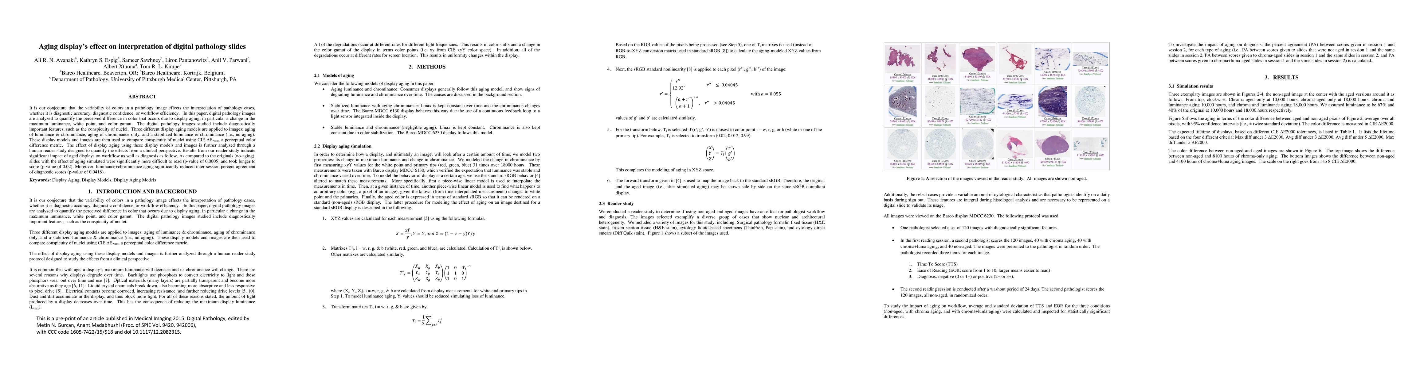
It is our conjecture that the variability of colors in a pathology image effects the interpretation of pathology cases, whether it is diagnostic accuracy, diagnostic confidence, or workflow efficiency. In this paper, digital pathology images are analyzed to quantify the perceived difference in color that occurs due to display aging, in particular a change in the maximum luminance, white point, and color gamut. The digital pathology images studied include diagnostically important features, such as the conspicuity of nuclei. Three different display aging models are applied to images: aging of luminance & chrominance, aging of chrominance only, and a stabilized luminance & chrominance (i.e., no aging). These display models and images are then used to compare conspicuity of nuclei using CIE deltaE2000, a perceptual color difference metric. The effect of display aging using these display models and images is further analyzed through a human reader study designed to quantify the effects from a clinical perspective. Results from our reader study indicate significant impact of aged displays on workflow as well as diagnosis as follow. As compared to the originals (no-aging), slides with the effect of aging simulated were significantly more difficult to read (p-value of 0.0005) and took longer to score (p-value of 0.02). Moreover, luminance+chrominance aging significantly reduced inter-session percent agreement of diagnostic scores (p-value of 0.0418).
This graph shows how this paper is connected to other research. The current paper is shown in dark gray, papers that cite it are in green, and papers it references are in blue. You can:
• Hover over any node to see the paper title
• Click a node to view that paper's details
• Drag nodes to rearrange the graph
• Use the zoom controls (bottom right) to zoom in/out
Title | Authors | Year | Actions |
---|---|---|---|
Digital Pathology Displays Under Pressure: Benchmarking Performance Across Market Grades.
|
S. Marletta, Alessandro Caputo, Gabriele Guidi, Liron Pantanowitz, F. Pagni, Iacopo Bavieri, Vincenzo L’Imperio, Matteo Brunelli, Angelo Paolo dei Tos, A. Eccher
|
2025
|
View Paper |
Not all stains are made equal: impact of stain normalization on prostate cancer diagnosis.
|
G. Cazzaniga, Alessandro Caputo, Vincenzo L'Imperio, Fabio Gibilisco, Manuela Scotto, Orazio Pennisi, Nicola Michielli, Alessandro Mogetta, Filippo Molinari, Filippo Fraggetta, M. Salvi
|
2024
|
View Paper |
Background removal for debiasing computer-aided cytological diagnosis
|
Keita Takeda, Tomoya Sakai, Eiji Mitate
|
2024
|
View Paper |
Whole Slide Imaging Technology and Its Applications: Current and Emerging Perspectives
|
E. Jain, Ankush U Patel, A. Parwani, S. Shafi, Zoya Brar, Shivani Sharma, S. Mohanty
|
2023
|
View Paper |
Background Subtraction Approach to Unsupervised Cell Segmentation: Toward Excluding Spurious Features in Degraded Cytology Slides
|
Keita Takeda, Eiji Mitate, Tomoya Sakai
|
2023
|
View Paper |
Whole-slide imaging, tissue image analysis, and artificial intelligence in veterinary pathology: An updated introduction and review
|
A. Zuraw, F. Aeffner
|
2021
|
View Paper |
The Application, Challenges, and Advancement Toward Regulatory Acceptance of Digital Toxicologic Pathology: Results of the 7th ESTP International Expert Workshop (September 20-21, 2019)
|
V. Schumacher, F. Aeffner, Erio Barale-Thomas, C. Botteron, Jonathan Carter, L. Elies, J. Engelhardt, P. Fant, T. Forest, Peter Hall, Deon Hildebrand, R. Klopfleisch, Thomas Lucotte, H. Marxfeld, LuAnn Mckinney, P. Moulin, E. Neyens, X. Palazzi, A. Piton, E. Riccardi, D. Roth, S. Rousselle, J. Vidal, B. Williams
|
2020
|
View Paper |
Display Characteristics and Their Impact on Digital Pathology: A Current Review of Pathologists’ Future “Microscope”
|
J. Abel, P. Ouillette, Christopher L. Williams, John L Blau, Jerome Cheng, Keluo Yao, Winston Y. Lee, Toby C. Cornish, Ulysses Balis, D. McClintock
|
2020
|
View Paper |
A Practical Guide to Whole Slide Imaging: A White Paper From the Digital Pathology Association.
|
M. Zarella, D. Bowman, F. Aeffner, Navid Farahani, A. Xthona, S. Absar, A. Parwani, M. Bui, D. Hartman
|
2018
|
View Paper |
Digital Pathology and Tissue Image Analysis
|
F. Aeffner, T. Forest, V. Schumacher, M. Zarella, A. Bradley
|
2022
|
View Paper |
Visualization of medical content on color display systems
|
2022
|
View Paper | |
Practical guidelines for color calibration and quality assurance of medical displays
|
T. Kimpe, J. Rostang, G. Hoey, A. Xthona
|
2017
|
View Paper |
INFLUENCE OF DISPLAY CHARACTERISTICS ON CLINICAL PERFORMANCE IN DIGITAL PATHOLOGY
|
T. Kimpe, A. Avanaki, K. Espig, J. Rostang, G. Hoey, A. Xthona
|
2016
|
View Paper |
Published: 2020-12-29
Jaime S. Cardoso, Diana Montezuma, Domingos Oliveira, Tomé Albuquerque, Arlindo L. Oliveira, João Monteiro, Liliana Ribeiro, Sofia Gonçalves, Isabel M. Pinto, Ana Beatriz Vieira, Maria Valente
Published: 2025-03-13
Comments (0)