DeepLIIF is an online platform that streamlines IHC scoring through multiplex immunofluorescence staining, improving efficiency and reproducibility in clinical pathology diagnosis.
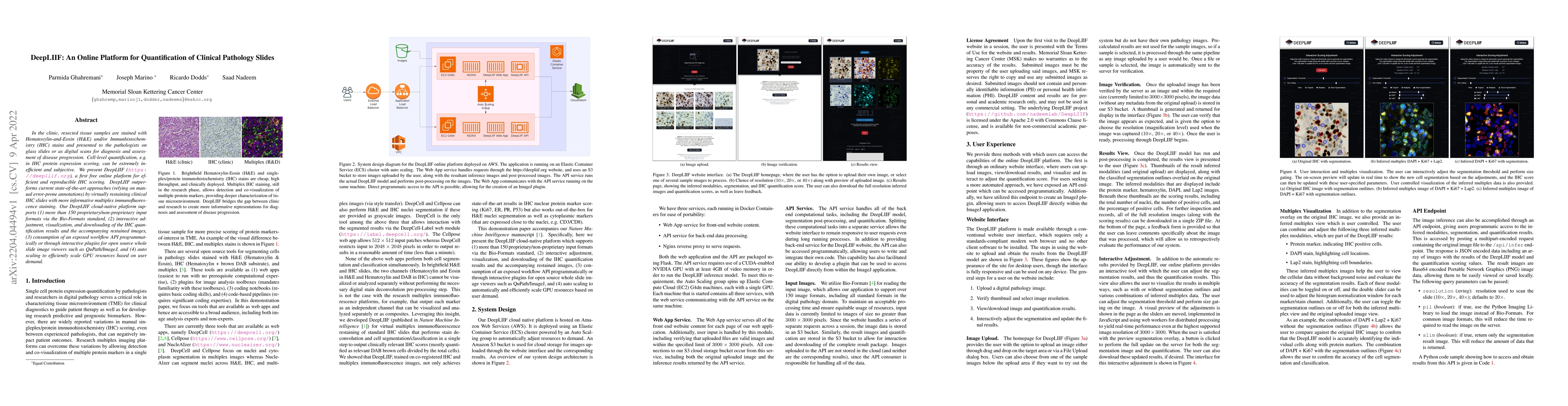
In the clinic, resected tissue samples are stained with Hematoxylin-and-Eosin (H&E) and/or Immunhistochemistry (IHC) stains and presented to the pathologists on glass slides or as digital scans for diagnosis and assessment of disease progression. Cell-level quantification, e.g. in IHC protein expression scoring, can be extremely inefficient and subjective. We present DeepLIIF (https://deepliif.org), a first free online platform for efficient and reproducible IHC scoring. DeepLIIF outperforms current state-of-the-art approaches (relying on manual error-prone annotations) by virtually restaining clinical IHC slides with more informative multiplex immunofluorescence staining. Our DeepLIIF cloud-native platform supports (1) more than 150 proprietary/non-proprietary input formats via the Bio-Formats standard, (2) interactive adjustment, visualization, and downloading of the IHC quantification results and the accompanying restained images, (3) consumption of an exposed workflow API programmatically or through interactive plugins for open source whole slide image viewers such as QuPath/ImageJ, and (4) auto scaling to efficiently scale GPU resources based on user demand.
Get AI-generated insights about this paper's methodology, results, and significance.
Current paper (gray), citations (green), references (blue)
Djork-Arné Clevert, Marco Bertolini, Van-Khoa Le et al.
Title | Authors | Year | Actions |
---|---|---|---|
Automatic Quantification of Ki-67 Labeling Index in Pediatric Brain Tumors Using QuPath
|
C. Spyretos, J. M. Pardo Ladino, H. Blomstrand, P. Nyman, O. Snodahl, A. Shamikh, N. O. Elander, N. Haj-Hosseini
|
2025
|
View Paper |
PD-L1 expression assessment in Angiosarcoma improves with artificial intelligence support
|
F.H. Reith, A. Jarosch, J.P. Albrecht, F. Ghoreschi, A. Flörcken, A. Dörr, S. Roohani, F.M. Schäfer, R. Öllinger, S. Märdian, K. Tielking, P. Bischoff, N. Frühauf, F. Brandes, D. Horst, C. Sers, D. Kainmueller
|
2025
|
View Paper |
CODAvision: best practices and a user-friendly interface for rapid, customizable segmentation of medical images
|
Valentina Matos-Romero, Jaime Gómez-Becerril, André Forjaz, Lucie Dequiedt, Tyler Newton, Saurabh Joshi, Yu Shen, Eban A. Hanna, P. Nair, Arrun Sivasubramanian, Jenny S. H. Wang, Emily L. Lasse-Opsahl, Alexander T. F. Bell, J. Czum, Charles Steenbergen, Dao-Fu Dai, Laura D. Wood, Luciane T. Kagohara, E. Fertig, M. Pasca di Magliano, J. Shatzel, O. McCarty, Jamie O. Lo, Avi Rosenberg, Ralph H. Hruban, A. Muñoz-Barrutia, Denis Wirtz, A. Kiemen
|
2025
|
View Paper |
The Diagnostic Utility of TRBC1 Immunohistochemistry in Mature T-Cell Lymphomas.
|
Ting Zhou, Rohan Sardana, Ozgur Can Eren, Melissa P Pulitzer, Achim A. Jungbluth, Ahmet Dogan, Megan S Lim
|
2025
|
View Paper |
StainDiffuser: MultiTask Dual Diffusion Model for Virtual Staining
|
Tushar Kataria, Beatrice Knudsen, Shireen Elhabian
|
2024
|
View Paper |
Computational pathology: A survey review and the way forward
|
M. S. Hosseini, B. Bejnordi, Vincent Quoc-Huy Trinh, Lyndon Chan, Danial Hasan, Xingwen Li, Stephen Yang, Taehyo Kim, Haochen Zhang, Theodore Wu, Kajanan Chinniah, Sina Maghsoudlou, Ryan Zhang, Jiadai Zhu, Samir Khaki, Andrei Buin, Fatemeh Chaji, Ala Salehi, Bich Ngoc Nguyen, Dimitris Samaras, K. Plataniotis
|
2024
|
View Paper |
Use of Novel Open-Source Deep Learning Platform for Quantification of Ki-67 in Neuroendocrine Tumors – Analytical Validation
|
Talat Zehra, Mahin Shams, Rabia Ali, A. Jafri, Amna Khurshid, Humaira Erum, Hanna Naqvi, Jamshid Abdul-Ghafar
|
2023
|
View Paper |
Use of a Novel Deep Learning Open-Source Model for Quantification of Ki-67 in Breast Cancer Patients in Pakistan: A Comparative Study between the Manual and Automated Methods
|
Talat Zehra, N. Jaffar, Mahin Shams, Q. Chundriger, Arsalan Ahmed, Fariha Anum, Najah Alsubaie, Zubair Ahmad
|
2023
|
View Paper |
Integrative Spatial Analysis of H&E and IHC Images Identifies Prognostic Immune Subtypes Correlated with Progression-Free Survival in Human Papillomavirus (HPV)-Related Oropharyngeal Squamous Cell Carcinoma
|
S. R. Nakkireddy, Inyeop Jang, Minji Kim, Linda X. Yin, Michael Rivera, Joaquin J Garcia, K. Bartemes, D. Routman, Eric J Moore, C. Abdel-Halim, Daniel J Ma, K. V. Van Abel, Tae Hyun Hwang
|
2023
|
View Paper |
Ki67 proliferation index in medullary thyroid carcinoma: a comparative study of multiple counting methods and validation of image analysis and deep learning platforms
|
Saad Nadeem, M. Hanna, K. Viswanathan, Joseph Marino, Mahsa S. Ahadi, Bayan A Alzumaili, M. Bani, Federico Chiarucci, Angela Chou, A. De Leo, T. Fuchs, Daniel J. Lubin, Catherine Luxford, K. Magliocca, Germán Martinez, Qiuying Shi, S. Sidhu, A. Al Ghuzlan, Anthony J. Gill, G. Tallini, R. Ghossein, Bin Xu
|
2023
|
View Paper |
DARC: Distribution-Aware Re-Coloring Model for Generalizable Nucleus Segmentation
|
Shengcong Chen, Changxing Ding, Dacheng Tao, Hao Chen
|
2023
|
View Paper |
Structural Cycle GAN for Virtual Immunohistochemistry Staining of Gland Markers in the Colon
|
Shikha Dubey, Tushar Kataria, B. Knudsen, Shireen Elhabian
|
2023
|
View Paper |
An AI-Ready Multiplex Staining Dataset for Reproducible and Accurate Characterization of Tumor Immune Microenvironment
|
Parmida Ghahremani, Joseph Marino, J. Hernandez-Prera, J. V. D. L. Iglesia, R. Slebos, C. Chung, S. Nadeem
|
2023
|
View Paper |
A review of open-source image analysis tools for mammalian cell culture: algorithms, features and implementations
|
Hafizi Malik, Ahmad Syahrin Idris, S. Toha, Izyan Mohd Idris, M. F. Daud, Nur Liyana Azmi
|
2023
|
View Paper |
Topology-Guided Multi-Class Cell Context Generation for Digital Pathology
|
Shahira Abousamra, Rajarsi R. Gupta, T. Kurç, D. Samaras, J. Saltz, Chao Chen
|
2023
|
View Paper |
DEPAS: De-novo Pathology Semantic Masks using a Generative Model
|
Ariel Larey, Nati Daniel, Eliel Aknin, Y. Fisher, Y. Savir
|
2023
|
View Paper |
Comments (0)