Air, bone and soft-tissue Segmentation on 3D brain MRI Using Semantic Classification Random Forest with Auto-Context Model
Publication
Metrics
AI Quick Summary
This paper proposes a Semantic Classification Random Forest with Auto-Context Model for segmenting air, bone, and soft tissue in 3D brain MRI. The method achieves superior segmentation accuracy compared to Random Forest and U-Net, with potential applications in PET/MRI attenuation correction, MRI-only radiation treatment planning, and MR-guided focused ultrasound surgery.
Paper Preview
Abstract
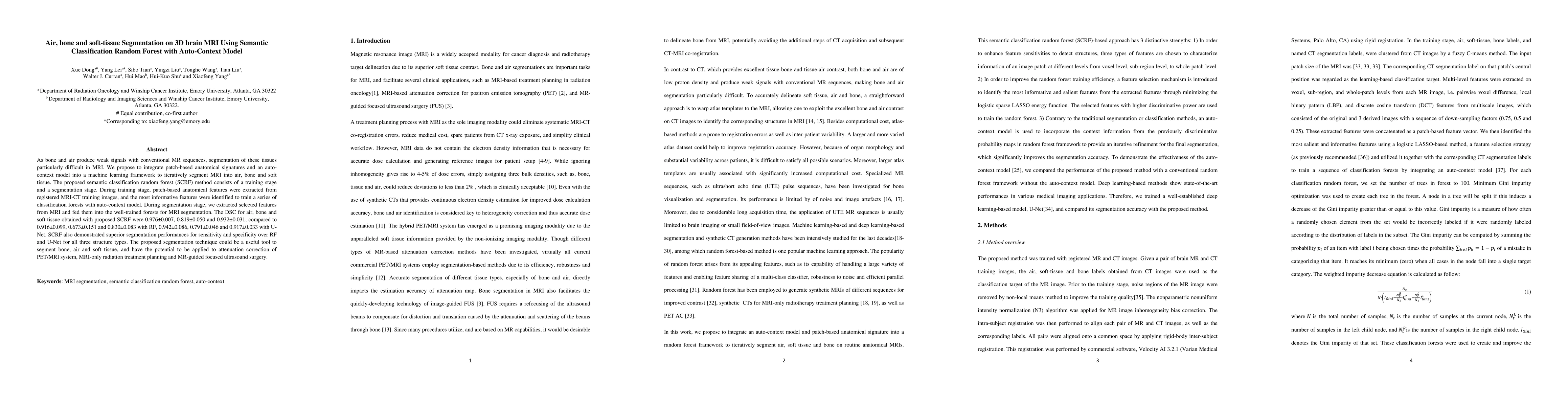
As bone and air produce weak signals with conventional MR sequences, segmentation of these tissues particularly difficult in MRI. We propose to integrate patch-based anatomical signatures and an auto-context model into a machine learning framework to iteratively segment MRI into air, bone and soft tissue. The proposed semantic classification random forest (SCRF) method consists of a training stage and a segmentation stage. During training stage, patch-based anatomical features were extracted from registered MRI-CT training images, and the most informative features were identified to train a series of classification forests with auto-context model. During segmentation stage, we extracted selected features from MRI and fed them into the well-trained forests for MRI segmentation. The DSC for air, bone and soft tissue obtained with proposed SCRF were 0.976, 0.819 and 0.932, compared to 0.916, 0.673 and 0.830 with RF, 0.942, 0.791 and 0.917 with U-Net. SCRF also demonstrated superior segmentation performances for sensitivity and specificity over RF and U-Net for all three structure types. The proposed segmentation technique could be a useful tool to segment bone, air and soft tissue, and have the potential to be applied to attenuation correction of PET/MRI system, MRI-only radiation treatment planning and MR-guided focused ultrasound surgery.
AI Key Findings
Get AI-generated insights about this paper's methodology, results, significance, and more — seven facets brought into focus.
Impact
Paper Details
PDF Preview
Key Terms
Citation Network
Current paper (gray), citations (green), references (blue)
Display is limited for performance on very large graphs.
Discussion 0