Summary
EEG slowing is reported in various neurological disorders including Alzheimer's, Parkinson's and Epilepsy. Here, we investigate alpha rhythm slowing in individuals with refractory temporal lobe epilepsy (TLE), compared to healthy controls, using scalp electroencephalography (EEG) and magnetoencephalography (MEG). We retrospectively analysed data from 17,(46) healthy controls and 22,(24) individuals with TLE who underwent scalp EEG and (MEG) recordings as part of presurgical evaluation. Resting-state, eyes-closed recordings were source reconstructed using the standardized low-resolution brain electrographic tomography (sLORETA) method. We extracted low (slow) 6-9 Hz and high (fast) 10-11 Hz alpha relative band power and calculated the alpha power ratio by dividing low (slow) alpha by high (fast) alpha. This ratio was computed for all brain regions in all individuals. Alpha oscillations were slower in individuals with TLE than controls (p<0.05). This effect was present in both the ipsilateral and contralateral hemispheres, and across widespread brain regions. Alpha slowing in TLE was found in both EEG and MEG recordings. We interpret greater low (slow)-alpha as greater deviation from health.
AI Key Findings
Generated Sep 02, 2025
Methodology
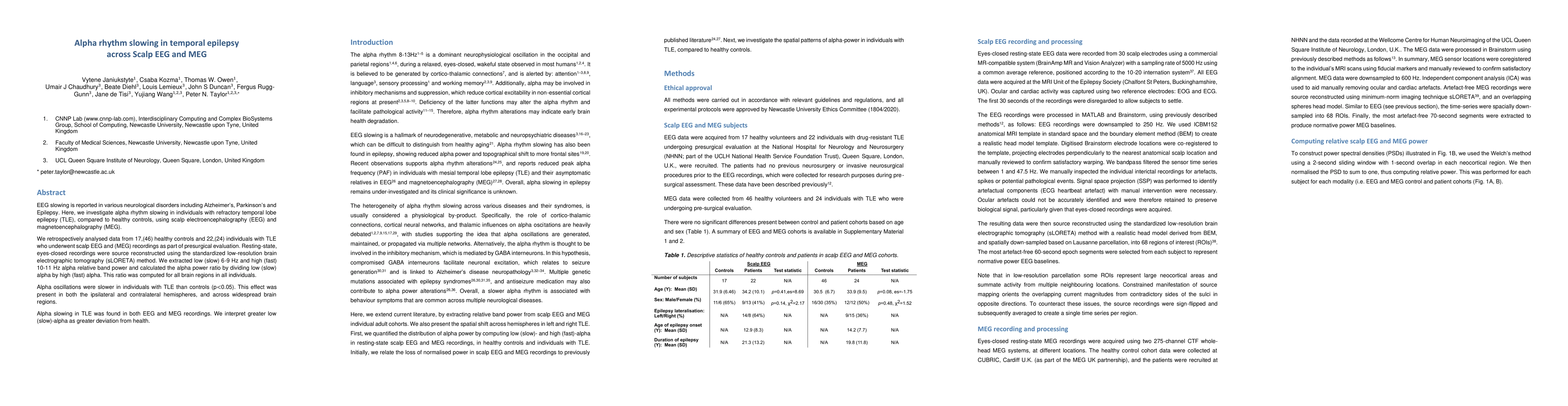
This study retrospectively analyzed data from 17 healthy controls and 22 individuals with refractory temporal lobe epilepsy (TLE) using scalp electroencephalography (EEG) and magnetoencephalography (MEG) recordings. Resting-state, eyes-closed recordings were source reconstructed using the standardized low-resolution brain electrographic tomography (sLORETA) method. The low (slow) 6-9 Hz and high (fast) 10-11 Hz alpha relative band power was extracted, and the alpha power ratio was computed by dividing low (slow) alpha by high (fast) alpha.
Key Results
- Alpha oscillations were slower in individuals with TLE than controls (p<0.05) in both the ipsilateral and contralateral hemispheres, and across widespread brain regions.
- Alpha slowing in TLE was found in both EEG and MEG recordings.
- Bilateral and widespread alpha slowing in TLE compared to controls was demonstrated, with a more pronounced effect seen in the left TLE on scalp EEG and in MEG for both left and right TLE.
Significance
This research reaffirms the alpha power shift to lower frequencies in TLE, supporting previous literature, and expands the epilepsy literature by showing significant alpha slowing in individuals with TLE compared to controls, and a forward spread of low-frequency (slow) alpha recorded with scalp EEG and MEG.
Technical Contribution
The study validates the reported alpha shift from higher to lower frequencies in TLE patients compared to healthy controls using both EEG and MEG, and demonstrates bilateral and widespread alpha slowing in TLE compared to controls.
Novelty
This research confirms and extends existing literature on alpha rhythm slowing in TLE by replicating findings across two independent cohorts with different modalities, and by illustrating the spatial alpha power shift from occipital to frontal regions.
Limitations
- Small sample sizes narrow the variability pool, and statistical significance should be interpreted with caution.
- Potential age effects on band power were not considered, as the sample consisted of adult subjects only.
- MEG data were collected at two different sites, which were not controlled for.
Future Work
- Investigate the relationship between alpha rhythm changes and anti-seizure medication (ASM) usage.
- Explore whether alpha rhythm slowing could be reversed in people with epilepsy and the implications of this.
Paper Details
PDF Preview
Key Terms
Citation Network
Current paper (gray), citations (green), references (blue)
Display is limited for performance on very large graphs.
Similar Papers
Found 4 papersNormative brain mapping using scalp EEG and potential clinical application
John S Duncan, Yujiang Wang, Vytene Janiukstyte et al.
Spatio-Temporal Adaptive Diffusion Models for EEG Super-Resolution in Epilepsy Diagnosis
Shuqiang Wang, Tong Zhou
No citations found for this paper.
Comments (0)